



Electrolyte imbalances and fluid status
Introduction
Electrolyte balance and fluid status are tightly linked to cardiac function, neurological integrity, renal perfusion, and cellular metabolism. Even mild electrolyte or fluid imbalances may contribute to clinical deterioration, especially in older adults, post-operative clients, and those receiving IV fluids or diuretics.
On the Next generation NCLEX (NGN), these concepts are frequently tested through trend analysis, prioritization, and clinical judgment, rather than isolated lab interpretation.
Learning objectives
By the end of this section, the learner should be able to:
- Identify common electrolyte imbalances and their clinical manifestations
- Interpret fluid status using labs, vital signs, and physical findings
- Recognize early vs late signs of imbalance
- Prioritize nursing actions based on severity and risk
Core concept: Fluid and electrolyte balance are interconnected
Fluid and electrolyte balance are closely interconnected. Changes in fluid volume frequently affect electrolyte concentrations.
Before interpreting electrolyte values, assess the client’s fluid status (fluid volume deficit, euvolemia, or fluid volume overload).
Regulation of fluid balance
Electrolyte and fluid disturbances are rarely random. They are usually the result of hormonal compensation mechanisms that become overwhelmed or dysregulated.
Key hormones
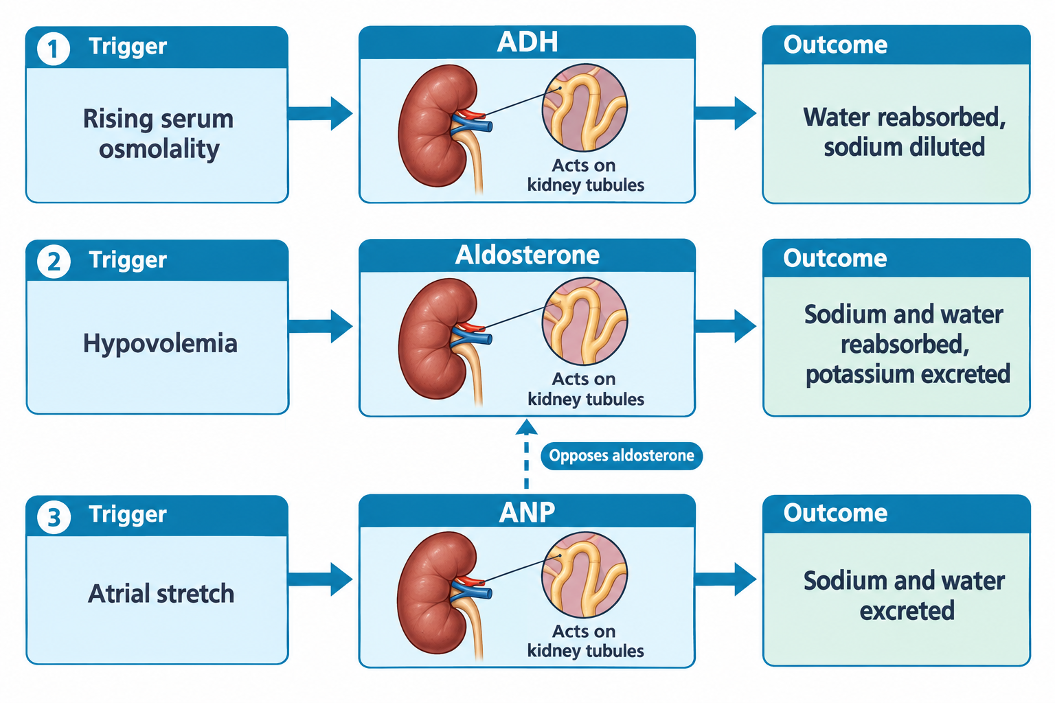
Antidiuretic hormone (ADH)
- Released when serum osmolality rises
- Increases water reabsorption in the kidneys
- Dilutes serum sodium concentration
- Excess ADH activity occurs in SIADH
Aldosterone
- Promotes sodium and water reabsorption
- Promotes potassium excretion
- Activated in hypovolemia
Atrial natriuretic peptide (ANP)
- Released when the atria stretch
- Promotes sodium and water excretion
- Opposes aldosterone
Fluid volume imbalances
Fluid volume deficit (dehydration)
Common causes
- Vomiting or diarrhea
- Poor oral intake
- Diuretics
- Hemorrhage
Assessment findings
- Dry mucous membranes
- Poor skin turgor (less reliable in older adults)
- Tachycardia
- Hypotension
- Decreased urine output
- Concentrated urine
Expected laboratory findings
- Elevated BUN, with a BUN-to-creatinine ratio above 20:1 (normally 10:1 to 20:1)
- Elevated hematocrit, from hemoconcentration
- Urine specific gravity above 1.020
- Serum osmolality above 295 mOsm/kg (normally 275 to 295)
Hematocrit is the exception in acute hemorrhage. Whole blood is lost, so the hematocrit can read normal at first and then fall as interstitial fluid shifts in or IV fluids are given. A normal hematocrit never rules out blood loss.
Fluid volume excess
Common causes
- Excessive IV fluids
- Heart failure
- Renal failure
- Cirrhosis
- Sodium retention
Assessment findings
- Edema
- Crackles in lungs
- Weight gain
- Jugular venous distention (JVD)
- Bounding pulses
- Elevated blood pressure
- Decreased oxygen saturation
Expected laboratory findings
- Decreased hematocrit, from hemodilution
- Decreased BUN, unless the overload is itself caused by kidney failure, where BUN and creatinine rise
- Urine specific gravity below 1.010
- Serum osmolality below 275 mOsm/kg
Sodium imbalances
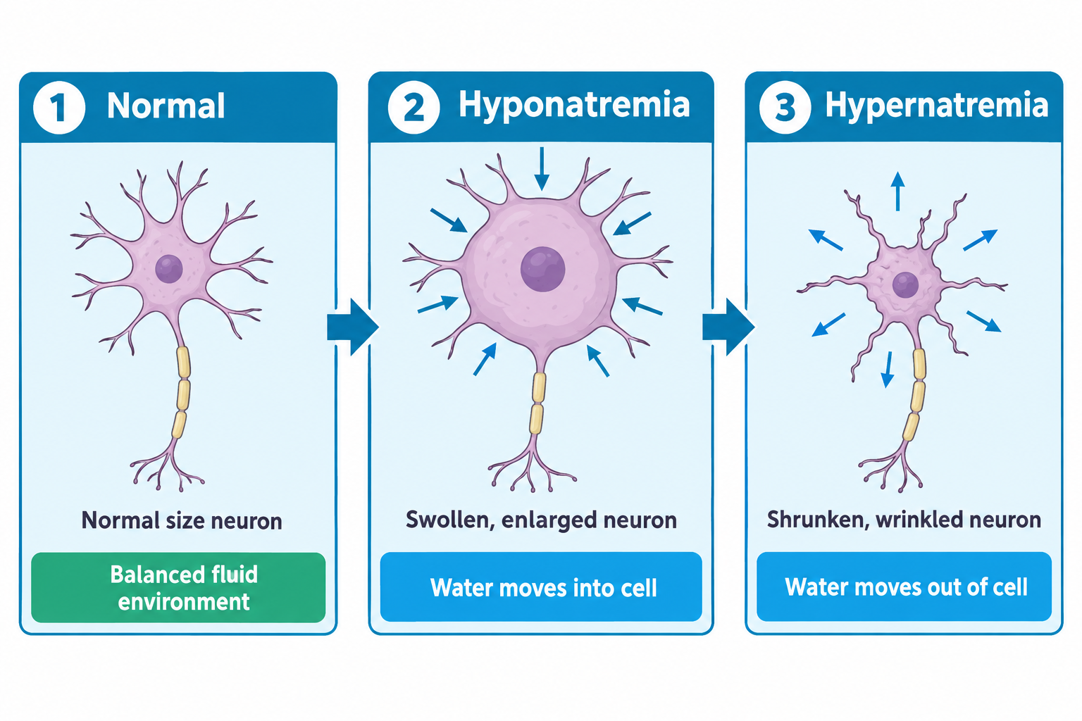
Hyponatremia (low sodium)
Common causes
- Excess free water
- SIADH
- Diuretics
- Heart failure
Key signs
- Headache
- Confusion
- Nausea
- Seizures (severe)
- Lethargy
Hypernatremia (high sodium)
Common causes
- Water loss (e.g., diarrhea, excessive sweating, high fever)
- Diabetes insipidus
- Excess sodium intake
Key signs
- Thirst
- Dry skin
- Restlessness
- Confusion
Potassium imbalances
Hypokalemia (low potassium)
Common causes
- Diuretics
- GI losses
- Insulin administration
Clinical manifestations
- Muscle weakness
- Ileus
- Cardiac dysrhythmias
Hyperkalemia (high potassium)
Common causes
- Renal failure
- Potassium-sparing diuretics
- Tissue breakdown
Clinical manifestations
- Muscle weakness
- Paresthesias
- Life-threatening dysrhythmias
Potassium & cardiac monitoring
Potassium imbalances are among the most dangerous electrolyte disturbances.
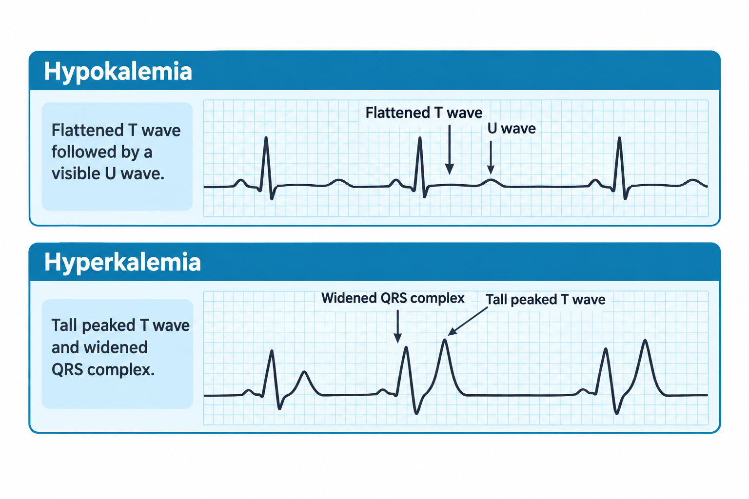
Hypokalemia ECG changes
- Flattened T waves
- Presence of U waves
- Increased risk for ventricular dysrhythmias
Hyperkalemia ECG changes
- Peaked T waves
- Widened QRS
- Risk for cardiac arrest
Acid-base implications
Electrolyte imbalances often occur alongside acid-base disorders. Most notable are potassium shifts with acid-base imbalances.
Potassium shifts opposite to pH:
- Acidosis → serum potassium rises (hyperkalemia)
- Alkalosis → serum potassium falls (hypokalemia)
Normal acid-base values
- pH: 7.35-7.45
- PaCO₂: 35-45 mmHg
- HCO₃⁻: 22-26 mEq/L
Calcium imbalances
Hypocalcemia
Signs
- Muscle spasms
- Tetany
- Positive Chvostek or Trousseau signs
Hypercalcemia
Signs
- Lethargy
- Constipation
- Kidney stones
- Bone pain
Magnesium imbalances (high-yield)
Hypomagnesemia
- Tremors
- Hyperreflexia
- Cardiac arrhythmias
Hypermagnesemia
- Hypotension
- Bradycardia
- Decreased deep tendon reflexes
- Respiratory depression
Medication interactions
Electrolyte shifts are frequently medication-induced. Some high-yield drug associations include:
- Loop diuretics → hypokalemia
- Thiazides → hyponatremia
- ACE inhibitors → hyperkalemia
- Corticosteroids → sodium retention
- Lithium → hyponatremia or sodium depletion increases the risk of lithium toxicity because the kidneys conserve sodium and reabsorb more lithium
Putting it together: lab values + assessment
NGN integration
- Labs confirm what the body is already showing
- Always assess mental status, heart rhythm, urine output, and lung sounds
Electrolyte questions often require choosing the most clinically dangerous imbalance, not the most abnormal number.
Clinical vignette 1
A post-operative client receiving IV fluids becomes confused. Labs show sodium of 128 mEq/L.
Nursing action: Perform a focused neurological assessment, notify the provider, implement prescribed treatment as ordered (e.g., fluid restriction for SIADH when appropriate), and monitor neurological status and serum sodium closely.
Rationale: Confusion with a serum sodium of 128 mEq/L suggests symptomatic hyponatremia with cerebral edema, requiring prompt evaluation and intervention.
Clinical vignette 2
A client with chronic kidney disease reports muscle weakness. ECG changes are noted, and potassium is 6.1 mEq/L.
Nursing action: Initiate continuous cardiac monitoring, notify the provider immediately, and anticipate prescribed treatment for hyperkalemia.
Rationale: Hyperkalemia with ECG changes places the client at high risk for life-threatening cardiac dysrhythmias.