


Achievable logoAchievable logo
CCMA
Sign in
Sign up
Purchase
Textbook
Practice exams
Support
How it works
Resources
Exam catalog
Mountain with a flag at the peak
Textbook
Introduction
1. Medical assistant
2. Electronic records
3. Medical terminology and anatomy
4. The fundamentals of infection control
5. Introduction to vital signs
6. The patient interview and history
7. The physical examination
8. Appointment scheduling
9. Insurance billing
10. Diagnostic coding and the ICD-10-CM System
11. Procedural coding
12. Medical billing and reimbursement essentials
13. Assisting with medical specialties
14. Assisting with the musculoskeletal system
15. Assisting with the cardiovascular system
16. Assisting with the respiratory system
17. Assisting with the nervous system
18. Anatomy and physiology of the urinary system
19. Assisting in obstetrics and gynecology
20. Assisting in endocrinology
21. Assisting in ophthalmology & otolaryngology
22. Assisting in gastroenterology
23. Assisting in the immune & lymphatic systems
24. Assisting in pediatrics: the developmental stages and care
25. The medical assistant’s role in caring for the older patient
26. The role of the medical assistant in physical therapy examination and assessment
27. Preparing for minor surgery: room, solutions, and supplies
27.1 Assisting in surgery
27.2 Surgical instruments: cutting and grasping
27.3 Surgical instruments: retracting, probing, and specialty
27.4 Drapes and sutures
27.5 Specialty instruments, needles, and wound closure
27.6 Autoclave loading/unloading, chemical sterilization, and professional responsibilities
27.7 Surgical asepsis, instrument care, and the utility room
27.8 The autoclave process: operation, wrapping, and quality assurance
27.9 Dressings, bandages, and professional conduct
27.10 Introduction to minor office surgery and technology
27.11 Specialized procedures and preoperative preparation
27.12 Sterile technique and intraoperative assistance
27.13 Completing surgery and postoperative care
27.14 Wound care and healing
28. Introduction to the clinical laboratory
29. Urinalysis
30. Blood collection
31. Analysis of blood
32. Electrocardiography and heart structure
33. The principles of pharmacology
34. Essential calculations and measurement systems
35. Solid, liquid, & solutions medication doses
36. Administering medications
37. Metabolism and core nutrient roles
38. Medical emergencies in the healthcare setting
Achievable logoAchievable logo
27.5 Specialty instruments, needles, and wound closure
Achievable CCMA
27. Preparing for minor surgery: room, solutions, and supplies

Specialty instruments, needles, and wound closure

10 min read
Font
Discuss
Share
Feedback

Ophthalmologic and otolaryngologic instruments

Instrument Description
Krause nasal snare
  • Wire loop at the tip that can be tightened
  • Used to remove polyps from the nares
Metal tongue depressor
  • Used to depress the tongue for oral examinations
Hartmann “alligator” ear forceps
  • 3-inch shaft; made in a variety of styles
  • Action of the jaw similar to that of an alligator’s jaws
  • Used to remove foreign bodies or polyps
Laryngeal mirror
  • May have a nonfogging surface
  • Used for examination of the larynx and postnasal area
Ivan laryngeal metal applicator
  • 6 to 9 inches long with curved end for use in throat or postnasal areas
  • Holds cotton in place with its roughened end; used to swab or sponge throat or postnasal tissue
  • Used to remove foreign bodies embedded in the pharynx
“Buck” ear curette
  • Available as disposable curettes in many different sizes
  • Has a stainless steel loop at the end
  • Made with sharp or blunt scraper ends
  • Used to remove foreign matter from the ear canals
Sharp ear dissector
  • Used to remove debris from the ear canal
Wilde ear forceps
  • Angled, with serrated tips
  • Provides easier access to the ear canal and nasal cavities
  • Used for packing after ear or nasal procedures
  • Can be used to remove foreign bodies

Biopsy instruments

Instrument Description
Cervical biopsy forceps
  • Available with or without teeth
  • Used to obtain cervical specimens for diagnostic examination
Rectal biopsy punch
  • Manufactured with interchangeable stems
  • Used through a proctoscope or sigmoidoscope
Silverman biopsy needle
  • Manufactured with a split cannula
  • Stylus is removed, and cannula is inserted to retrieve the specimen
  • Needle biopsy can eliminate the need for surgical incision

Needles

Surgical needles are chosen according to the area in which they are to be used and the depth and width of the desired suture. They are classified according to shape, which may be straight or curved. Most sutures are applied with curved needles because they allow the provider to penetrate the surface and then come up on the other side. The sharper the curve of the needle, the deeper the provider can pass it into the tissue. The point of a needle can be a taper or a cutting edge. A taper is used on delicate tissues. The cutting-edge needle is used on the skin. It lacerates the skin as the needle is passed through. This is advantageous for tougher tissues, such as connective tissue.

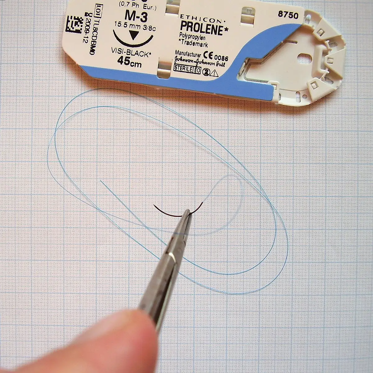
Needles are manufactured with the suture material attached, or swaged, to the needle. These atraumatic needles do not have an eyelet and cause the least amount of trauma as they are passed through the tissue. Manufacturers package suture strands with the suture needle attached in peel-apart sterile, disposable packages. These may be obtained as single, individually packed, or multipack sutures in a variety of needle types and sizes with a wide range of suture materials and lengths. The most common needle type for minor skin repair is the curved, cutting-edge, swaged needle.

Genitourinary instruments

Instrument Description
Foley catheter with inflated balloon
  • Manufactured in sizes 8 to 32 French with a double rubber lining toward the tip (each French unit is equal to about 0.33 mm, so the diameter in millimeters is the French size divided by 3; the higher the number, the larger the lumen)
  • After insertion, sterile solution is injected into the inner lining (inflating the balloon) to hold it in the bladder
  • Used as an indwelling catheter
Robinson (red rubber) catheter
  • Soft rubber urethral catheter in sizes 8 to 32 French
  • Inserted temporarily into the bladder for drainage or to obtain a specimen
Coudé-tip catheter
  • Slightly curved catheter tip
  • Designed to allow it to navigate past obstructions in the urinary tract, such as a swollen prostate in men
12-mL Luer-Lok syringe
  • Typically used to inject sterile saline into a catheter to inflate the balloon at the tip of an indwelling catheter
  • Used for injecting amounts greater than 5 mL
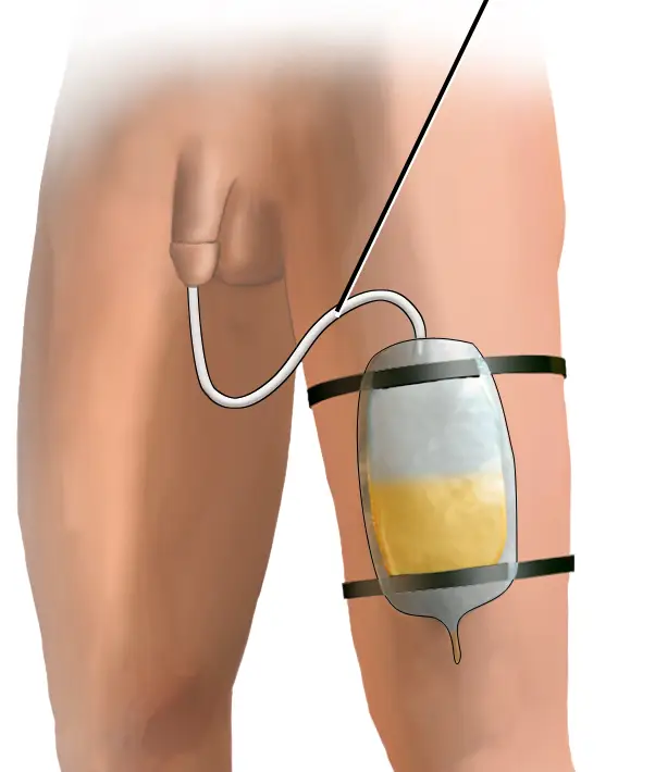
Foley catheter with drainage bag displayed
Foley bag
Wikimedia Commons
/
CC BY-SA 4.0

The clinical need drives which catheter is used: a Robinson catheter is chosen for brief, one-time drainage or to collect a specimen; a Foley catheter is chosen when the catheter must stay in place (indwelling) because its inflated balloon holds it in the bladder; and a Coudé-tip catheter is chosen when an obstruction, such as an enlarged prostate, blocks the path of a straight-tipped catheter.

Wound care with sutures

Nonabsorbable sutures that are on the face should be removed in 5 to 7 days, on the neck, 7 days; on the scalp, 10 days; on the trunk and upper extremities, 10 to 14 days; and on the lower extremities, 14 to 21 days. Sources vary on these intervals, so the provider’s order sets the removal date for each patient. The patient will need to have instructions on how to care for the site while the sutures are still in place. Those instructions should include the following guidelines:

  • Keep the area clean and dry for the first 24 to 48 hours after the sutures have been placed.
  • After 24 to 48 hours, gently wash around the site one to two times daily with cool water and soap. Do not wash or rub the sutures directly.
  • Dab the site dry. Do not rub the area, and avoid using the towel directly on the stitches.
  • If there was a bandage, replace it with a new clean bandage and antibiotic ointment if instructed to do so.
  • Return for suture removal as directed by the provider.

Other closure materials

Surgical staples can also be used for skin closure. They are made of stainless steel or titanium and are available in different sizes. Surgical staples usually remain in place for 7 to 14 days. They are applied and removed with specific staple instruments. Care for a surgical site closed with surgical staples is the same as the care for a site closed with sutures.

Suture packet and needle used for wound closure
Suture packet and needle
Wikimedia Commons
/
Public Domain

Another technique for wound closure is the use of adhesive skin closure strips, which are self-adhesive tapes that are placed over the wound, pulling the wound edges together. Using a tincture of benzoin prior to the application of adhesive skin closure strips can help them remain in place longer. A tincture of benzoin is applied parallel to the edges of the wound. Care should be taken so that it does not go into the wound because it can cause burning pain. The compound becomes tacky, and the adhesive skin strips stay on longer. Adhesive skin strips can be used to support a wound if there is potential tension at the site or for superficial wounds.

Frequently, small, clean lacerations may be closed with adhesive skin strips. These strips reduce the chance of infection and do not leave suture scars. Adhesive skin strips are used on areas of the body that are protected from movement and stress. They often are used on the face. A medical assistant can place adhesive skin strips as ordered by the provider. They are placed on the wound in the same sequence and at the same intervals as interrupted sutures and are left in place until they fall off or the wound heals. The patient can wash or shower with them in place but should take care to pat dry with a clean towel instead of rubbing.

Scope of practice: What a medical assistant may perform is set by state law and board rule and delegated by the supervising provider - there is no single national list. Whether a medical assistant may place adhesive skin strips, handle a given instrument, or assist with a closure is decided by the state’s rules and the provider’s order, so confirm both before beginning. See Standards of care for the full scope-of-practice framework.

Tissue adhesives, similar to glue, can also be used for superficial wounds. Examples of tissue adhesives are Histoacryl, Dermabond, and SurgiSeal. Tissue adhesives form a strong bond across wound edges, allowing normal healing to occur below. Skin adhesives are also an alternative to sutures. They are frequently used for the closure of facial lacerations, especially in pediatric patients, because they are effective for wound closure without requiring the use of needles and sutures.

Advantages of tissue adhesive

  • Saves time during wound repair and eliminates the need for suture removal
  • Creates a flexible, water-resistant coating that results in better cosmetic outcomes, since there’s no suture-entry scarring
  • May be used on larger wounds only after subcutaneous (deep) sutures relieve tension, so the adhesive closes just the skin surface

Sign up for free to take 12 quiz questions on this topic

Previous
Next  | 27.6 Autoclave loading/unloading, chemical sterilization, and professional responsibilities
All rights reserved ©2016 - 2026 Achievable, Inc.

Specialty instruments, needles, and wound closure

Ophthalmologic and otolaryngologic instruments

Instrument Description
Krause nasal snare
  • Wire loop at the tip that can be tightened
  • Used to remove polyps from the nares
Metal tongue depressor
  • Used to depress the tongue for oral examinations
Hartmann “alligator” ear forceps
  • 3-inch shaft; made in a variety of styles
  • Action of the jaw similar to that of an alligator’s jaws
  • Used to remove foreign bodies or polyps
Laryngeal mirror
  • May have a nonfogging surface
  • Used for examination of the larynx and postnasal area
Ivan laryngeal metal applicator
  • 6 to 9 inches long with curved end for use in throat or postnasal areas
  • Holds cotton in place with its roughened end; used to swab or sponge throat or postnasal tissue
  • Used to remove foreign bodies embedded in the pharynx
“Buck” ear curette
  • Available as disposable curettes in many different sizes
  • Has a stainless steel loop at the end
  • Made with sharp or blunt scraper ends
  • Used to remove foreign matter from the ear canals
Sharp ear dissector
  • Used to remove debris from the ear canal
Wilde ear forceps
  • Angled, with serrated tips
  • Provides easier access to the ear canal and nasal cavities
  • Used for packing after ear or nasal procedures
  • Can be used to remove foreign bodies

Biopsy instruments

Instrument Description
Cervical biopsy forceps
  • Available with or without teeth
  • Used to obtain cervical specimens for diagnostic examination
Rectal biopsy punch
  • Manufactured with interchangeable stems
  • Used through a proctoscope or sigmoidoscope
Silverman biopsy needle
  • Manufactured with a split cannula
  • Stylus is removed, and cannula is inserted to retrieve the specimen
  • Needle biopsy can eliminate the need for surgical incision

Needles

Surgical needles are chosen according to the area in which they are to be used and the depth and width of the desired suture. They are classified according to shape, which may be straight or curved. Most sutures are applied with curved needles because they allow the provider to penetrate the surface and then come up on the other side. The sharper the curve of the needle, the deeper the provider can pass it into the tissue. The point of a needle can be a taper or a cutting edge. A taper is used on delicate tissues. The cutting-edge needle is used on the skin. It lacerates the skin as the needle is passed through. This is advantageous for tougher tissues, such as connective tissue.

Needles are manufactured with the suture material attached, or swaged, to the needle. These atraumatic needles do not have an eyelet and cause the least amount of trauma as they are passed through the tissue. Manufacturers package suture strands with the suture needle attached in peel-apart sterile, disposable packages. These may be obtained as single, individually packed, or multipack sutures in a variety of needle types and sizes with a wide range of suture materials and lengths. The most common needle type for minor skin repair is the curved, cutting-edge, swaged needle.

Genitourinary instruments

Instrument Description
Foley catheter with inflated balloon
  • Manufactured in sizes 8 to 32 French with a double rubber lining toward the tip (each French unit is equal to about 0.33 mm, so the diameter in millimeters is the French size divided by 3; the higher the number, the larger the lumen)
  • After insertion, sterile solution is injected into the inner lining (inflating the balloon) to hold it in the bladder
  • Used as an indwelling catheter
Robinson (red rubber) catheter
  • Soft rubber urethral catheter in sizes 8 to 32 French
  • Inserted temporarily into the bladder for drainage or to obtain a specimen
Coudé-tip catheter
  • Slightly curved catheter tip
  • Designed to allow it to navigate past obstructions in the urinary tract, such as a swollen prostate in men
12-mL Luer-Lok syringe
  • Typically used to inject sterile saline into a catheter to inflate the balloon at the tip of an indwelling catheter
  • Used for injecting amounts greater than 5 mL

The clinical need drives which catheter is used: a Robinson catheter is chosen for brief, one-time drainage or to collect a specimen; a Foley catheter is chosen when the catheter must stay in place (indwelling) because its inflated balloon holds it in the bladder; and a Coudé-tip catheter is chosen when an obstruction, such as an enlarged prostate, blocks the path of a straight-tipped catheter.

Wound care with sutures

Nonabsorbable sutures that are on the face should be removed in 5 to 7 days, on the neck, 7 days; on the scalp, 10 days; on the trunk and upper extremities, 10 to 14 days; and on the lower extremities, 14 to 21 days. Sources vary on these intervals, so the provider’s order sets the removal date for each patient. The patient will need to have instructions on how to care for the site while the sutures are still in place. Those instructions should include the following guidelines:

  • Keep the area clean and dry for the first 24 to 48 hours after the sutures have been placed.
  • After 24 to 48 hours, gently wash around the site one to two times daily with cool water and soap. Do not wash or rub the sutures directly.
  • Dab the site dry. Do not rub the area, and avoid using the towel directly on the stitches.
  • If there was a bandage, replace it with a new clean bandage and antibiotic ointment if instructed to do so.
  • Return for suture removal as directed by the provider.

Other closure materials

Surgical staples can also be used for skin closure. They are made of stainless steel or titanium and are available in different sizes. Surgical staples usually remain in place for 7 to 14 days. They are applied and removed with specific staple instruments. Care for a surgical site closed with surgical staples is the same as the care for a site closed with sutures.

Another technique for wound closure is the use of adhesive skin closure strips, which are self-adhesive tapes that are placed over the wound, pulling the wound edges together. Using a tincture of benzoin prior to the application of adhesive skin closure strips can help them remain in place longer. A tincture of benzoin is applied parallel to the edges of the wound. Care should be taken so that it does not go into the wound because it can cause burning pain. The compound becomes tacky, and the adhesive skin strips stay on longer. Adhesive skin strips can be used to support a wound if there is potential tension at the site or for superficial wounds.

Frequently, small, clean lacerations may be closed with adhesive skin strips. These strips reduce the chance of infection and do not leave suture scars. Adhesive skin strips are used on areas of the body that are protected from movement and stress. They often are used on the face. A medical assistant can place adhesive skin strips as ordered by the provider. They are placed on the wound in the same sequence and at the same intervals as interrupted sutures and are left in place until they fall off or the wound heals. The patient can wash or shower with them in place but should take care to pat dry with a clean towel instead of rubbing.

Scope of practice: What a medical assistant may perform is set by state law and board rule and delegated by the supervising provider - there is no single national list. Whether a medical assistant may place adhesive skin strips, handle a given instrument, or assist with a closure is decided by the state’s rules and the provider’s order, so confirm both before beginning. See Standards of care for the full scope-of-practice framework.

Tissue adhesives, similar to glue, can also be used for superficial wounds. Examples of tissue adhesives are Histoacryl, Dermabond, and SurgiSeal. Tissue adhesives form a strong bond across wound edges, allowing normal healing to occur below. Skin adhesives are also an alternative to sutures. They are frequently used for the closure of facial lacerations, especially in pediatric patients, because they are effective for wound closure without requiring the use of needles and sutures.

Advantages of tissue adhesive

  • Saves time during wound repair and eliminates the need for suture removal
  • Creates a flexible, water-resistant coating that results in better cosmetic outcomes, since there’s no suture-entry scarring
  • May be used on larger wounds only after subcutaneous (deep) sutures relieve tension, so the adhesive closes just the skin surface

More from Preparing for minor surgery: room, solutions, and supplies

  • Assisting in surgery
  • Surgical instruments: cutting and grasping
  • Surgical instruments: retracting, probing, and specialty
  • Drapes and sutures
  • Autoclave loading/unloading, chemical sterilization, and professional responsibilities