Parvovirus
Parvovirus is the only single-stranded DNA virus. It’s transmitted by the respiratory route, transplacentally, or via blood. The virus replicates in the host cell nucleus.
Replication can occur only during the S phase of the cell cycle. For this reason, it doesn’t infect mature RBCs. Instead, it mainly infects erythroblasts and endothelial cells.
The virus requires the P blood antigen receptor (also known as globoside) to enter the cell. Rare individuals who lack the P antigen are immune to parvovirus B19 infection.
Deposition of immune complexes composed of virus particles and IgM or IgG occurs in endothelial cells and joints.
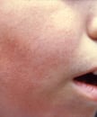
Clinical features: It causes erythema infectiosum, also called “slapped cheek disease.” It’s typically seen in children. It presents with fever, malaise, headache, myalgia, nausea, and rhinorrhea, followed by a bright red macular exanthem on the cheeks that’s often associated with circumoral pallor. A diffuse maculopapular rash can appear 1-4 days later and fade to a lacy erythematous rash, which may be pruritic and gradually spread toward the distal extremities.
Slapped cheek rash
In patients with hemoglobinopathies or hemolytic anemias, parvovirus may precipitate aplastic crisis. The bone marrow reveals an absence of erythroid precursors and the presence of striking giant pronormoblasts.
Cases of immune thrombocytopenic purpura, Henoch-Schönlein purpura, and hemophagocytic syndrome have been attributed to parvovirus B19. It may also cause polyarthropathy syndrome, mainly affecting the small joints of the hands and feet and mimicking rheumatoid arthritis.
Intrauterine infections may cause hydrops fetalis. This occurs when a nonimmune woman is infected, usually in the first 20 weeks of pregnancy. There is generalized swelling of the fetus from heart failure secondary to severe anemia from parvovirus infection.
Diagnosis of parvovirus infections: IgG and IgM antibodies can be detected by ELISA, RIA, or immunofluorescence. DNA hybridization or PCR can be used for viral detection. PCR of amniotic fluid can be done in fetal hydrops fetalis.