


This browser is too old to run Achievable.

Update it, or open this page in a current version of Chrome, Safari, Firefox or Edge. On an iPhone or iPad, update iOS: every browser there runs on the version of Safari that comes with it.

Achievable logoAchievable logo
NPTE-PTA
Sign in
Sign up
Purchase
Textbook
Practice exams
Support
How it works
Resources
Exam catalog
Mountain with a flag at the peak
Textbook
Introduction
1. Cardiovascular system
2. Pulmonary system
2.1 Anatomy, ventilation, and pulmonary function tests
2.2 Auscultation and differential diagnosis of pulmonary system, lines and tubes
2.3 Understanding arterial blood gases
2.4 Interventions for pulmonary system
3. Neuromuscular system
4. Pediatrics
5. Musculoskeletal system
6. Other systems
7. Non-systems
Wrapping up
Achievable logoAchievable logo
2.4 Interventions for pulmonary system
Achievable NPTE-PTA
2. Pulmonary system

Interventions for pulmonary system

11 min read
Font
Discuss
Share
Feedback

Clinical safety note: For every technique in this chapter, the PTA carries out the plan of care established by the supervising PT - collecting data, recognizing a change in the patient’s status, and communicating findings to the PT rather than independently evaluating or modifying the plan of care. Monitor dyspnea or RPE during exercise, and stop the intervention and notify the supervising PT if the patient shows an abnormal vital sign response, new or worsening chest pain, increased dyspnea, hemoptysis, or a drop in oxygen saturation (SpO2 below about 88-90%).

Activity pacing and energy conservation

  • Pacing
    • Rationale: breathing technique used to spread the metabolic demands of an activity to improve performance
      • Example: An individual running on a treadmill attempting not to become short of breath with the activity
    • How to perform:
      • Break the activity into manageable components
      • Inhale at rest or during eccentric (easier) activation
      • Exhale through pursed lips (to slow breathing) during concentric (exertion) activation
      • Repeat until the activity is complete
  • Energy conservation
    • Rationale: strategic planning of normal activities during the day to lower their energy cost, reducing dyspnea and fatigue. This is used primarily with individuals who have a pulmonary disorder, such as COPD, though it can benefit others as well.
    • How to perform:
      • Recognize activities that are performed with ease and those with increased difficulty
      • Schedule the most demanding tasks for the time of day when energy is highest, alternate heavy and light tasks, plan rest breaks, and simplify or sit for tasks where possible

Breathing exercises for pulmonary function

Breathing exercises assist with improving ventilation through the facilitation of the muscles of respiration.

  • Diaphragmatic breathing
    • Rationale for use: increased ventilation, decreased work of breathing, and increased relaxation
    • Patient population: individuals with restrictive or obstructive lung disease, postoperative patients
    • How to perform:
      • Position the patient in semi-Fowler’s position
      • Place a hand over the rectus abdominis just below the anterior costal margin
      • Have the patient inhale slowly through the nose so the abdomen rises against the hand while the upper chest stays relaxed, then exhale slowly
      • Apply light resistance with the hand during inhalation, progressing the resistance over successive cycles
      • Progress from semi-Fowler’s position to sitting and standing
  • Segmental breathing
    • Rationale: increased ventilation to a segment of hypoventilated lung, maintain or improve functional residual capacity, or prevent pulmonary compromise
    • Patient population: individuals with immobility of part of the thorax, such as from rib fractures, thorax incisions, or partial lung removal
    • How to perform:
      • Position the individual to provide access to the lung segment, such as postural drainage positions (see later in this chapter)
      • Apply firm hand pressure over the segment at the end of exhalation
      • Ask the patient to breathe in against the hand, lightening the pressure during inhalation so the segment can fully expand
      • Encourage a 2-3 second hold at the end of inspiration
  • Pursed lip breathing
    • Rationale: provide pressure support to allow for bronchioles to remain dilated during exhalation, decrease respiratory rate, and improve gas exchange for those with COPD
    • Patient population: primarily used with individuals with obstructive lung disease
    • How to perform:
      • Inhale through the nose
      • Exhale slowly through pursed lips

Airway clearance techniques

The goal of airway clearance techniques is to remove secretions from the pulmonary system to allow for adequate oxygenation. Can be performed independently or with assistance.

  • Cough
    • Rationale: remove secretions from all the major and central airways
      • Performed independently by the patient
  • Assisted cough
    • Rationale: used when the patient’s abdominal muscles cannot generate sufficient pressure to aid in effective cough
    • How to perform:
      • Individual positioned supine or semi-Fowler’s with hand placement at the subcostal angle
      • Individual inhales
      • During exhalation, the hand is forcefully pushed up and in to aid in rapid exhalation, thus causing a cough
  • Huffing
    • Rationale: forced expiration with an open glottis moves secretions while causing less airway collapse than a cough; useful in COPD, cystic fibrosis, and postoperative pain
    • How to perform:
      • The individual inhales deeply
      • Upon exhalation, the individual then says “ha ha” to allow the airways to remain open and remove secretions
  • Endotracheal suctioning
    • Rationale: typically utilized when a patient is unable to perform an independent cough, an assisted cough, or huffing to provide airway clearance. Likely to perform when the patient is on mechanical ventilation
    • How to perform: A suctioning catheter is used to remove secretions either through an artificial airway, oral airway, or nares for 10-15 seconds

Independent secretion removal techniques

The goal of independent secretion removal techniques is to remove secretions from the pulmonary system for those diagnosed with chronic pulmonary diseases. These techniques are all performed independently by the patient.

  • Active cycle of breathing technique
    • Rationale: removal of peripheral secretions for those in which a combination of techniques must be utilized to remove secretions
    • Patient population: Obstructive lung disease
    • How to perform:
      • An individual performs diaphragmatic breathing
      • Followed by thoracic expansion exercises
      • Lastly, forced expiration with a series of huffing
      • Repeat cycle until the airway is clear
  • Autogenic drainage
    • Rationale: moves peripheral secretions toward the central airways by controlling the rate, depth, and lung volume of breathing, without the airway compression and irritation of repeated forceful coughing
    • How to perform:
      • Unstick phase: breathe at low lung volume (exhaling into the expiratory reserve volume), with a 2-3 second hold at the end of each inhalation, to loosen mucus in the peripheral airways
      • Collect phase: breathe at low-to-mid lung volume (around tidal volume) to gather the loosened secretions in the middle airways
      • Evacuation phase: breathe at mid-to-high lung volume (into the inspiratory reserve volume) to move secretions into the central airways, then clear them with a high-lung-volume huff; coughing is held back and used only if a huff does not clear them
      • Repeat these cycles until all secretions are removed
  • Flutter or Acapella device (oscillatory PEP)
    • Rationale: an oscillatory positive expiratory pressure (PEP) device that provides intermittent positive airway support and airway vibration to allow for adequate secretion clearance
    • How to perform:
      • Inhale through the nose or around the device
      • Exhale through the device
      • Perform huffing or coughing as appropriate
      • Repeat until all secretions are removed
  • Low-pressure positive expiratory pressure mask
    • Rationale: prevent lung collapse by providing a low amount of pressure to allow for adequate secretion removal in diagnoses of COPD, cystic fibrosis, or bronchiectasis
    • How to perform:
      • Seated or supine, a mask is placed over the face with inspiration hold for 2-3 seconds before expiration (~10 breaths)
      • Remove the mask for coughing or huffing
      • Repeat until all secretions are removed
  • High-pressure positive expiratory pressure mask
    • Rationale: splint unstable, collapsible airways open during a forced expiration against a high resistance (about 60 cm H2O on average in the original method) to allow for adequate secretion removal; developed for cystic fibrosis
    • Not a treatment for acute respiratory failure or pulmonary edema, which are managed medically with positive airway pressure such as CPAP or noninvasive ventilation
    • How to perform:
      • Seated or supine with normal breathing performed with a mask in place (~10 breaths)
      • Forced expirations (huffs) are performed through the mask against the resistance
      • Repeat until all secretions are removed

Other pulmonary interventions

  • Incentive spirometry
    • Rationale: visual feedback device that encourages a slow, deep, sustained inhalation to prevent or reverse atelectasis, commonly used after thoracic or abdominal surgery
    • How to perform: seated upright, exhale normally, then inhale slowly through the mouthpiece to raise the indicator to the target volume, hold for at least 3-5 seconds, then exhale; repeat 5-10 times per hour while awake
  • Inspiratory muscle training
    • Rationale: strengthens the diaphragm and accessory inspiratory muscles using a handheld resistive or threshold device, improving ventilatory endurance in individuals with COPD or neuromuscular weakness
    • How to perform: inhale against a set resistance for a prescribed number of breaths or minutes, progressing the resistance as tolerated

Manual secretion removal techniques

The goal of manual secretion removal techniques is to remove secretions from the pulmonary system for those diagnosed with acute pulmonary disease, from mucus plugs, respiratory failure, and on mechanical ventilation, weakness of the respiratory system without neurological origin, or acute exacerbations of cystic fibrosis.

  • Postural bronchial drainage
    • Rationale: provides removal of secretions by positioning the lung (i.e., lung segments) to be assisted by gravity to remove secretions
    • How to perform:
      • Specific to the lung segment
Postural drainage positions
Achievable
  • Shaking (vibration)
    • Rationale: used in conjunction with postural drainage positions to aid in the removal of secretions from the tracheobronchial tree
    • How to perform:
      • Place in appropriate postural drainage position
      • The individual inhales, and the therapist’s hands are placed over the appropriate segment parallel to the ribs
      • At exhalation, the therapist provides a fast, rhythmic bouncing motion to the ribcage at the frequency and duration of the patient’s pulmonary needs
  • Percussion
    • Rationale: used in conjunction with postural drainage positions to loosen and mobilize secretions in the lungs.
      • Chest physiotherapy such as percussion is indicated for patients whose cough is insufficient to clear thick, tenacious, copious, or loculated secretions.
      • Percussion is typically performed at a rate of 100 to 480 beats per minute (BPM), commonly around 150 to 200 BPM (about 2.5 to 3.3 claps per second).
    • How to perform:
      • Place in appropriate postural drainage position
      • Place a barrier between the hand and the segment that will be percussed
      • Percuss over the identified segment with a cupped hand by the therapist at a rhythmic rate. Percussion is applied during inhalation and exhalation at the frequency determined by the patient’s pulmonary needs.
Definitions
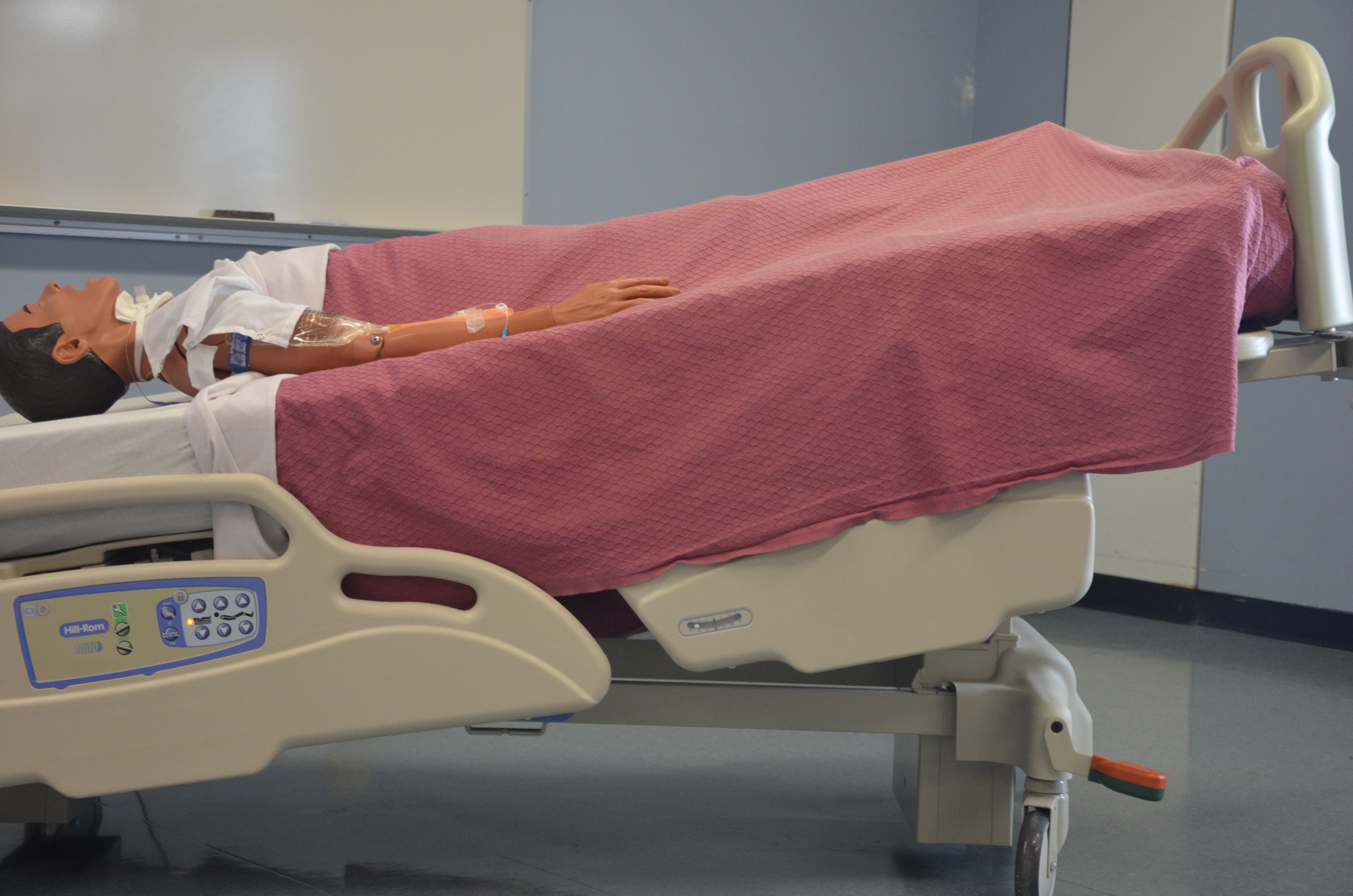
Trendelenburg
The Trendelenburg position, where a patient lies head-down, is used in postural drainage to help clear secretions from the lower parts of the lungs using gravity.
Trendelenburg position
Glynda Rees Doyle and Jodie Anita McCutcheon
/
Wikimedia Commons
/
CC BY 4.0

Precautions for postural drainage positions

  • Placement in Trendelenburg position
    • Pulmonary edema
    • Congestive heart failure
    • Obesity
    • Recent food consumption
    • Recent neurosurgery
    • Increased intracranial pressure <20 mmHg
    • Shortness of breath
    • Esophageal surgery
    • General discomfort from the position
  • Placement in side-lying
    • Axillo-femoral graft
    • Humeral fractures
    • Arthritis
    • Shoulder or hip bursitis
    • General discomfort from the position

Contraindications for all postural drainage positions

  • Active hemorrhage
  • Intracranial pressure >20 mmHg
  • Embolism
  • Large pleural effusion
  • Bullous emphysema
  • Recent spinal surgery
  • Non-stabilized head or neck injury

Precautions for percussion and shaking

  • Pain increased with the techniques
  • Decreased platelet count
  • Increased clotting time - on anti-coagulation medication
  • Fractured rib
  • Degenerative bone disease
  • Bone metastases

Do not percuss directly over a rib fracture, bone metastasis, or recent incision; report these findings to the supervising PT before treatment.

Pulmonary medications

  • Bronchodilators
    • Mechanism of action: medications that assist with improving ventilation by providing relaxation of bronchioles (bronchodilation) in either short-acting or long acting
    • Appropriate diagnoses: asthma, chronic obstructive pulmonary disease
    • Physical therapy considerations: trembling, palpitations, headache, muscle cramps
    • Physical therapy management: schedule airway clearance or exercise shortly after a dose, when the airways are most open; make sure a rescue inhaler is available during exercise; monitor heart rate, since tachycardia is possible
    • Common name: albuterol
  • Expectorants
    • Mechanism of action: increases the volume and hydration of respiratory secretions, thinning the mucus and making cough more productive
    • Appropriate diagnoses: pneumonia, flu, cold, cystic fibrosis, and any other diagnoses that produce increased mucus
    • Physical therapy considerations: rash, difficulty breathing, dizziness
    • Physical therapy management: schedule airway clearance techniques for when secretions have been mobilized by the medication
    • Common name: Guaifenesin
  • Corticosteroids
    • Mechanism of action: an anti-inflammatory that reduces the inflammation that is present in the bronchial lining
    • Appropriate diagnoses: chronic obstructive pulmonary disease (COPD), asthma, interstitial lung disease (ILD), sarcoidosis, and severe acute respiratory illness.
    • Physical therapy considerations: typically, long-term use can cause osteoporosis, increased blood sugar, increased risk for infections, and development of glaucoma or cataract
    • Physical therapy management: watch for steroid myopathy and fragile skin, check blood glucose in patients with diabetes, and use fall and fracture precautions given the osteoporosis risk
    • Common names: prednisone (oral), the main source of the long-term effects above; inhaled corticosteroids such as fluticasone and budesonide, used for daily control, have far fewer systemic effects but can cause hoarseness and oral thrush, so the patient rinses the mouth after each dose

Safety and PTA role

  • PTA implements PT’s plan of care: collects data, recognizes status changes, reports to PT
  • Stop intervention/notify PT for: abnormal vital signs, new/worsening chest pain, increased dyspnea, hemoptysis, SpO2 drop (~below 88-90%)

Pacing and energy conservation

  • Pacing: break activity into parts, inhale at rest/eccentric phase, exhale pursed-lips during exertion
  • Energy conservation (esp. COPD): schedule demanding tasks during high-energy times, alternate heavy/light tasks, plan rest breaks

Breathing exercises

  • Diaphragmatic breathing: semi-Fowler’s position, hand below costal margin, abdomen rises on inhale; used for restrictive/obstructive disease, postoperative patients
  • Segmental breathing: targets hypoventilated lung segment via hand pressure released on inhalation; used for thoracic immobility (rib fractures, incisions)
  • Pursed lip breathing: inhale nose, exhale through pursed lips; keeps bronchioles open, mainly for COPD

Airway clearance techniques

  • Cough (independent), assisted cough (manual pressure at subcostal angle during exhalation for weak abdominals)
  • Huffing: forced exhale saying “ha ha,” keeps airway open, less collapse than cough; used in COPD, cystic fibrosis, post-op
  • Endotracheal suctioning: for patients unable to cough/huff, often on ventilation; 10-15 second catheter suction

Independent secretion removal techniques

  • Active cycle of breathing: diaphragmatic breathing → thoracic expansion → huffing, repeated until clear (obstructive disease)
  • Autogenic drainage: three phases (unstick→collect→evacuate) controlling breath volume/location to move secretions centrally
  • Flutter/Acapella (oscillatory PEP): inhale around device, exhale through it, creates vibration for clearance
  • Low-pressure PEP mask: ~10 breaths with inspiratory hold, for COPD/CF/bronchiectasis
  • High-pressure PEP mask: forced exhalation against high resistance (~60 cm H2O) to splint open airways; not for acute respiratory failure/pulmonary edema

Other pulmonary interventions

  • Incentive spirometry: slow deep sustained inhale to target volume, hold 3-5 sec, prevents/reverses atelectasis post-surgery; 5-10 reps/hour
  • Inspiratory muscle training: breathing against resistance device, strengthens diaphragm/accessory muscles, progressive resistance

Manual secretion removal techniques

  • Postural drainage: gravity-assisted positioning by lung segment (includes Trendelenburg for lower lobes)
  • Shaking/vibration: rhythmic bouncing on exhale in drainage position
  • Percussion: cupped-hand clapping over segment (~100-480 BPM, commonly 150-200) during in/exhalation, with barrier over skin

Precautions/contraindications for postural drainage & percussion

  • Trendelenburg precautions: pulmonary edema, CHF, obesity, recent eating, neurosurgery, increased ICP <20mmHg, dyspnea
  • Side-lying precautions: axillo-femoral graft, humeral fractures, arthritis, shoulder/hip bursitis
  • Absolute contraindications: active hemorrhage, ICP >20mmHg, embolism, large pleural effusion, bullous emphysema, recent spinal surgery, unstable head/neck injury
  • Percussion/shaking precautions: increased pain, low platelets, anticoagulation, rib fracture, bone disease/metastases — never percuss directly over fracture, metastasis, or recent incision

Pulmonary medications

  • Bronchodilators (e.g., albuterol): relax bronchioles; time exercise/airway clearance soon after dose; watch for tachycardia, tremor
  • Expectorants (e.g., Guaifenesin): thin mucus for productive cough; schedule clearance techniques after secretions mobilize
  • Corticosteroids (e.g., prednisone): reduce airway inflammation; long-term risks include osteoporosis, hyperglycemia, infection, myopathy — use fall/fracture precautions; inhaled forms (fluticasone, budesonide): hoarseness, oral thrush—rinse mouth

Sign up for free to take 28 quiz questions on this topic

Previous
Next  | 3.1 Central nervous system
All rights reserved ©2016 - 2026 Achievable, Inc.

Interventions for pulmonary system

Clinical safety note: For every technique in this chapter, the PTA carries out the plan of care established by the supervising PT - collecting data, recognizing a change in the patient’s status, and communicating findings to the PT rather than independently evaluating or modifying the plan of care. Monitor dyspnea or RPE during exercise, and stop the intervention and notify the supervising PT if the patient shows an abnormal vital sign response, new or worsening chest pain, increased dyspnea, hemoptysis, or a drop in oxygen saturation (SpO2 below about 88-90%).

Activity pacing and energy conservation

  • Pacing
    • Rationale: breathing technique used to spread the metabolic demands of an activity to improve performance
      • Example: An individual running on a treadmill attempting not to become short of breath with the activity
    • How to perform:
      • Break the activity into manageable components
      • Inhale at rest or during eccentric (easier) activation
      • Exhale through pursed lips (to slow breathing) during concentric (exertion) activation
      • Repeat until the activity is complete
  • Energy conservation
    • Rationale: strategic planning of normal activities during the day to lower their energy cost, reducing dyspnea and fatigue. This is used primarily with individuals who have a pulmonary disorder, such as COPD, though it can benefit others as well.
    • How to perform:
      • Recognize activities that are performed with ease and those with increased difficulty
      • Schedule the most demanding tasks for the time of day when energy is highest, alternate heavy and light tasks, plan rest breaks, and simplify or sit for tasks where possible

Breathing exercises for pulmonary function

Breathing exercises assist with improving ventilation through the facilitation of the muscles of respiration.

  • Diaphragmatic breathing
    • Rationale for use: increased ventilation, decreased work of breathing, and increased relaxation
    • Patient population: individuals with restrictive or obstructive lung disease, postoperative patients
    • How to perform:
      • Position the patient in semi-Fowler’s position
      • Place a hand over the rectus abdominis just below the anterior costal margin
      • Have the patient inhale slowly through the nose so the abdomen rises against the hand while the upper chest stays relaxed, then exhale slowly
      • Apply light resistance with the hand during inhalation, progressing the resistance over successive cycles
      • Progress from semi-Fowler’s position to sitting and standing
  • Segmental breathing
    • Rationale: increased ventilation to a segment of hypoventilated lung, maintain or improve functional residual capacity, or prevent pulmonary compromise
    • Patient population: individuals with immobility of part of the thorax, such as from rib fractures, thorax incisions, or partial lung removal
    • How to perform:
      • Position the individual to provide access to the lung segment, such as postural drainage positions (see later in this chapter)
      • Apply firm hand pressure over the segment at the end of exhalation
      • Ask the patient to breathe in against the hand, lightening the pressure during inhalation so the segment can fully expand
      • Encourage a 2-3 second hold at the end of inspiration
  • Pursed lip breathing
    • Rationale: provide pressure support to allow for bronchioles to remain dilated during exhalation, decrease respiratory rate, and improve gas exchange for those with COPD
    • Patient population: primarily used with individuals with obstructive lung disease
    • How to perform:
      • Inhale through the nose
      • Exhale slowly through pursed lips

Airway clearance techniques

The goal of airway clearance techniques is to remove secretions from the pulmonary system to allow for adequate oxygenation. Can be performed independently or with assistance.

  • Cough
    • Rationale: remove secretions from all the major and central airways
      • Performed independently by the patient
  • Assisted cough
    • Rationale: used when the patient’s abdominal muscles cannot generate sufficient pressure to aid in effective cough
    • How to perform:
      • Individual positioned supine or semi-Fowler’s with hand placement at the subcostal angle
      • Individual inhales
      • During exhalation, the hand is forcefully pushed up and in to aid in rapid exhalation, thus causing a cough
  • Huffing
    • Rationale: forced expiration with an open glottis moves secretions while causing less airway collapse than a cough; useful in COPD, cystic fibrosis, and postoperative pain
    • How to perform:
      • The individual inhales deeply
      • Upon exhalation, the individual then says “ha ha” to allow the airways to remain open and remove secretions
  • Endotracheal suctioning
    • Rationale: typically utilized when a patient is unable to perform an independent cough, an assisted cough, or huffing to provide airway clearance. Likely to perform when the patient is on mechanical ventilation
    • How to perform: A suctioning catheter is used to remove secretions either through an artificial airway, oral airway, or nares for 10-15 seconds

Independent secretion removal techniques

The goal of independent secretion removal techniques is to remove secretions from the pulmonary system for those diagnosed with chronic pulmonary diseases. These techniques are all performed independently by the patient.

  • Active cycle of breathing technique
    • Rationale: removal of peripheral secretions for those in which a combination of techniques must be utilized to remove secretions
    • Patient population: Obstructive lung disease
    • How to perform:
      • An individual performs diaphragmatic breathing
      • Followed by thoracic expansion exercises
      • Lastly, forced expiration with a series of huffing
      • Repeat cycle until the airway is clear
  • Autogenic drainage
    • Rationale: moves peripheral secretions toward the central airways by controlling the rate, depth, and lung volume of breathing, without the airway compression and irritation of repeated forceful coughing
    • How to perform:
      • Unstick phase: breathe at low lung volume (exhaling into the expiratory reserve volume), with a 2-3 second hold at the end of each inhalation, to loosen mucus in the peripheral airways
      • Collect phase: breathe at low-to-mid lung volume (around tidal volume) to gather the loosened secretions in the middle airways
      • Evacuation phase: breathe at mid-to-high lung volume (into the inspiratory reserve volume) to move secretions into the central airways, then clear them with a high-lung-volume huff; coughing is held back and used only if a huff does not clear them
      • Repeat these cycles until all secretions are removed
  • Flutter or Acapella device (oscillatory PEP)
    • Rationale: an oscillatory positive expiratory pressure (PEP) device that provides intermittent positive airway support and airway vibration to allow for adequate secretion clearance
    • How to perform:
      • Inhale through the nose or around the device
      • Exhale through the device
      • Perform huffing or coughing as appropriate
      • Repeat until all secretions are removed
  • Low-pressure positive expiratory pressure mask
    • Rationale: prevent lung collapse by providing a low amount of pressure to allow for adequate secretion removal in diagnoses of COPD, cystic fibrosis, or bronchiectasis
    • How to perform:
      • Seated or supine, a mask is placed over the face with inspiration hold for 2-3 seconds before expiration (~10 breaths)
      • Remove the mask for coughing or huffing
      • Repeat until all secretions are removed
  • High-pressure positive expiratory pressure mask
    • Rationale: splint unstable, collapsible airways open during a forced expiration against a high resistance (about 60 cm H2O on average in the original method) to allow for adequate secretion removal; developed for cystic fibrosis
    • Not a treatment for acute respiratory failure or pulmonary edema, which are managed medically with positive airway pressure such as CPAP or noninvasive ventilation
    • How to perform:
      • Seated or supine with normal breathing performed with a mask in place (~10 breaths)
      • Forced expirations (huffs) are performed through the mask against the resistance
      • Repeat until all secretions are removed

Other pulmonary interventions

  • Incentive spirometry
    • Rationale: visual feedback device that encourages a slow, deep, sustained inhalation to prevent or reverse atelectasis, commonly used after thoracic or abdominal surgery
    • How to perform: seated upright, exhale normally, then inhale slowly through the mouthpiece to raise the indicator to the target volume, hold for at least 3-5 seconds, then exhale; repeat 5-10 times per hour while awake
  • Inspiratory muscle training
    • Rationale: strengthens the diaphragm and accessory inspiratory muscles using a handheld resistive or threshold device, improving ventilatory endurance in individuals with COPD or neuromuscular weakness
    • How to perform: inhale against a set resistance for a prescribed number of breaths or minutes, progressing the resistance as tolerated

Manual secretion removal techniques

The goal of manual secretion removal techniques is to remove secretions from the pulmonary system for those diagnosed with acute pulmonary disease, from mucus plugs, respiratory failure, and on mechanical ventilation, weakness of the respiratory system without neurological origin, or acute exacerbations of cystic fibrosis.

  • Postural bronchial drainage
    • Rationale: provides removal of secretions by positioning the lung (i.e., lung segments) to be assisted by gravity to remove secretions
    • How to perform:
      • Specific to the lung segment
  • Shaking (vibration)
    • Rationale: used in conjunction with postural drainage positions to aid in the removal of secretions from the tracheobronchial tree
    • How to perform:
      • Place in appropriate postural drainage position
      • The individual inhales, and the therapist’s hands are placed over the appropriate segment parallel to the ribs
      • At exhalation, the therapist provides a fast, rhythmic bouncing motion to the ribcage at the frequency and duration of the patient’s pulmonary needs
  • Percussion
    • Rationale: used in conjunction with postural drainage positions to loosen and mobilize secretions in the lungs.
      • Chest physiotherapy such as percussion is indicated for patients whose cough is insufficient to clear thick, tenacious, copious, or loculated secretions.
      • Percussion is typically performed at a rate of 100 to 480 beats per minute (BPM), commonly around 150 to 200 BPM (about 2.5 to 3.3 claps per second).
    • How to perform:
      • Place in appropriate postural drainage position
      • Place a barrier between the hand and the segment that will be percussed
      • Percuss over the identified segment with a cupped hand by the therapist at a rhythmic rate. Percussion is applied during inhalation and exhalation at the frequency determined by the patient’s pulmonary needs.
Definitions
Trendelenburg
The Trendelenburg position, where a patient lies head-down, is used in postural drainage to help clear secretions from the lower parts of the lungs using gravity.

Precautions for postural drainage positions

  • Placement in Trendelenburg position
    • Pulmonary edema
    • Congestive heart failure
    • Obesity
    • Recent food consumption
    • Recent neurosurgery
    • Increased intracranial pressure <20 mmHg
    • Shortness of breath
    • Esophageal surgery
    • General discomfort from the position
  • Placement in side-lying
    • Axillo-femoral graft
    • Humeral fractures
    • Arthritis
    • Shoulder or hip bursitis
    • General discomfort from the position

Contraindications for all postural drainage positions

  • Active hemorrhage
  • Intracranial pressure >20 mmHg
  • Embolism
  • Large pleural effusion
  • Bullous emphysema
  • Recent spinal surgery
  • Non-stabilized head or neck injury

Precautions for percussion and shaking

  • Pain increased with the techniques
  • Decreased platelet count
  • Increased clotting time - on anti-coagulation medication
  • Fractured rib
  • Degenerative bone disease
  • Bone metastases

Do not percuss directly over a rib fracture, bone metastasis, or recent incision; report these findings to the supervising PT before treatment.

Pulmonary medications

  • Bronchodilators
    • Mechanism of action: medications that assist with improving ventilation by providing relaxation of bronchioles (bronchodilation) in either short-acting or long acting
    • Appropriate diagnoses: asthma, chronic obstructive pulmonary disease
    • Physical therapy considerations: trembling, palpitations, headache, muscle cramps
    • Physical therapy management: schedule airway clearance or exercise shortly after a dose, when the airways are most open; make sure a rescue inhaler is available during exercise; monitor heart rate, since tachycardia is possible
    • Common name: albuterol
  • Expectorants
    • Mechanism of action: increases the volume and hydration of respiratory secretions, thinning the mucus and making cough more productive
    • Appropriate diagnoses: pneumonia, flu, cold, cystic fibrosis, and any other diagnoses that produce increased mucus
    • Physical therapy considerations: rash, difficulty breathing, dizziness
    • Physical therapy management: schedule airway clearance techniques for when secretions have been mobilized by the medication
    • Common name: Guaifenesin
  • Corticosteroids
    • Mechanism of action: an anti-inflammatory that reduces the inflammation that is present in the bronchial lining
    • Appropriate diagnoses: chronic obstructive pulmonary disease (COPD), asthma, interstitial lung disease (ILD), sarcoidosis, and severe acute respiratory illness.
    • Physical therapy considerations: typically, long-term use can cause osteoporosis, increased blood sugar, increased risk for infections, and development of glaucoma or cataract
    • Physical therapy management: watch for steroid myopathy and fragile skin, check blood glucose in patients with diabetes, and use fall and fracture precautions given the osteoporosis risk
    • Common names: prednisone (oral), the main source of the long-term effects above; inhaled corticosteroids such as fluticasone and budesonide, used for daily control, have far fewer systemic effects but can cause hoarseness and oral thrush, so the patient rinses the mouth after each dose
Key points

Safety and PTA role

  • PTA implements PT’s plan of care: collects data, recognizes status changes, reports to PT
  • Stop intervention/notify PT for: abnormal vital signs, new/worsening chest pain, increased dyspnea, hemoptysis, SpO2 drop (~below 88-90%)

Pacing and energy conservation

  • Pacing: break activity into parts, inhale at rest/eccentric phase, exhale pursed-lips during exertion
  • Energy conservation (esp. COPD): schedule demanding tasks during high-energy times, alternate heavy/light tasks, plan rest breaks

Breathing exercises

  • Diaphragmatic breathing: semi-Fowler’s position, hand below costal margin, abdomen rises on inhale; used for restrictive/obstructive disease, postoperative patients
  • Segmental breathing: targets hypoventilated lung segment via hand pressure released on inhalation; used for thoracic immobility (rib fractures, incisions)
  • Pursed lip breathing: inhale nose, exhale through pursed lips; keeps bronchioles open, mainly for COPD

Airway clearance techniques

  • Cough (independent), assisted cough (manual pressure at subcostal angle during exhalation for weak abdominals)
  • Huffing: forced exhale saying “ha ha,” keeps airway open, less collapse than cough; used in COPD, cystic fibrosis, post-op
  • Endotracheal suctioning: for patients unable to cough/huff, often on ventilation; 10-15 second catheter suction

Independent secretion removal techniques

  • Active cycle of breathing: diaphragmatic breathing → thoracic expansion → huffing, repeated until clear (obstructive disease)
  • Autogenic drainage: three phases (unstick→collect→evacuate) controlling breath volume/location to move secretions centrally
  • Flutter/Acapella (oscillatory PEP): inhale around device, exhale through it, creates vibration for clearance
  • Low-pressure PEP mask: ~10 breaths with inspiratory hold, for COPD/CF/bronchiectasis
  • High-pressure PEP mask: forced exhalation against high resistance (~60 cm H2O) to splint open airways; not for acute respiratory failure/pulmonary edema

Other pulmonary interventions

  • Incentive spirometry: slow deep sustained inhale to target volume, hold 3-5 sec, prevents/reverses atelectasis post-surgery; 5-10 reps/hour
  • Inspiratory muscle training: breathing against resistance device, strengthens diaphragm/accessory muscles, progressive resistance

Manual secretion removal techniques

  • Postural drainage: gravity-assisted positioning by lung segment (includes Trendelenburg for lower lobes)
  • Shaking/vibration: rhythmic bouncing on exhale in drainage position
  • Percussion: cupped-hand clapping over segment (~100-480 BPM, commonly 150-200) during in/exhalation, with barrier over skin

Precautions/contraindications for postural drainage & percussion

  • Trendelenburg precautions: pulmonary edema, CHF, obesity, recent eating, neurosurgery, increased ICP <20mmHg, dyspnea
  • Side-lying precautions: axillo-femoral graft, humeral fractures, arthritis, shoulder/hip bursitis
  • Absolute contraindications: active hemorrhage, ICP >20mmHg, embolism, large pleural effusion, bullous emphysema, recent spinal surgery, unstable head/neck injury
  • Percussion/shaking precautions: increased pain, low platelets, anticoagulation, rib fracture, bone disease/metastases — never percuss directly over fracture, metastasis, or recent incision

Pulmonary medications

  • Bronchodilators (e.g., albuterol): relax bronchioles; time exercise/airway clearance soon after dose; watch for tachycardia, tremor
  • Expectorants (e.g., Guaifenesin): thin mucus for productive cough; schedule clearance techniques after secretions mobilize
  • Corticosteroids (e.g., prednisone): reduce airway inflammation; long-term risks include osteoporosis, hyperglycemia, infection, myopathy — use fall/fracture precautions; inhaled forms (fluticasone, budesonide): hoarseness, oral thrush—rinse mouth

More from Pulmonary system

  • Anatomy, ventilation, and pulmonary function tests
  • Auscultation and differential diagnosis of pulmonary system, lines and tubes
  • Understanding arterial blood gases