


This browser is too old to run Achievable.

Update it, or open this page in a current version of Chrome, Safari, Firefox or Edge. On an iPhone or iPad, update iOS: every browser there runs on the version of Safari that comes with it.

Achievable logoAchievable logo
NPTE-PTA
Sign in
Sign up
Purchase
Textbook
Practice exams
Support
How it works
Resources
Exam catalog
Mountain with a flag at the peak
Textbook
Introduction
1. Cardiovascular system
2. Pulmonary system
2.1 Anatomy, ventilation, and pulmonary function tests
2.2 Auscultation and differential diagnosis of pulmonary system, lines and tubes
2.3 Understanding arterial blood gases
2.4 Interventions for pulmonary system
3. Neuromuscular system
4. Pediatrics
5. Musculoskeletal system
6. Other systems
7. Non-systems
Wrapping up
Achievable logoAchievable logo
2.1 Anatomy, ventilation, and pulmonary function tests
Achievable NPTE-PTA
2. Pulmonary system

Anatomy, ventilation, and pulmonary function tests

10 min read
Font
Discuss
Share
Feedback

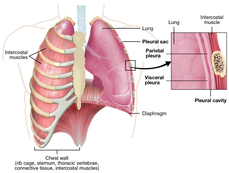
External structures

  • Provide the external borders for the lungs
  • Borders composed of:
    • Sternum
      • Provides anatomical landmarks for auscultation
    • Rib cage
      • 12 pairs of ribs and adjacent intercostal musculature assist with movement of air
      • Provide anatomical landmarks for auscultation
    • Vertebral column
      • Provides posterior border
    • Shoulder girdle
      • Provides attachment for accessory muscles to support in movement of air
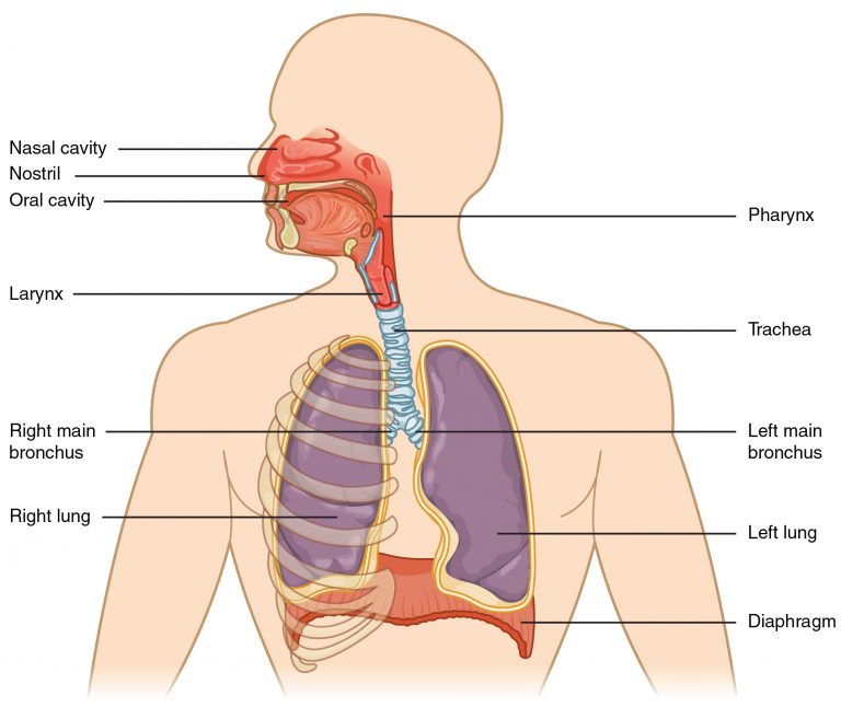
Diagram of pulmonary system

Internal structures

  • Upper airways

    • Entry point for air
    • Composed of nose, mouth, pharynx, and larynx
    • The pharynx (throat) receives air from the nasal cavity and air, food and water from the oral cavity, and opens below into the larynx and esophagus, so it is a passageway shared by the respiratory and digestive systems.
  • Lower airways

    • Conduct air (trachea, bronchi, bronchioles) to the alveolar ducts and sacs, where gas exchange occurs
    • Composed of trachea, bronchi, bronchioles, alveolar ducts, and alveolar sacs
  • Internal supporting structures

    • Each lung is divided into lobes and further subdivided into bronchopulmonary segments.
      • The right lung has three lobes - upper, middle, and lower - and a total of 10 bronchopulmonary segments:
        • Upper lobe: 3 segments - apical, posterior, anterior
        • Middle lobe: 2 segments - lateral, medial
        • Lower lobe: 5 segments - superior, medial basal, anterior basal, lateral basal, posterior basal
      • The left lung has two lobes - upper and lower - and typically 8 bronchopulmonary segments:
        • Upper lobe: 4 segments - apicoposterior, anterior, superior lingular, inferior lingular
        • Lower lobe: 4 segments - superior, anteromedial basal, lateral basal, posterior basal
  • Pleura

    • Parietal pleura located on the outer surface and provides barrier to thoracic cage, diaphragm, and mediastinal border
    • Visceral pleura located on the inner surface and provides barrier to lung
    • Pleurisy, inflammation of the pleura, causes sharp chest pain with breathing.
    • Pneumothorax is air between the layers of the pleura that partly or completely collapses the lung; it causes dyspnea and pleuritic chest pain, with decreased breath sounds and hyperresonance to percussion over the affected side.
    • Clamping a chest tube for pneumothorax adds the risk of a tension pneumothorax.
External lungs
External lungs
Anatomy & Physiology by Lindsay M. Biga, Sierra Dawson, Amy Harwell, Robin Hopkins, Joel Kaufmann, Mike LeMaster, Philip Matern, Katie Morrison-Graham, Devon Quick & Jon Runyeon
/
Oregon State University
/
CC BY 4.0

Muscles and mechanics of ventilation

Ventilation cycle
Ventilation cycle
Anatomy & Physiology by Lindsay M. Biga, Sierra Dawson, Amy Harwell, Robin Hopkins, Joel Kaufmann, Mike LeMaster, Philip Matern, Katie Morrison-Graham, Devon Quick & Jon Runyeon
/
Oregon State University
/
CC BY 4.0

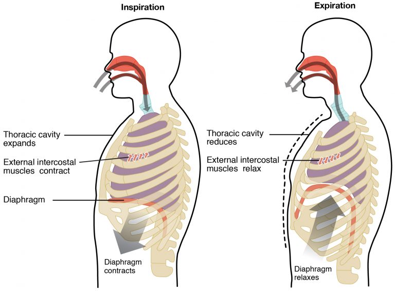
Primary muscles of inspiration

  • Diaphragm
    • When contracted, muscle is pulled downward - inhalation
      • Leads to protruded abdominal cavity
    • Relaxes and rises back into a dome beneath the lower ribs - exhalation
  • External intercostals
    • Lift the rib cage to aid inspiration

Accessory muscles of inspiration

  • Not used during normal, quiet breathing; recruited during exercise or respiratory distress
    • Scalenes
    • Sternocleidomastoid
    • Trapezius
    • Serratus anterior
    • Pectoralis muscles
  • Visible accessory muscle use at rest is a sign of increased work of breathing

Exhalation

  • Quiet exhalation is passive: the inspiratory muscles relax, and the elastic recoil of the lungs and chest wall drives air out
  • Internal intercostals and abdominal muscles are accessory muscles recruited for forced exhalation during a disease state or exercise
    • Quadratus lumborum
    • Abdominal muscles
    • Internal intercostals

Mechanism of breathing

  • Inspiration
    • Inspiratory muscles enlarge the thoracic cavity; the larger volume lowers the pressure inside the lungs below atmospheric pressure, so air flows in
    • Causes the movement of bony thorax outward
  • Exhalation
    • Thoracic volume shrinks as the inspiratory muscles relax; pressure inside the lungs rises above atmospheric pressure, so air flows out
    • Causes the movement of bony thorax inward

Breathing pattern and chest wall excursion

  • The PTA measures chest wall excursion with a tape measure at the axilla, the xiphoid process, and the lower ribs, comparing the circumference at full inhalation to full exhalation at each level
  • Upper ribs move in a pump-handle motion (anterior-posterior), and lower ribs move in a bucket-handle motion (lateral)
  • Normal adult respiratory rate at rest is 12-20 breaths per minute
  • The PTA observes the breathing pattern, noting diaphragmatic breathing (normal) versus accessory muscle use or paradoxical breathing (the abdomen moves inward during inspiration), which can signal respiratory distress or diaphragm weakness
  • These findings are documented and reported to the supervising PT as part of the pulmonary assessment
  • Biot respirations are clusters of deep, regular breaths interspersed with periods of apnea, caused by damage to the medulla from stroke, trauma, or uncal herniation; Kussmaul respirations, in contrast, compensate for metabolic acidosis

Pulmonary function test - ventilation

Definitions
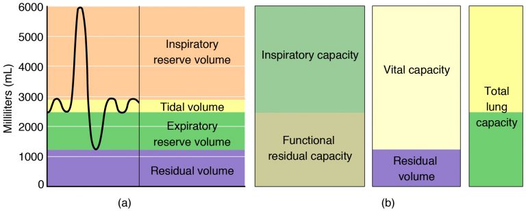
Tidal volume (TV)
Volume of air inhaled and exhaled during normal breathing
Inspiratory reserve volume (IRV)
Maximal volume of air that can be inhaled beyond a normal tidal inspiration
Expiratory reserve volume (ERV)
Maximal amount of air that can be forcefully exhaled after completing a normal, passive breath out
Residual volume (RV)
Volume of air that remains in the lungs after maximal exhalation
Inspiratory capacity (IRV + TV)
The amount of air that can be inhaled beginning at normal exhalation and ending at maximal inspiration
Vital capacity (IRV + TV + ERV)
Maximal volume of air that can be exhaled after a maximal inspiration. Forced vital capacity (FVC) is vital capacity measured with an exhalation that is as fast and forceful as possible.
Functional residual capacity (ERV + RV)
The amount of air left in the lungs at the end of a normal, passive exhalation
Total lung capacity (IRV + TV + ERV + RV)
Total volume of air in the lungs after a maximal inspiration; includes the residual volume that cannot be exhaled.
Forced expiratory volume in 1 second (FEV1)
The volume of air exhaled during the first second of a forced exhalation, starting from maximal inspiration.
FEV1/FVC ratio
The percentage of the forced vital capacity (FVC) that is exhaled in the first second; normally about 75-80%, with a ratio below 70% suggesting an obstructive pattern.
Pulmonary function tests
Anatomy & Physiology by Lindsay M. Biga, Sierra Dawson, Amy Harwell, Robin Hopkins, Joel Kaufmann, Mike LeMaster, Philip Matern, Katie Morrison-Graham, Devon Quick & Jon Runyeon
/
Oregon State University
/
CC BY 4.0

Obstructive vs. restrictive pulmonary disease patterns

Pulmonary function test results are used to sort disease processes into two broad patterns:

  • Obstructive pattern: airflow out of the lungs is limited, so FEV1 drops more than FVC and the FEV1/FVC ratio falls below 70%. RV and TLC are often increased from air trapping. Common causes include chronic obstructive pulmonary disease (COPD), asthma, and bronchiectasis.
    • During an acute asthma exacerbation the patient has wheezing, tachypnea, tachycardia and visible effort to breathe, and in severe cases a silent chest with no audible wheezing can signal impending respiratory failure.
    • Chronic asthma is controlled with medications that modulate the allergic and immune response, usually inhaled glucocorticoids, while a short-acting beta-2 agonist is the quick-relief (rescue) inhaler.
    • Vocal cord dysfunction is paradoxical closing of the vocal cords on inspiration, causing inspiratory stridor and throat tightness that is often mistaken for asthma.
  • Restrictive pattern: lung expansion itself is limited, so TLC and vital capacity are reduced while the FEV1/FVC ratio stays normal or increases. Common causes include pulmonary fibrosis, chest wall or neuromuscular restriction, and atelectasis.

Example: interpreting a spirometry result

A patient’s spirometry shows an FEV1/FVC ratio of 62%, along with an increased RV. Which disease pattern does this suggest?

Answer: An obstructive pattern, such as COPD. The FEV1/FVC ratio is below 70%, and the increased RV reflects air trapping. The PTA notes the documented PFT results in the medical record and communicates relevant findings to the supervising PT, who considers them when establishing or modifying the plan of care; the medical diagnosis itself is made by the physician.

Safety tip: stop testing or treatment and notify the supervising PT if a patient’s SpO2 drops below about 88-90%, blood pressure rises or falls sharply, dyspnea reaches a high rating on a dyspnea scale (or RPE becomes excessive), or the patient develops chest pain - these signs indicate the patient can’t tolerate the current activity level.

Other pulmonary findings

  • Clubbing is a change in the tissue under and around the fingernails and toenails; lung cancer is its most common cause, and chronic lung infections (bronchiectasis, cystic fibrosis, lung abscess), interstitial lung disease, and conditions that lower blood oxygen, such as some congenital heart defects, can also cause it.
  • In the early stages of pulmonary hypertension, a person may have shortness of breath only with exercise, and breathlessness becomes more frequent as the disease progresses.
  • Tactile fremitus is increased over pulmonary consolidation, such as lobar pneumonia, and decreased in pleural effusion and pneumothorax; bronchial breath sounds are heard over areas of consolidation.
  • With some pneumonias a patient coughs up greenish or yellow mucus, or even bloody mucus; in pulmonary edema the sputum is frothy and blood-tinged instead.

Pulmonary rehabilitation, postural drainage, and mucolytics

  • Pulmonary rehabilitation is a supervised program of exercise training and education for people with lung disease such as COPD; oxygen level, blood pressure and heart rate are tracked during exercise.
  • In postural drainage, the right middle lobe is drained with the patient in Trendelenburg position rotated a quarter turn to the left with the knees flexed, the lingula with the patient in Trendelenburg position rotated a quarter turn to the right with the knees flexed, and the lateral basal segment of a lower lobe with the patient lying on the opposite side, rotated a quarter turn toward prone, with the foot of the bed raised.
  • Mucolytics such as N-acetylcysteine and dornase alfa break down the chemical bonds or DNA polymers that hold mucus together, whereas expectorants increase the water content of secretions.

External structures of the lungs

  • Borders formed by sternum, rib cage (12 pairs + intercostals), vertebral column, and shoulder girdle
  • Sternum/ribs serve as auscultation landmarks; shoulder girdle anchors accessory muscles

Internal structures

  • Upper airways (nose, mouth, pharynx, larynx) = air entry; lower airways (trachea, bronchi, bronchioles, alveolar ducts/sacs) = air conduction and gas exchange
  • Right lung: 3 lobes, 10 bronchopulmonary segments; left lung: 2 lobes, typically 8 segments
  • Pleura: parietal (outer, lines thoracic cavity) vs. visceral (inner, covers lung)

Muscles and mechanics of ventilation

  • Primary inspiration muscles: diaphragm (contracts/flattens to inhale) and external intercostals (lift rib cage)
  • Accessory inspiratory muscles (scalenes, SCM, trapezius, serratus anterior, pectoralis) only used with exertion/distress; visible use at rest = increased work of breathing
  • Quiet exhalation is passive (elastic recoil); forced exhalation recruits internal intercostals, abdominals, quadratus lumborum

Mechanism of breathing

  • Inspiration: thoracic volume increases, pressure drops below atmospheric, air flows in, thorax expands outward
  • Exhalation: thoracic volume decreases, pressure rises above atmospheric, air flows out, thorax moves inward

Breathing pattern and chest wall excursion

  • PTA measures excursion at axilla, xiphoid, and lower ribs (inhalation vs. exhalation circumference)
  • Upper ribs: pump-handle (A-P) motion; lower ribs: bucket-handle (lateral) motion
  • Normal adult RR at rest: 12-20 breaths/min
  • Watch for diaphragmatic breathing (normal) vs. accessory muscle use or paradoxical breathing (signs of distress/diaphragm weakness); document and report to supervising PT

Pulmonary function test volumes/capacities

  • Key volumes: TV (normal breath), IRV (extra inhale), ERV (extra exhale), RV (air remaining after max exhale, cannot be measured by spirometry)
  • Key capacities (combinations): Inspiratory capacity (IRV+TV), Vital capacity (IRV+TV+ERV; FVC = forced version), Functional residual capacity (ERV+RV), Total lung capacity (IRV+TV+ERV+RV)
  • FEV1 = volume exhaled in first second of forced exhalation; FEV1/FVC ratio normally ~75-80%, <70% suggests obstruction

Obstructive vs. restrictive patterns

  • Obstructive: FEV1/FVC <70%, RV and TLC often increased (air trapping) — e.g., COPD, asthma, bronchiectasis
  • Restrictive: TLC and vital capacity reduced, FEV1/FVC normal/increased — e.g., pulmonary fibrosis, chest wall/neuromuscular restriction, atelectasis
  • PTA documents/reports PFT findings; physician makes diagnosis, PT adjusts plan of care

Safety considerations

  • Stop testing/treatment and notify supervising PT if SpO2 <88-90%, significant BP changes, high dyspnea/RPE, or chest pain occur

Sign up for free to take 34 quiz questions on this topic

Previous
Next  | 2.2 Auscultation and differential diagnosis of pulmonary system, lines and tubes
All rights reserved ©2016 - 2026 Achievable, Inc.

Anatomy, ventilation, and pulmonary function tests

External structures

  • Provide the external borders for the lungs
  • Borders composed of:
    • Sternum
      • Provides anatomical landmarks for auscultation
    • Rib cage
      • 12 pairs of ribs and adjacent intercostal musculature assist with movement of air
      • Provide anatomical landmarks for auscultation
    • Vertebral column
      • Provides posterior border
    • Shoulder girdle
      • Provides attachment for accessory muscles to support in movement of air

Internal structures

  • Upper airways

    • Entry point for air
    • Composed of nose, mouth, pharynx, and larynx
    • The pharynx (throat) receives air from the nasal cavity and air, food and water from the oral cavity, and opens below into the larynx and esophagus, so it is a passageway shared by the respiratory and digestive systems.
  • Lower airways

    • Conduct air (trachea, bronchi, bronchioles) to the alveolar ducts and sacs, where gas exchange occurs
    • Composed of trachea, bronchi, bronchioles, alveolar ducts, and alveolar sacs
  • Internal supporting structures

    • Each lung is divided into lobes and further subdivided into bronchopulmonary segments.
      • The right lung has three lobes - upper, middle, and lower - and a total of 10 bronchopulmonary segments:
        • Upper lobe: 3 segments - apical, posterior, anterior
        • Middle lobe: 2 segments - lateral, medial
        • Lower lobe: 5 segments - superior, medial basal, anterior basal, lateral basal, posterior basal
      • The left lung has two lobes - upper and lower - and typically 8 bronchopulmonary segments:
        • Upper lobe: 4 segments - apicoposterior, anterior, superior lingular, inferior lingular
        • Lower lobe: 4 segments - superior, anteromedial basal, lateral basal, posterior basal
  • Pleura

    • Parietal pleura located on the outer surface and provides barrier to thoracic cage, diaphragm, and mediastinal border
    • Visceral pleura located on the inner surface and provides barrier to lung
    • Pleurisy, inflammation of the pleura, causes sharp chest pain with breathing.
    • Pneumothorax is air between the layers of the pleura that partly or completely collapses the lung; it causes dyspnea and pleuritic chest pain, with decreased breath sounds and hyperresonance to percussion over the affected side.
    • Clamping a chest tube for pneumothorax adds the risk of a tension pneumothorax.

Muscles and mechanics of ventilation

Primary muscles of inspiration

  • Diaphragm
    • When contracted, muscle is pulled downward - inhalation
      • Leads to protruded abdominal cavity
    • Relaxes and rises back into a dome beneath the lower ribs - exhalation
  • External intercostals
    • Lift the rib cage to aid inspiration

Accessory muscles of inspiration

  • Not used during normal, quiet breathing; recruited during exercise or respiratory distress
    • Scalenes
    • Sternocleidomastoid
    • Trapezius
    • Serratus anterior
    • Pectoralis muscles
  • Visible accessory muscle use at rest is a sign of increased work of breathing

Exhalation

  • Quiet exhalation is passive: the inspiratory muscles relax, and the elastic recoil of the lungs and chest wall drives air out
  • Internal intercostals and abdominal muscles are accessory muscles recruited for forced exhalation during a disease state or exercise
    • Quadratus lumborum
    • Abdominal muscles
    • Internal intercostals

Mechanism of breathing

  • Inspiration
    • Inspiratory muscles enlarge the thoracic cavity; the larger volume lowers the pressure inside the lungs below atmospheric pressure, so air flows in
    • Causes the movement of bony thorax outward
  • Exhalation
    • Thoracic volume shrinks as the inspiratory muscles relax; pressure inside the lungs rises above atmospheric pressure, so air flows out
    • Causes the movement of bony thorax inward

Breathing pattern and chest wall excursion

  • The PTA measures chest wall excursion with a tape measure at the axilla, the xiphoid process, and the lower ribs, comparing the circumference at full inhalation to full exhalation at each level
  • Upper ribs move in a pump-handle motion (anterior-posterior), and lower ribs move in a bucket-handle motion (lateral)
  • Normal adult respiratory rate at rest is 12-20 breaths per minute
  • The PTA observes the breathing pattern, noting diaphragmatic breathing (normal) versus accessory muscle use or paradoxical breathing (the abdomen moves inward during inspiration), which can signal respiratory distress or diaphragm weakness
  • These findings are documented and reported to the supervising PT as part of the pulmonary assessment
  • Biot respirations are clusters of deep, regular breaths interspersed with periods of apnea, caused by damage to the medulla from stroke, trauma, or uncal herniation; Kussmaul respirations, in contrast, compensate for metabolic acidosis

Pulmonary function test - ventilation

Definitions
Tidal volume (TV)
Volume of air inhaled and exhaled during normal breathing
Inspiratory reserve volume (IRV)
Maximal volume of air that can be inhaled beyond a normal tidal inspiration
Expiratory reserve volume (ERV)
Maximal amount of air that can be forcefully exhaled after completing a normal, passive breath out
Residual volume (RV)
Volume of air that remains in the lungs after maximal exhalation
Inspiratory capacity (IRV + TV)
The amount of air that can be inhaled beginning at normal exhalation and ending at maximal inspiration
Vital capacity (IRV + TV + ERV)
Maximal volume of air that can be exhaled after a maximal inspiration. Forced vital capacity (FVC) is vital capacity measured with an exhalation that is as fast and forceful as possible.
Functional residual capacity (ERV + RV)
The amount of air left in the lungs at the end of a normal, passive exhalation
Total lung capacity (IRV + TV + ERV + RV)
Total volume of air in the lungs after a maximal inspiration; includes the residual volume that cannot be exhaled.
Forced expiratory volume in 1 second (FEV1)
The volume of air exhaled during the first second of a forced exhalation, starting from maximal inspiration.
FEV1/FVC ratio
The percentage of the forced vital capacity (FVC) that is exhaled in the first second; normally about 75-80%, with a ratio below 70% suggesting an obstructive pattern.

Obstructive vs. restrictive pulmonary disease patterns

Pulmonary function test results are used to sort disease processes into two broad patterns:

  • Obstructive pattern: airflow out of the lungs is limited, so FEV1 drops more than FVC and the FEV1/FVC ratio falls below 70%. RV and TLC are often increased from air trapping. Common causes include chronic obstructive pulmonary disease (COPD), asthma, and bronchiectasis.
    • During an acute asthma exacerbation the patient has wheezing, tachypnea, tachycardia and visible effort to breathe, and in severe cases a silent chest with no audible wheezing can signal impending respiratory failure.
    • Chronic asthma is controlled with medications that modulate the allergic and immune response, usually inhaled glucocorticoids, while a short-acting beta-2 agonist is the quick-relief (rescue) inhaler.
    • Vocal cord dysfunction is paradoxical closing of the vocal cords on inspiration, causing inspiratory stridor and throat tightness that is often mistaken for asthma.
  • Restrictive pattern: lung expansion itself is limited, so TLC and vital capacity are reduced while the FEV1/FVC ratio stays normal or increases. Common causes include pulmonary fibrosis, chest wall or neuromuscular restriction, and atelectasis.

Example: interpreting a spirometry result

A patient’s spirometry shows an FEV1/FVC ratio of 62%, along with an increased RV. Which disease pattern does this suggest?

Answer: An obstructive pattern, such as COPD. The FEV1/FVC ratio is below 70%, and the increased RV reflects air trapping. The PTA notes the documented PFT results in the medical record and communicates relevant findings to the supervising PT, who considers them when establishing or modifying the plan of care; the medical diagnosis itself is made by the physician.

Safety tip: stop testing or treatment and notify the supervising PT if a patient’s SpO2 drops below about 88-90%, blood pressure rises or falls sharply, dyspnea reaches a high rating on a dyspnea scale (or RPE becomes excessive), or the patient develops chest pain - these signs indicate the patient can’t tolerate the current activity level.

Other pulmonary findings

  • Clubbing is a change in the tissue under and around the fingernails and toenails; lung cancer is its most common cause, and chronic lung infections (bronchiectasis, cystic fibrosis, lung abscess), interstitial lung disease, and conditions that lower blood oxygen, such as some congenital heart defects, can also cause it.
  • In the early stages of pulmonary hypertension, a person may have shortness of breath only with exercise, and breathlessness becomes more frequent as the disease progresses.
  • Tactile fremitus is increased over pulmonary consolidation, such as lobar pneumonia, and decreased in pleural effusion and pneumothorax; bronchial breath sounds are heard over areas of consolidation.
  • With some pneumonias a patient coughs up greenish or yellow mucus, or even bloody mucus; in pulmonary edema the sputum is frothy and blood-tinged instead.

Pulmonary rehabilitation, postural drainage, and mucolytics

  • Pulmonary rehabilitation is a supervised program of exercise training and education for people with lung disease such as COPD; oxygen level, blood pressure and heart rate are tracked during exercise.
  • In postural drainage, the right middle lobe is drained with the patient in Trendelenburg position rotated a quarter turn to the left with the knees flexed, the lingula with the patient in Trendelenburg position rotated a quarter turn to the right with the knees flexed, and the lateral basal segment of a lower lobe with the patient lying on the opposite side, rotated a quarter turn toward prone, with the foot of the bed raised.
  • Mucolytics such as N-acetylcysteine and dornase alfa break down the chemical bonds or DNA polymers that hold mucus together, whereas expectorants increase the water content of secretions.
Key points

External structures of the lungs

  • Borders formed by sternum, rib cage (12 pairs + intercostals), vertebral column, and shoulder girdle
  • Sternum/ribs serve as auscultation landmarks; shoulder girdle anchors accessory muscles

Internal structures

  • Upper airways (nose, mouth, pharynx, larynx) = air entry; lower airways (trachea, bronchi, bronchioles, alveolar ducts/sacs) = air conduction and gas exchange
  • Right lung: 3 lobes, 10 bronchopulmonary segments; left lung: 2 lobes, typically 8 segments
  • Pleura: parietal (outer, lines thoracic cavity) vs. visceral (inner, covers lung)

Muscles and mechanics of ventilation

  • Primary inspiration muscles: diaphragm (contracts/flattens to inhale) and external intercostals (lift rib cage)
  • Accessory inspiratory muscles (scalenes, SCM, trapezius, serratus anterior, pectoralis) only used with exertion/distress; visible use at rest = increased work of breathing
  • Quiet exhalation is passive (elastic recoil); forced exhalation recruits internal intercostals, abdominals, quadratus lumborum

Mechanism of breathing

  • Inspiration: thoracic volume increases, pressure drops below atmospheric, air flows in, thorax expands outward
  • Exhalation: thoracic volume decreases, pressure rises above atmospheric, air flows out, thorax moves inward

Breathing pattern and chest wall excursion

  • PTA measures excursion at axilla, xiphoid, and lower ribs (inhalation vs. exhalation circumference)
  • Upper ribs: pump-handle (A-P) motion; lower ribs: bucket-handle (lateral) motion
  • Normal adult RR at rest: 12-20 breaths/min
  • Watch for diaphragmatic breathing (normal) vs. accessory muscle use or paradoxical breathing (signs of distress/diaphragm weakness); document and report to supervising PT

Pulmonary function test volumes/capacities

  • Key volumes: TV (normal breath), IRV (extra inhale), ERV (extra exhale), RV (air remaining after max exhale, cannot be measured by spirometry)
  • Key capacities (combinations): Inspiratory capacity (IRV+TV), Vital capacity (IRV+TV+ERV; FVC = forced version), Functional residual capacity (ERV+RV), Total lung capacity (IRV+TV+ERV+RV)
  • FEV1 = volume exhaled in first second of forced exhalation; FEV1/FVC ratio normally ~75-80%, <70% suggests obstruction

Obstructive vs. restrictive patterns

  • Obstructive: FEV1/FVC <70%, RV and TLC often increased (air trapping) — e.g., COPD, asthma, bronchiectasis
  • Restrictive: TLC and vital capacity reduced, FEV1/FVC normal/increased — e.g., pulmonary fibrosis, chest wall/neuromuscular restriction, atelectasis
  • PTA documents/reports PFT findings; physician makes diagnosis, PT adjusts plan of care

Safety considerations

  • Stop testing/treatment and notify supervising PT if SpO2 <88-90%, significant BP changes, high dyspnea/RPE, or chest pain occur

More from Pulmonary system

  • Auscultation and differential diagnosis of pulmonary system, lines and tubes
  • Understanding arterial blood gases
  • Interventions for pulmonary system