


Achievable logoAchievable logo
NCLEX
Sign in
Sign up
Purchase
Textbook
Practice exams
Support
How it works
Exam catalog
Mountain with a flag at the peak
Textbook
Introduction
1. Safe and effective care environment
2. Health promotion and maintenance
3. Psychosocial Integrity
4. Physiological Integrity
4.1 Basic care and comfort
4.2 Pharmacological and parenteral therapies
4.3 Reduction of risk potential
4.4 Physiological adaptation
4.4.1 Medical emergencies
4.4.2 Fluid and electrolyte disorders
4.4.3 Acute and chronic conditions management
4.4.4 Alterations in body systems
4.4.5 Organ transplants and end of life changes
4.4.6 Hemodynamic monitoring and critical care concepts
Wrapping up
Achievable logoAchievable logo
4.4.6 Hemodynamic monitoring and critical care concepts
Achievable NCLEX
4. Physiological Integrity
4.4. Physiological adaptation
Our NCLEX course is currently in development and is a work-in-progress.

Hemodynamic monitoring and critical care concepts

7 min read
Font
Discuss
Share
Feedback

Introduction

Hemodynamic monitoring is used in critical care settings to assess circulation, perfusion, and oxygen delivery in unstable clients. These measurements help clinicians evaluate cardiac function, fluid status, and vascular tone, guiding timely interventions in shock, sepsis, heart failure, and postoperative complications.

On the Next Generation NCLEX (NGN), this content is tested through trend interpretation, prioritization, and clinical judgment, not memorization of device mechanics.

Learning objectives

By the end of this section, the learner should be able to:

  • Understand the purpose of hemodynamic monitoring
  • Interpret basic hemodynamic parameters
  • Recognize dangerous trends indicating decompensation
  • Prioritize nursing actions in critical care scenarios

Core concept: Numbers support, clients decide

Hemodynamic values support clinical decisions, but the client’s clinical presentation always comes first. The nurse must always correlate numbers with the client’s clinical presentation, including:

  • Level of consciousness
  • Skin color and temperature
  • Urine output
  • Work of breathing
  • Vital sign trends
NGN insight:
NGN questions often reward nurses who interpret monitor data in the context of the client’s clinical presentation.

What is hemodynamic monitoring

Hemodynamic monitoring involves the measurement of variables that reflect blood flow, pressure, and oxygen delivery to tissues.

alt_text
//////Caption: Overview of Common Hemodynamic Parameters
Illustration type: Concept diagram
Illustration note: Illustrate the relationship between common hemodynamic parameters—including blood pressure, MAP, CVP, cardiac output, cardiac index, and oxygen saturation—and what each reflects about circulation, perfusion, or cardiac function.///////

Commonly monitored parameters

  • Blood pressure (invasive and noninvasive)
  • Heart rate and rhythm
  • Mean arterial pressure (MAP)
  • Central venous pressure (CVP)
  • Cardiac output (CO) and cardiac index (CI)
  • Oxygen saturation (SpO₂, sometimes ScvO₂)
Definitions
Hemodynamics
The study of blood flow and the forces involved in circulation
Arterial line
A catheter placed in an artery to allow continuous blood pressure monitoring and blood sampling
CVP
Pressure in the thoracic vena cava reflects right ventricular preload
Mean arterial pressure (MAP)
Average pressure in the arteries during one cardiac cycle
Cardiac output
Volume of blood pumped by the heart per minute

Noninvasive hemodynamic monitoring

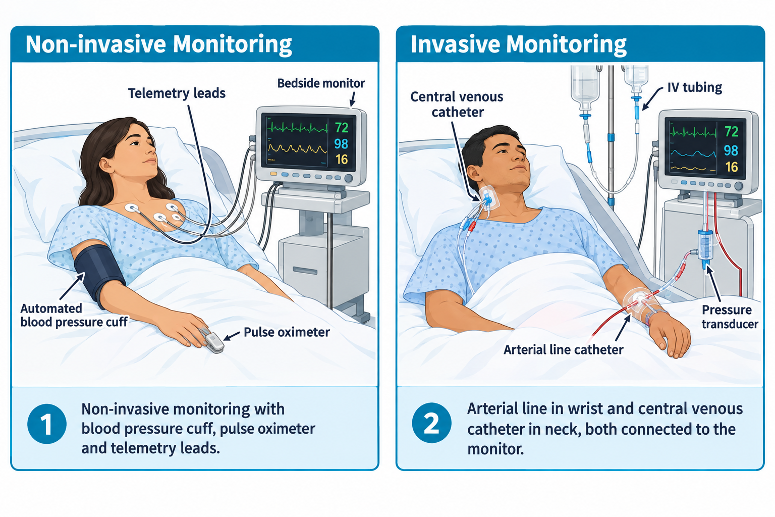
Noninvasive monitoring is commonly used in stable or moderately ill clients and serves as the first line of assessment.

alt_text Oximetry //////📷 Image suggestion: “Noninvasive vs. invasive monitoring setup” — side-by-side comparison showing automated BP cuff/pulse oximeter vs. an arterial line/central line setup, helps visually anchor the distinction the chapter is teaching.//////

Examples

  • Automated blood pressure cuffs
  • Pulse oximetry
  • Cardiac telemetry

Key nursing considerations

  • Verify abnormal readings manually
  • Ensure proper cuff size and probe placement
  • Trend values rather than react to single readings
NGN tip:
Unexpected values should always prompt client assessment before intervention.

Invasive hemodynamic monitoring

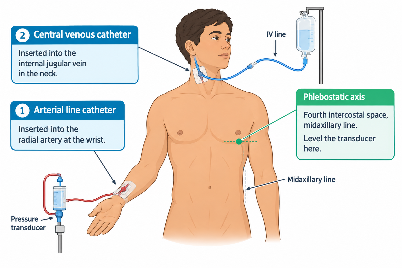
alt_text //////📷 Image suggestion: “Arterial line and central venous catheter placement” — anatomical diagram showing typical insertion sites (radial artery, subclavian/internal jugular vein) with labeled transducer setup.//////

Invasive monitoring provides continuous, real-time data in critically ill clients.

Common invasive devices

  • Arterial lines
  • Central venous catheters
  • Pulmonary artery catheters (less common)

Key hemodynamic parameters (high-yield)

Blood pressure and mean arterial pressure (MAP)

MAP ≥65 mmHg is generally required for adequate organ perfusion

MAP = (SBP + 2 × DBP) ÷ 3

SBP: Systolic Blood Pressure; DBP: Diastolic Blood Pressure

  • Diastole counts twice because the heart spends more time there.
  • Low MAP suggests inadequate tissue perfusion.
  • A normal systolic BP does not guarantee adequate perfusion.
NGN tip:
BP with low MAP may still indicate shock.
alt_text
//////Caption: Understanding Mean Arterial Pressure (MAP)
Illustration type: Clinical Concept diagram
Illustration note: Illustrate how MAP is calculated using systolic and diastolic blood pressure, emphasizing that MAP ≥65 mmHg is generally required for adequate organ perfusion and that a normal systolic blood pressure does not always indicate adequate perfusion.///////

Central venous pressure (CVP)

The CVP reflects the right ventricular preload (how much blood is returning to the heart). It is measured directly via a central venous catheter and reported in mmHg (or cm H₂O)

  • Low CVP → hypovolemia
  • High CVP → fluid overload or right-sided heart failure
NGN :
CVP trends matter more than absolute numbers.
alt_text
////////Caption: Interpreting Central Venous Pressure (CVP)
Illustration type: Comparison diagram
Illustration note: Compare low versus high CVP, showing common causes such as hypovolemia, fluid overload, and right-sided heart failure, along with the corresponding effects on preload and tissue perfusion.////////

Cardiac output and cardiac index

Cardiac output (CO) = heart rate × stroke volume

Where:

  • Heart rate (HR) = beats per minute
  • Stroke volume (SV) = amount of blood ejected per beat
NGN :
Anything that lowers HR or SV will lower CO.

Cardiac index (CI) = CO ÷ body surface area (BSA)

The cardiac index adjusts cardiac output for client size and provides a more accurate comparison between clients.

Low CO/CI leads to:

  • Hypotension
  • Decreased urine output (Oliguria)
  • Altered mental status
  • Cool extremities

Hemodynamic patterns in shock

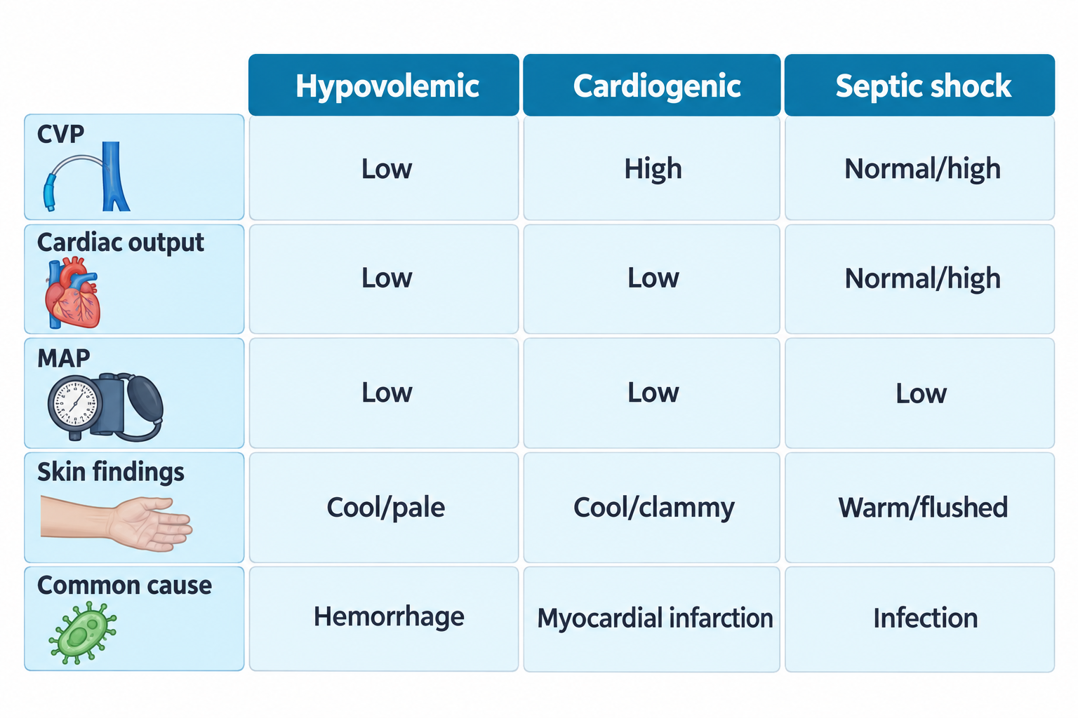
alt_text //////📷 Image suggestion: “Hemodynamic profiles by shock type” — comparison table/chart showing CVP, CO, and MAP patterns across hypovolemic, cardiogenic, and septic shock, this is one of the most testable, easily-visualized concepts in the whole chapter.//////

Hypovolemic shock

  • Low CVP
  • Low cardiac output
  • Low MAP

Common causes: Hemorrhage, dehydration, burns

Cardiogenic shock

  • High CVP
  • Low cardiac output
  • Pulmonary congestion

Common causes: Myocardial infarction, severe heart failure

Septic shock

  • Early (compensated):
    • Normal or high cardiac output
    • Low systemic vascular resistance (SVR)
    • Warm, flushed skin
  • Late (decompensated):
    • Low cardiac output
    • Hypotension
    • Oliguria
NGN tip:
Early septic shock may present with warm, flushed skin and normal blood pressure despite impaired tissue perfusion.
alt_text
////////Caption: Hemodynamic Patterns in Common Types of Shock
Illustration type: Comparison table
Illustration note: Compare hypovolemic, cardiogenic, and septic shock using CVP, cardiac output, MAP, skin findings, and common causes to reinforce recognition of characteristic hemodynamic patterns.////////

Critical care nursing priorities

NGN insight:
In critical care, time equals tissue survival.

Priority nursing actions

  • Continuous monitoring and reassessment
  • Early recognition of deterioration
  • Rapid escalation of care
  • Prevention of complications (infection, bleeding)

Complications of hemodynamic monitoring

alt_text //////📷 Image suggestion: “Central line complications” — quick-reference diagram showing infection, bleeding, air embolism, and thrombosis risk points along an invasive line, reinforces this same content taught earlier in the device complications chapter.//////

Potential complications

  • Infection
  • Bleeding
  • Air embolism
  • Thrombosis
NGN tip:
Sudden dyspnea or hypotension in a client with invasive lines suggests air embolism.
alt_text
////////Caption: Complications of Invasive Hemodynamic Monitoring
Illustration type: Comparison table
Illustration note: Compare common complications of invasive hemodynamic monitoring—including infection, bleeding, air embolism, and thrombosis—with their early warning signs and priority nursing responses.////////

Clinical vignette 1

A critically ill client has a MAP of 58 mmHg, decreasing urine output, and increasing confusion.

(spoiler)

Nursing action: Notify the provider promptly, continue close monitoring, and anticipate prescribed fluid resuscitation or vasopressor therapy.

Rationale: A low MAP with signs of poor perfusion suggests inadequate tissue perfusion requiring prompt intervention.

Clinical vignette 2

A client with septic shock initially has warm skin and bounding pulses but later develops hypotension and oliguria.

(spoiler)

Nursing action: Escalate care immediately and continue close monitoring.

Rationale: These findings indicate progression from compensated to decompensated septic shock.

Common NCLEX pitfalls

  • Treating monitor numbers without assessing the client
  • Ignoring MAP in favor of systolic BP
  • Reacting to single readings instead of trends
  • Missing early compensated shock
  • Hemodynamic monitoring guides critical care decisions
  • MAP is a key indicator of perfusion
  • Trends matter more than single values
  • Shock presents differently depending on type and stage
  • Nursing vigilance saves lives

Sign up for free to take 5 quiz questions on this topic

Previous
Next  | Wrapping up
All rights reserved ©2016 - 2026 Achievable, Inc.

Hemodynamic monitoring and critical care concepts

Introduction

Hemodynamic monitoring is used in critical care settings to assess circulation, perfusion, and oxygen delivery in unstable clients. These measurements help clinicians evaluate cardiac function, fluid status, and vascular tone, guiding timely interventions in shock, sepsis, heart failure, and postoperative complications.

On the Next Generation NCLEX (NGN), this content is tested through trend interpretation, prioritization, and clinical judgment, not memorization of device mechanics.

Learning objectives

By the end of this section, the learner should be able to:

  • Understand the purpose of hemodynamic monitoring
  • Interpret basic hemodynamic parameters
  • Recognize dangerous trends indicating decompensation
  • Prioritize nursing actions in critical care scenarios

Core concept: Numbers support, clients decide

Hemodynamic values support clinical decisions, but the client’s clinical presentation always comes first. The nurse must always correlate numbers with the client’s clinical presentation, including:

  • Level of consciousness
  • Skin color and temperature
  • Urine output
  • Work of breathing
  • Vital sign trends
NGN insight:
NGN questions often reward nurses who interpret monitor data in the context of the client’s clinical presentation.

What is hemodynamic monitoring

Hemodynamic monitoring involves the measurement of variables that reflect blood flow, pressure, and oxygen delivery to tissues.

alt_text
//////Caption: Overview of Common Hemodynamic Parameters
Illustration type: Concept diagram
Illustration note: Illustrate the relationship between common hemodynamic parameters—including blood pressure, MAP, CVP, cardiac output, cardiac index, and oxygen saturation—and what each reflects about circulation, perfusion, or cardiac function.///////

Commonly monitored parameters

  • Blood pressure (invasive and noninvasive)
  • Heart rate and rhythm
  • Mean arterial pressure (MAP)
  • Central venous pressure (CVP)
  • Cardiac output (CO) and cardiac index (CI)
  • Oxygen saturation (SpO₂, sometimes ScvO₂)
Definitions
Hemodynamics
The study of blood flow and the forces involved in circulation
Arterial line
A catheter placed in an artery to allow continuous blood pressure monitoring and blood sampling
CVP
Pressure in the thoracic vena cava reflects right ventricular preload
Mean arterial pressure (MAP)
Average pressure in the arteries during one cardiac cycle
Cardiac output
Volume of blood pumped by the heart per minute

Noninvasive hemodynamic monitoring

Noninvasive monitoring is commonly used in stable or moderately ill clients and serves as the first line of assessment.

alt_text Oximetry //////📷 Image suggestion: “Noninvasive vs. invasive monitoring setup” — side-by-side comparison showing automated BP cuff/pulse oximeter vs. an arterial line/central line setup, helps visually anchor the distinction the chapter is teaching.//////

Examples

  • Automated blood pressure cuffs
  • Pulse oximetry
  • Cardiac telemetry

Key nursing considerations

  • Verify abnormal readings manually
  • Ensure proper cuff size and probe placement
  • Trend values rather than react to single readings
NGN tip:
Unexpected values should always prompt client assessment before intervention.

Invasive hemodynamic monitoring

alt_text //////📷 Image suggestion: “Arterial line and central venous catheter placement” — anatomical diagram showing typical insertion sites (radial artery, subclavian/internal jugular vein) with labeled transducer setup.//////

Invasive monitoring provides continuous, real-time data in critically ill clients.

Common invasive devices

  • Arterial lines
  • Central venous catheters
  • Pulmonary artery catheters (less common)

Key hemodynamic parameters (high-yield)

Blood pressure and mean arterial pressure (MAP)

MAP ≥65 mmHg is generally required for adequate organ perfusion

MAP = (SBP + 2 × DBP) ÷ 3

SBP: Systolic Blood Pressure; DBP: Diastolic Blood Pressure

  • Diastole counts twice because the heart spends more time there.
  • Low MAP suggests inadequate tissue perfusion.
  • A normal systolic BP does not guarantee adequate perfusion.
NGN tip:
BP with low MAP may still indicate shock.
alt_text
//////Caption: Understanding Mean Arterial Pressure (MAP)
Illustration type: Clinical Concept diagram
Illustration note: Illustrate how MAP is calculated using systolic and diastolic blood pressure, emphasizing that MAP ≥65 mmHg is generally required for adequate organ perfusion and that a normal systolic blood pressure does not always indicate adequate perfusion.///////

Central venous pressure (CVP)

The CVP reflects the right ventricular preload (how much blood is returning to the heart). It is measured directly via a central venous catheter and reported in mmHg (or cm H₂O)

  • Low CVP → hypovolemia
  • High CVP → fluid overload or right-sided heart failure
NGN :
CVP trends matter more than absolute numbers.
alt_text
////////Caption: Interpreting Central Venous Pressure (CVP)
Illustration type: Comparison diagram
Illustration note: Compare low versus high CVP, showing common causes such as hypovolemia, fluid overload, and right-sided heart failure, along with the corresponding effects on preload and tissue perfusion.////////

Cardiac output and cardiac index

Cardiac output (CO) = heart rate × stroke volume

Where:

  • Heart rate (HR) = beats per minute
  • Stroke volume (SV) = amount of blood ejected per beat
NGN :
Anything that lowers HR or SV will lower CO.

Cardiac index (CI) = CO ÷ body surface area (BSA)

The cardiac index adjusts cardiac output for client size and provides a more accurate comparison between clients.

Low CO/CI leads to:

  • Hypotension
  • Decreased urine output (Oliguria)
  • Altered mental status
  • Cool extremities

Hemodynamic patterns in shock

alt_text //////📷 Image suggestion: “Hemodynamic profiles by shock type” — comparison table/chart showing CVP, CO, and MAP patterns across hypovolemic, cardiogenic, and septic shock, this is one of the most testable, easily-visualized concepts in the whole chapter.//////

Hypovolemic shock

  • Low CVP
  • Low cardiac output
  • Low MAP

Common causes: Hemorrhage, dehydration, burns

Cardiogenic shock

  • High CVP
  • Low cardiac output
  • Pulmonary congestion

Common causes: Myocardial infarction, severe heart failure

Septic shock

  • Early (compensated):
    • Normal or high cardiac output
    • Low systemic vascular resistance (SVR)
    • Warm, flushed skin
  • Late (decompensated):
    • Low cardiac output
    • Hypotension
    • Oliguria
NGN tip:
Early septic shock may present with warm, flushed skin and normal blood pressure despite impaired tissue perfusion.
alt_text
////////Caption: Hemodynamic Patterns in Common Types of Shock
Illustration type: Comparison table
Illustration note: Compare hypovolemic, cardiogenic, and septic shock using CVP, cardiac output, MAP, skin findings, and common causes to reinforce recognition of characteristic hemodynamic patterns.////////

Critical care nursing priorities

NGN insight:
In critical care, time equals tissue survival.

Priority nursing actions

  • Continuous monitoring and reassessment
  • Early recognition of deterioration
  • Rapid escalation of care
  • Prevention of complications (infection, bleeding)

Complications of hemodynamic monitoring

alt_text //////📷 Image suggestion: “Central line complications” — quick-reference diagram showing infection, bleeding, air embolism, and thrombosis risk points along an invasive line, reinforces this same content taught earlier in the device complications chapter.//////

Potential complications

  • Infection
  • Bleeding
  • Air embolism
  • Thrombosis
NGN tip:
Sudden dyspnea or hypotension in a client with invasive lines suggests air embolism.
alt_text
////////Caption: Complications of Invasive Hemodynamic Monitoring
Illustration type: Comparison table
Illustration note: Compare common complications of invasive hemodynamic monitoring—including infection, bleeding, air embolism, and thrombosis—with their early warning signs and priority nursing responses.////////

Clinical vignette 1

A critically ill client has a MAP of 58 mmHg, decreasing urine output, and increasing confusion.

(spoiler)

Nursing action: Notify the provider promptly, continue close monitoring, and anticipate prescribed fluid resuscitation or vasopressor therapy.

Rationale: A low MAP with signs of poor perfusion suggests inadequate tissue perfusion requiring prompt intervention.

Clinical vignette 2

A client with septic shock initially has warm skin and bounding pulses but later develops hypotension and oliguria.

(spoiler)

Nursing action: Escalate care immediately and continue close monitoring.

Rationale: These findings indicate progression from compensated to decompensated septic shock.

Common NCLEX pitfalls

  • Treating monitor numbers without assessing the client
  • Ignoring MAP in favor of systolic BP
  • Reacting to single readings instead of trends
  • Missing early compensated shock
Key points
  • Hemodynamic monitoring guides critical care decisions
  • MAP is a key indicator of perfusion
  • Trends matter more than single values
  • Shock presents differently depending on type and stage
  • Nursing vigilance saves lives

More from Physiological adaptation

  • Medical emergencies
  • Fluid and electrolyte disorders
  • Acute and chronic conditions management
  • Alterations in body systems
  • Organ transplants and end of life changes