



Alterations in body systems
Introduction
Alterations in body systems occur when physiological compensation fails, leading to dysfunction across one or more organ systems. These changes may arise from acute illness, chronic disease progression, trauma, infection, or metabolic disturbances. The body initially attempts to maintain homeostasis through compensation (increased heart rate, increased respiratory rate, vasoconstriction, and hormone release). When compensation is no longer effective, decompensation occurs, leading to rapid deterioration.
On the Next generation NCLEX (NGN), this content is tested through system-based pattern recognition, prioritization, and the ability to link symptoms across systems rather than evaluating each finding in isolation.
Learning objectives
By the end of this section, the learner should be able to:
- recognize system-specific signs of deterioration
- identify early vs late manifestations of system failure
- correlate findings across multiple body systems
- prioritize nursing interventions using clinical judgment
- interpret basic lab abnormalities
- recognize when to escalate care
Core principle: Systems fail in patterns
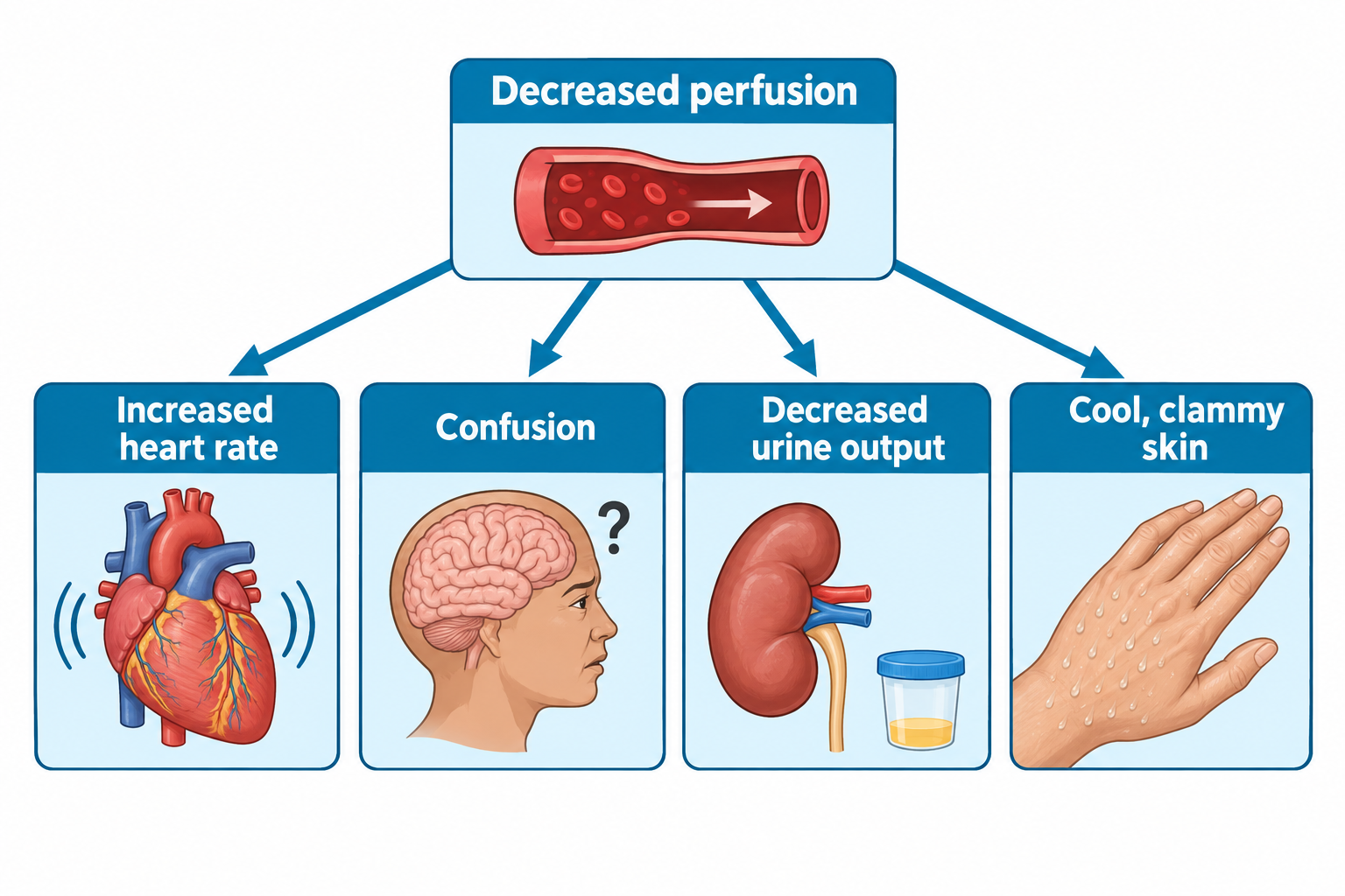
Body systems rarely fail in isolation. When perfusion decreases:
- The brain shows confusion.
- The kidneys reduce urine output.
- The skin becomes cool and clammy.
- The heart rate increases to compensate.
NGN rewards recognizing connected changes, not isolated symptoms.
Cardiovascular system alterations
Common causes
- Heart failure
- Myocardial infarction
- Shock
- Dysrhythmias
Compensation pattern
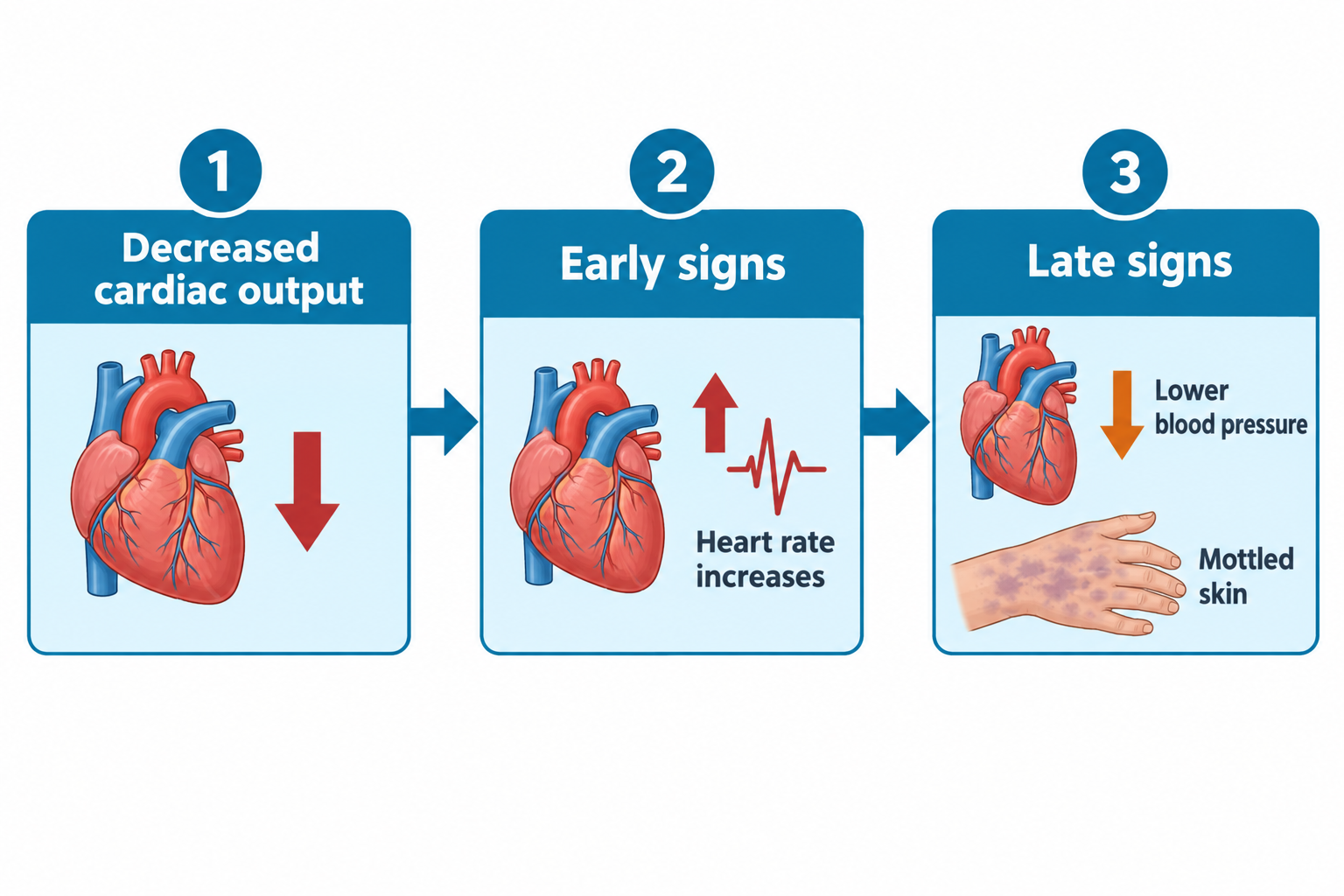
When cardiac output drops, the body:
- increases heart rate
- constricts peripheral vessels
- activates RAAS (fluid retention)
Key assessment findings
Early signs
- Tachycardia
- Restlessness
- Narrow pulse pressure
- Slight decrease in urine output
Late signs
- Hypotension
- Cool, mottled skin
- Altered mental status
- Significant oliguria
Labs to watch
- Elevated troponin (MI)
- BNP (heart failure)
- Lactate (poor perfusion)
- Rising creatinine (renal impact)
Respiratory system alterations
Common causes
- Pneumonia
- Pulmonary edema
- COPD exacerbation
- Pulmonary embolism
Compensation pattern
The body increases respiratory rate to improve oxygenation. Carbon dioxide retention leads to acidosis if untreated.
Key assessment findings
Early signs
- Increased respiratory rate (Tachypnea)
- Restlessness
- Anxiety
- Mild desaturation
Late signs
- Cyanosis
- Decreased LOC
- Bradypnea (impending failure)
- Silent chest (severe obstruction)
ABG clues
- ↑ CO₂ = respiratory acidosis
- ↓ O₂ = hypoxemia
- Low pH = decompensation
Neurological system alterations
Common causes
- Stroke
- Hypoxia
- Infection
- Electrolyte imbalance
- Increased intracranial pressure
The brain is extremely sensitive to oxygen deprivation. Neurological changes are often the first indicators of systemic decline.
Key assessment findings
- Confusion or agitation
- Decreased level of consciousness
- Unequal pupils
- New motor deficits
- Seizure activity
Renal system alterations
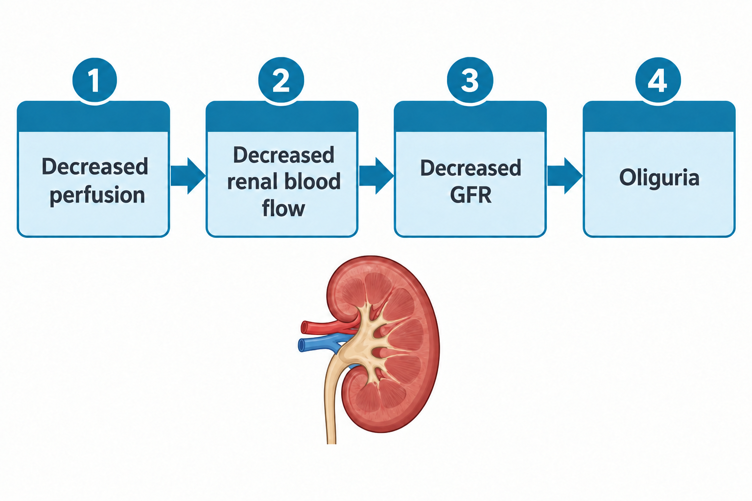
The kidneys are highly sensitive to perfusion changes, so decreased urine output is often an early sign of shock.
Common causes
- Hypoperfusion
- Sepsis
- Nephrotoxic medications
- Acute-on-chronic kidney disease
Key assessment findings
- Oliguria (<30 mL/hr)
- Rising creatinine
- Fluid overload
- Electrolyte disturbances
- Hypertension (fluid retention)
Gastrointestinal system alterations
Common causes
- Ileus
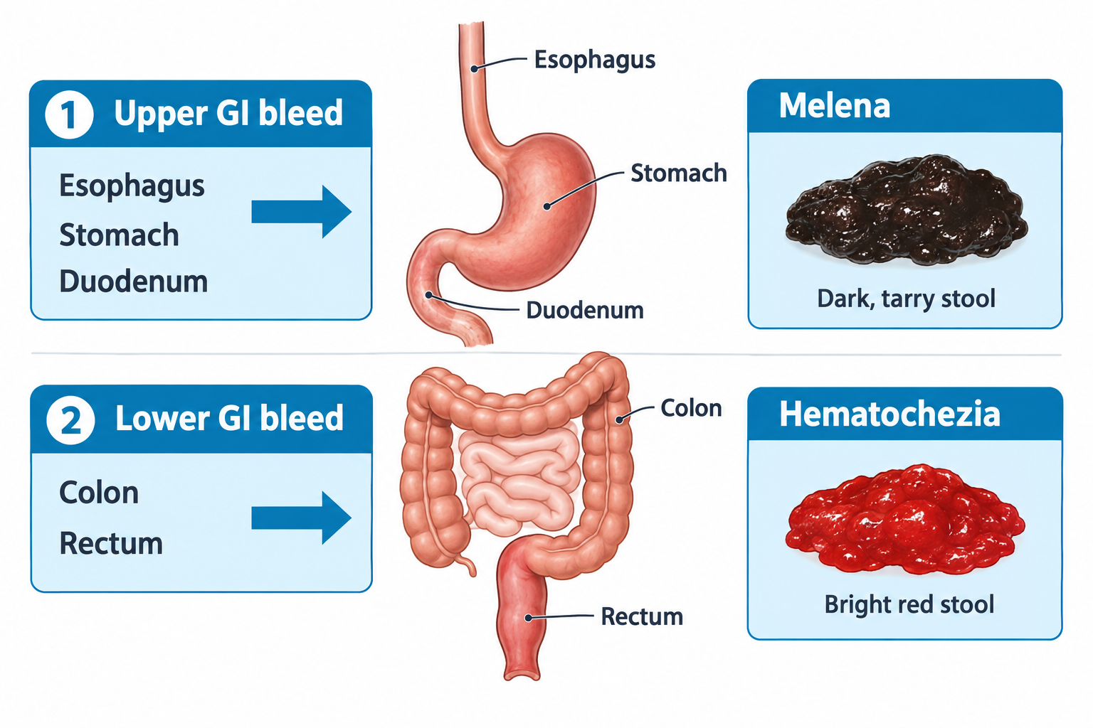
- GI bleeding
- Bowel obstruction
- Infection
Key assessment findings
- Abdominal distention
- Absent or hyperactive bowel sounds
- Nausea and vomiting
- Melena or hematemesis
Endocrine and metabolic alterations
Common causes
- Hypoglycemia
- Hyperglycemia
- Thyroid disorders
- Adrenal insufficiency
Metabolic instability often presents with neurological changes first.
Key assessment findings
- Altered mental status
- Diaphoresis
- Tremors
- Abnormal glucose levels
- Hemodynamic instability
Procedural and postoperative care skills
NGN items also test hands-on interventions, not just recognizing deterioration. Key skills:
Wound and drain care
- Assess wounds/drains for infection (redness, warmth, purulent drainage, fever) and track drainage amount, color, and consistency.
- Use sterile technique for dressing changes and drain-site care.
- Report drainage that suddenly increases, changes color, or stops - this can mean infection, a blocked drain, or bleeding.
Chest tubes and negative-pressure wound therapy
- Keep the chest tube system below chest level and watch for fluctuation (“tidaling”) in the water-seal chamber; continuous bubbling suggests an air leak.
- Never clamp a chest tube without a provider order - this can cause a tension pneumothorax.
- A wound vac (negative-pressure wound therapy) needs an airtight seal.
Suctioning, ventilator care, and pulmonary hygiene
- Suction only as needed, pre-oxygenate first, and limit each pass to reduce hypoxia.
- Ventilator care includes head-of-bed elevation (- degrees) and oral care to reduce ventilator-associated pneumonia risk.
- Chest physiotherapy and incentive spirometry mobilize secretions and prevent atelectasis, especially after surgery.
Ostomy care
- A healthy stoma is pink to red and moist; pale, dusky, or black tissue means poor perfusion and needs immediate reporting.
- Empty the pouch when about a third full, and protect peristomal skin from irritation.
Postoperative care
- Monitor airway, vital signs, the surgical site, and pain frequently right after surgery.
- Encourage early ambulation, deep breathing, and leg exercises to prevent pneumonia, DVT, and ileus.
Seizure and increased intracranial pressure (ICP) care
- During a seizure: protect from injury, turn to the side if possible, don’t restrain or place anything in the mouth, and time it.
- After a seizure: assess airway, place in a side-lying recovery position, reorient, and document.
- For increased ICP: elevate the head of the bed, keep the head midline, minimize stimulation, and watch for Cushing’s triad (widening pulse pressure, bradycardia, irregular respirations) as a late sign.
Other supportive therapies
- Peritoneal dialysis: warm the dialysate, watch for cloudy outflow (a sign of peritonitis), and use sterile technique at the catheter site.
- Phototherapy (neonatal hyperbilirubinemia): protect the eyes, keep skin exposed, and monitor hydration and temperature.
Infectious disease and precautions
Caring for a client with an infectious disease means knowing how the organism spreads and where the client is in the incubation period - the time between exposure and first symptoms, when the client may already be contagious.
Monitor response to treatment - fever trend, symptom resolution, repeat labs - to confirm the infection is actually clearing.
Multisystem involvement
When one system fails, others often follow.
Warning signs
- Hypotension + confusion
- Decreased urine output + edema
- Hypoxia + tachycardia
- Fever + tachycardia + hypotension (possible sepsis)
Clinical judgment and prioritization
NGN case studies measure clinical judgment using the NCSBN’s Clinical Judgment Measurement Model (CJMM), a six-step cycle:
- Recognize cues - identify the relevant assessment data.
- Analyze cues - determine which findings are relevant and how they relate.
- Prioritize hypotheses - rank the possible explanations by urgency.
- Generate solutions - identify expected outcomes and interventions.
- Take action - implement the highest-priority intervention(s).
- Evaluate outcomes - determine whether the client improved.
Prioritize in this order:
- Unstable over stable
- Acute over chronic
- Unexpected over expected
- Airway > breathing > circulation
Clinical vignette 1
A hospitalized client develops tachycardia, decreasing urine output, and confusion.
Nursing action: Perform a rapid ABC assessment, monitor vital signs and urine output, administer oxygen as indicated, and notify the provider or activate the rapid response team as appropriate.
Rationale: Tachycardia, decreasing urine output, and confusion suggest worsening systemic hypoperfusion affecting multiple organ systems.
Clinical vignette 2
An older adult with pneumonia becomes increasingly lethargic with falling oxygen saturation.
Nursing action: Administer oxygen as indicated, perform a focused respiratory assessment, monitor oxygen saturation closely, and notify the provider promptly.
Rationale: Increasing lethargy with falling oxygen saturation suggests worsening respiratory failure requiring immediate intervention.