


Achievable logoAchievable logo
NCLEX
Sign in
Sign up
Purchase
Textbook
Practice exams
Support
How it works
Resources
Exam catalog
Mountain with a flag at the peak
Textbook
Introduction
1. Safe and effective care environment
2. Health promotion and maintenance
3. Psychosocial Integrity
4. Physiological Integrity
4.1 Basic care and comfort
4.2 Pharmacological and parenteral therapies
4.3 Reduction of risk potential
4.4 Physiological adaptation
4.4.1 Medical emergencies
4.4.2 Fluid and electrolyte disorders
4.4.3 Acute and chronic conditions management
4.4.4 Alterations in body systems
4.4.5 Organ transplants and end of life changes
4.4.6 Hemodynamic monitoring and critical care concepts
Wrapping up
Achievable logoAchievable logo
4.4.2 Fluid and electrolyte disorders
Achievable NCLEX
4. Physiological Integrity
4.4. Physiological adaptation
Our NCLEX course is currently in development and is a work-in-progress.

Fluid and electrolyte disorders

8 min read
Font
Discuss
Share
Feedback

Introduction

Fluid and electrolyte balance is essential for cellular function, cardiac conduction, neurological stability, and tissue perfusion. Disruptions can lead to rapid physiological decompensation, particularly in older adults, critically ill clients, and those receiving IV therapy, diuretics, or renal replacement therapy.

On the Next generation NCLEX (NGN), these disorders are tested through pattern recognition, prioritization, and integration of labs with assessment findings, rather than memorization alone.

Learning objectives

By the end of this section, the learner should be able to:

  • Differentiate fluid volume disorders from electrolyte abnormalities
  • Recognize early and late clinical manifestations
  • Interpret laboratory values in clinical context
  • Prioritize nursing interventions based on physiological risk

Core principle: water moves first

Water shifts before cells fail.

Electrolyte symptoms often reflect fluid movement, not just lab abnormalities.

NGN tip: Always assess fluid status before reacting to electrolyte values.

Fluid volume disorders

Fluid volume deficit

Fluid volume deficit occurs when fluid losses exceed intake, reducing circulating volume and tissue perfusion.

Common causes

  • Vomiting or diarrhea
  • Diuretics
  • Hemorrhage
  • Poor oral intake

Assessment findings

  • Dry mucous membranes
  • Poor skin turgor
  • Tachycardia
  • Hypotension (late sign)
  • Decreased urine output

NGN tip: Tachycardia and decreased urine output often appear before hypotension in fluid volume deficit.

Definitions
Fluid volume deficit
Reduction in intravascular, interstitial, or intracellular fluid
Fluid volume excess
Excess isotonic fluid in the intravascular space

Fluid volume excess

Fluid volume excess results from retention of sodium and water, overwhelming cardiac and renal compensatory mechanisms.

Common causes

  • Excessive IV fluids
  • Heart failure
  • Renal failure
  • Corticosteroid therapy

Assessment findings

  • Peripheral edema
  • Crackles in lungs
  • Weight gain
  • Bounding pulses
  • Elevated blood pressure

NGN insight: Older adults are more susceptible to fluid overload and may deteriorate rapidly because of reduced cardiac and renal reserve.

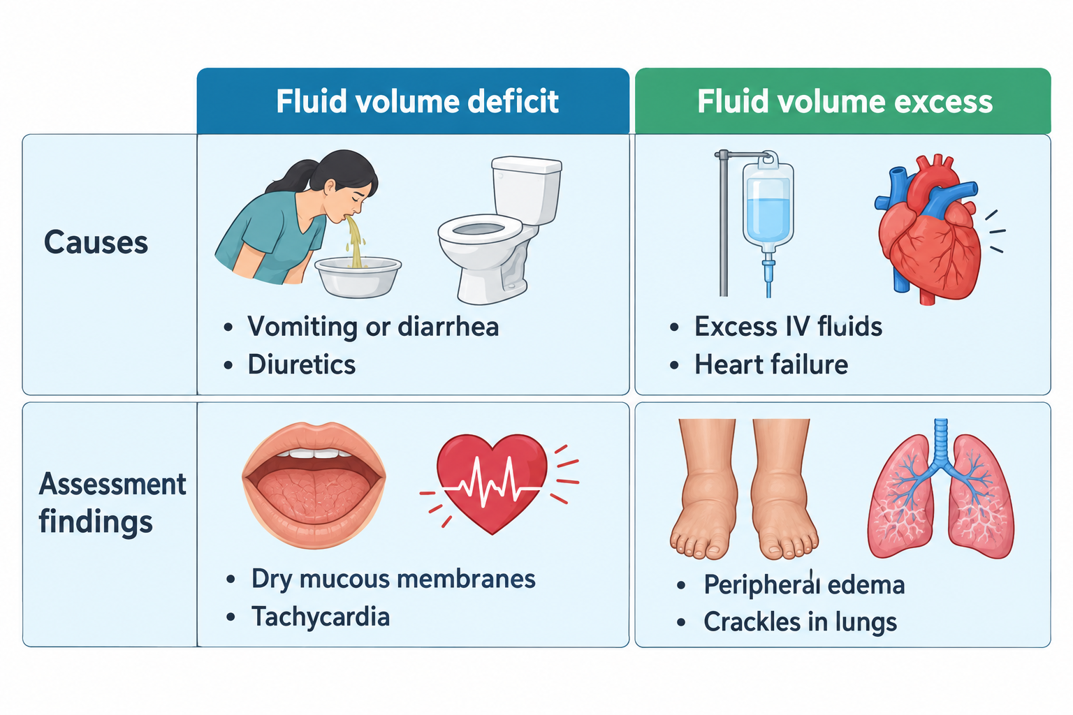
A four-quadrant comparison chart contrasting fluid volume deficit and fluid volume excess. Left column (fluid volume deficit): causes shown as a person vomiting into a basin and a toilet, with text 'Vomiting or diarrhea' and 'Diuretics'; assessment findings shown as a dry mouth/tongue and a heart with a rapid pulse trace, labeled 'Dry mucous membranes' and 'Tachycardia'. Right column (fluid volume excess): causes shown as an IV fluid bag and an enlarged heart, labeled 'Excess IV fluids' and 'Heart failure'; assessment findings shown as swollen feet and lungs, labeled 'Peripheral edema' and 'Crackles in lungs'.
Fluid volume deficit vs fluid volume excess
Achievable

Sodium disorders

Definitions
Hyponatremia
Serum sodium below the normal range.

Hyponatremia

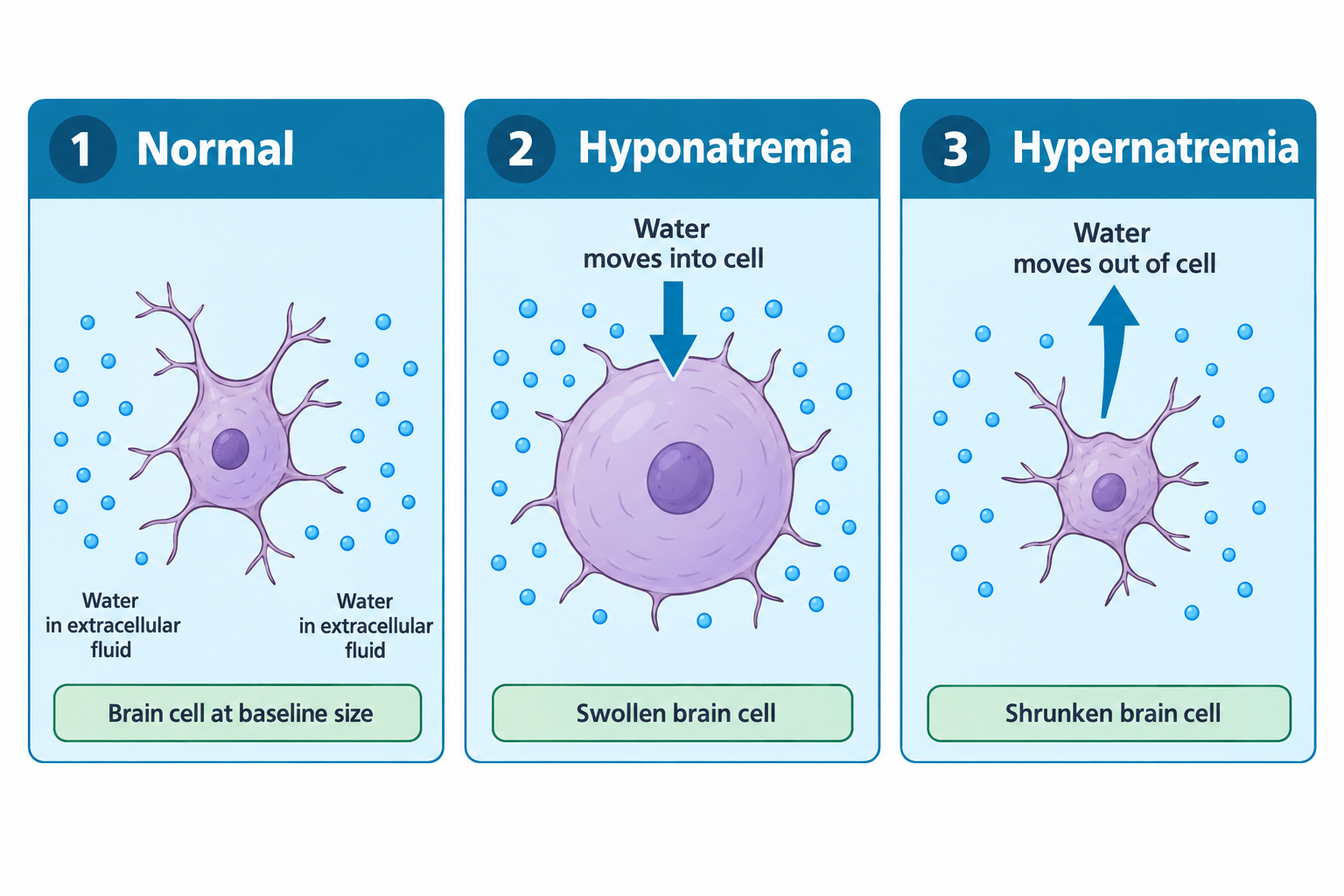
Hyponatremia causes water to shift into brain cells, increasing intracranial pressure.

Key manifestations

  • Headache
  • Confusion
  • Nausea
  • Seizures (severe)

Hypernatremia

Hypernatremia reflects water loss, not excess sodium.

Key manifestations

  • Thirst
  • Dry skin and mucosa
  • Restlessness
  • Confusion

NGN tip: Treat hypernatremia by replacing water, not removing sodium.

Sodium correction risk

Electrolytes should be corrected gradually unless the client is unstable.

Rapid correction of chronic hyponatremia can cause osmotic demyelination syndrome, resulting in permanent neurological injury.

Rapid correction of hypernatremia can cause cerebral edema.

Three-panel diagram comparing brain cell size under different sodium conditions. Panel 1 shows a normal brain cell with balanced water dots in the extracellular fluid on both sides, labeled 'Brain cell at baseline size.' Panel 2 shows hyponatremia, with an arrow indicating water moves into the cell, resulting in a swollen, rounded brain cell. Panel 3 shows hypernatremia, with an arrow indicating water moves out of the cell, resulting in a shrunken brain cell with reduced volume.
Effects of sodium disorders on brain cells
Achievable

Potassium disorders

Definitions
Hypokalemia
Serum potassium below the normal range.

Hypokalemia

Low potassium impairs muscle contraction and GI motility.

Manifestations

  • Muscle weakness
  • Ileus
  • Dysrhythmias

Hyperkalemia

High potassium alters cardiac electrical conduction and can be fatal.

Manifestations

  • Muscle weakness
  • Paresthesias
  • ECG changes
  • Cardiac arrest

NGN insight: Potassium abnormalities threaten the heart first.

ECG correlations

Hypokalemia

  • Flattened T waves
  • Presence of U waves
  • ST depression

Hyperkalemia

  • Peaked T waves
  • Widened QRS complex
  • Prolonged PR interval
  • Sine-wave pattern (pre-arrest)
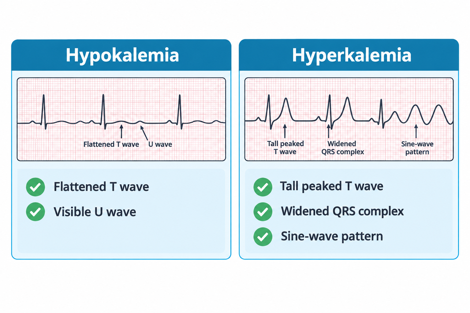
Two side-by-side ECG strips compare potassium imbalances. The hypokalemia strip shows a flattened T wave and a following U wave labeled with arrows, with checkmarked bullets for 'Flattened T wave' and 'Visible U wave'. The hyperkalemia strip shows a tall peaked T wave, a widened QRS complex, and a smooth sine-wave pattern labeled with arrows, with checkmarked bullets for 'Tall peaked T wave', 'Widened QRS complex', and 'Sine-wave pattern'.
ECG changes in potassium disorders
Achievable

Calcium disorders

Definitions
Hypocalcemia
Serum calcium below the normal range.

Hypocalcemia

Low calcium increases neuromuscular excitability.

Manifestations

  • Tetany
  • Muscle cramps
  • Positive Chvostek or Trousseau signs

Hypercalcemia

High calcium reduces neuromuscular activity and kidney function.

Manifestations

  • Lethargy
  • Constipation
  • Kidney stones
  • Bone pain

NGN tip: To remember hypercalcemia, rhyme → stones, bones, groans, psychiatric overtones.

Magnesium disorders

Hypomagnesemia

  • Tremors
  • Hyperreflexia
  • Dysrhythmias

Hypermagnesemia

  • Hypotension
  • Bradycardia
  • Decreased reflexes
  • Respiratory depression

Magnesium toxicity management

Severe hypermagnesemia may require:

  • IV calcium gluconate (antidote)
  • Loop diuretics
  • Dialysis in renal failure

NGN tip: Loss of deep tendon reflexes is an early sign of magnesium toxicity.

Laboratory interpretation in clinical contexts

Interpreting labs safely

Electrolyte values should never be interpreted in isolation. Always correlate with:

  • Recent IV fluids
  • Renal function (BUN, creatinine)
  • Medication profile
  • Urine output trends
  • Acid-base status

Normal reference ranges (adult)

  • Sodium: 135-145 mEq/L
  • Potassium: 3.5-5.0 mEq/L
  • Calcium: 8.5-10.5 mg/dL
  • Magnesium: 1.5-2.5 mEq/L

NGN insight: Always interpret laboratory values in the context of the client’s clinical condition. A “mildly abnormal” result with symptoms may require more urgent intervention than a severely abnormal value in a stable client.

Acid-base and electrolyte connection

Electrolyte disorders often accompany acid-base imbalance.

  • Metabolic acidosis → hyperkalemia (potassium shifts out of cells)
  • Metabolic alkalosis → hypokalemia
  • NG suctioning → metabolic alkalosis + hypokalemia
  • Diabetic ketoacidosis (DKA) → total body potassium deficit despite normal or elevated serum potassium
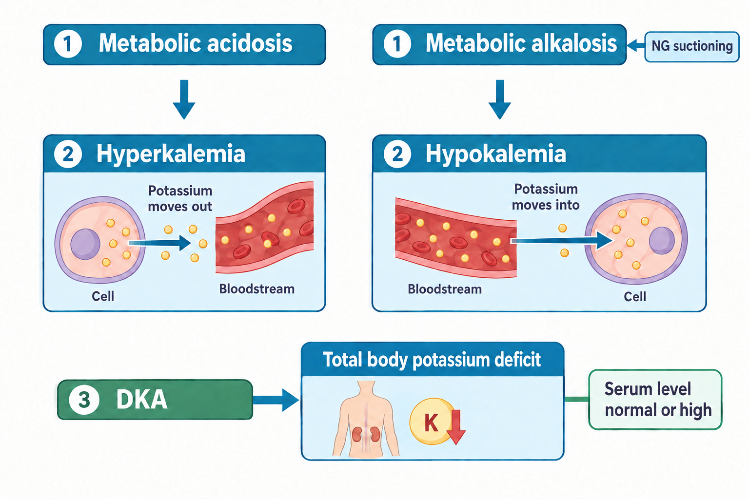
A flowchart in two columns showing potassium shifts with acid-base disorders. Left: metabolic acidosis leads to hyperkalemia, illustrated by potassium ions moving out of a cell into the bloodstream. Right: metabolic alkalosis (also triggered by NG suctioning) leads to hypokalemia, illustrated by potassium ions moving from the bloodstream into a cell. Below, a third pathway shows DKA leading to a total body potassium deficit, depicted with kidneys and a downward-pointing K icon, with a side note that serum potassium level may be normal or high despite the deficit.
Acid-base disorders and potassium shifts
Achievable

Putting it together: NGN clinical judgment

NGN integration

  • Assess the client before reacting to labs
  • Neurological and cardiac symptoms override numeric values
  • Identify the most immediately dangerous imbalance

Clinical vignette 1

A client on hypotonic IV fluids becomes confused. The sodium level is 126 mEq/L.

(spoiler)

Nursing action: Perform a focused neurological assessment, notify the provider promptly, implement fluid restriction as prescribed, and monitor neurological status and serum sodium closely.

Rationale: Confusion with a sodium level of 126 mEq/L suggests symptomatic hyponatremia with cerebral edema requiring prompt intervention.

Clinical vignette 2

A client with renal failure reports weakness and has ECG changes. Potassium is 6.2 mEq/L.

(spoiler)

Nursing action: Initiate continuous cardiac monitoring, notify the provider immediately, and prepare for treatment of hyperkalemia as prescribed.

Rationale: Muscle weakness, ECG changes, and a potassium level of 6.2 mEq/L indicate hyperkalemia with a high risk of life-threatening dysrhythmias.

Common NCLEX pitfalls:

  • Applying adult reference ranges to a client whose age isn’t stated, instead of confirming whether age-specific norms apply
  • Treating an “as prescribed” intervention as something the nurse can initiate independently, rather than confirming an actual prescription exists before acting
  • Fluid balance drives electrolyte balance
  • Neurological and cardiac changes are early warning signs
  • Trend recognition is central to NGN success
  • Older adults decompensate faster
  • Nursing judgment prevents life-threatening outcomes

Sign up for free to take 5 quiz questions on this topic

Previous
Next  | 4.4.3 Acute and chronic conditions management
All rights reserved ©2016 - 2026 Achievable, Inc.

Fluid and electrolyte disorders

Introduction

Fluid and electrolyte balance is essential for cellular function, cardiac conduction, neurological stability, and tissue perfusion. Disruptions can lead to rapid physiological decompensation, particularly in older adults, critically ill clients, and those receiving IV therapy, diuretics, or renal replacement therapy.

On the Next generation NCLEX (NGN), these disorders are tested through pattern recognition, prioritization, and integration of labs with assessment findings, rather than memorization alone.

Learning objectives

By the end of this section, the learner should be able to:

  • Differentiate fluid volume disorders from electrolyte abnormalities
  • Recognize early and late clinical manifestations
  • Interpret laboratory values in clinical context
  • Prioritize nursing interventions based on physiological risk

Core principle: water moves first

Water shifts before cells fail.

Electrolyte symptoms often reflect fluid movement, not just lab abnormalities.

NGN tip: Always assess fluid status before reacting to electrolyte values.

Fluid volume disorders

Fluid volume deficit

Fluid volume deficit occurs when fluid losses exceed intake, reducing circulating volume and tissue perfusion.

Common causes

  • Vomiting or diarrhea
  • Diuretics
  • Hemorrhage
  • Poor oral intake

Assessment findings

  • Dry mucous membranes
  • Poor skin turgor
  • Tachycardia
  • Hypotension (late sign)
  • Decreased urine output

NGN tip: Tachycardia and decreased urine output often appear before hypotension in fluid volume deficit.

Definitions
Fluid volume deficit
Reduction in intravascular, interstitial, or intracellular fluid
Fluid volume excess
Excess isotonic fluid in the intravascular space

Fluid volume excess

Fluid volume excess results from retention of sodium and water, overwhelming cardiac and renal compensatory mechanisms.

Common causes

  • Excessive IV fluids
  • Heart failure
  • Renal failure
  • Corticosteroid therapy

Assessment findings

  • Peripheral edema
  • Crackles in lungs
  • Weight gain
  • Bounding pulses
  • Elevated blood pressure

NGN insight: Older adults are more susceptible to fluid overload and may deteriorate rapidly because of reduced cardiac and renal reserve.

Sodium disorders

Definitions
Hyponatremia
Serum sodium below the normal range.

Hyponatremia

Hyponatremia causes water to shift into brain cells, increasing intracranial pressure.

Key manifestations

  • Headache
  • Confusion
  • Nausea
  • Seizures (severe)

Hypernatremia

Hypernatremia reflects water loss, not excess sodium.

Key manifestations

  • Thirst
  • Dry skin and mucosa
  • Restlessness
  • Confusion

NGN tip: Treat hypernatremia by replacing water, not removing sodium.

Sodium correction risk

Electrolytes should be corrected gradually unless the client is unstable.

Rapid correction of chronic hyponatremia can cause osmotic demyelination syndrome, resulting in permanent neurological injury.

Rapid correction of hypernatremia can cause cerebral edema.

Potassium disorders

Definitions
Hypokalemia
Serum potassium below the normal range.

Hypokalemia

Low potassium impairs muscle contraction and GI motility.

Manifestations

  • Muscle weakness
  • Ileus
  • Dysrhythmias

Hyperkalemia

High potassium alters cardiac electrical conduction and can be fatal.

Manifestations

  • Muscle weakness
  • Paresthesias
  • ECG changes
  • Cardiac arrest

NGN insight: Potassium abnormalities threaten the heart first.

ECG correlations

Hypokalemia

  • Flattened T waves
  • Presence of U waves
  • ST depression

Hyperkalemia

  • Peaked T waves
  • Widened QRS complex
  • Prolonged PR interval
  • Sine-wave pattern (pre-arrest)

Calcium disorders

Definitions
Hypocalcemia
Serum calcium below the normal range.

Hypocalcemia

Low calcium increases neuromuscular excitability.

Manifestations

  • Tetany
  • Muscle cramps
  • Positive Chvostek or Trousseau signs

Hypercalcemia

High calcium reduces neuromuscular activity and kidney function.

Manifestations

  • Lethargy
  • Constipation
  • Kidney stones
  • Bone pain

NGN tip: To remember hypercalcemia, rhyme → stones, bones, groans, psychiatric overtones.

Magnesium disorders

Hypomagnesemia

  • Tremors
  • Hyperreflexia
  • Dysrhythmias

Hypermagnesemia

  • Hypotension
  • Bradycardia
  • Decreased reflexes
  • Respiratory depression

Magnesium toxicity management

Severe hypermagnesemia may require:

  • IV calcium gluconate (antidote)
  • Loop diuretics
  • Dialysis in renal failure

NGN tip: Loss of deep tendon reflexes is an early sign of magnesium toxicity.

Laboratory interpretation in clinical contexts

Interpreting labs safely

Electrolyte values should never be interpreted in isolation. Always correlate with:

  • Recent IV fluids
  • Renal function (BUN, creatinine)
  • Medication profile
  • Urine output trends
  • Acid-base status

Normal reference ranges (adult)

  • Sodium: 135-145 mEq/L
  • Potassium: 3.5-5.0 mEq/L
  • Calcium: 8.5-10.5 mg/dL
  • Magnesium: 1.5-2.5 mEq/L

NGN insight: Always interpret laboratory values in the context of the client’s clinical condition. A “mildly abnormal” result with symptoms may require more urgent intervention than a severely abnormal value in a stable client.

Acid-base and electrolyte connection

Electrolyte disorders often accompany acid-base imbalance.

  • Metabolic acidosis → hyperkalemia (potassium shifts out of cells)
  • Metabolic alkalosis → hypokalemia
  • NG suctioning → metabolic alkalosis + hypokalemia
  • Diabetic ketoacidosis (DKA) → total body potassium deficit despite normal or elevated serum potassium

Putting it together: NGN clinical judgment

NGN integration

  • Assess the client before reacting to labs
  • Neurological and cardiac symptoms override numeric values
  • Identify the most immediately dangerous imbalance

Clinical vignette 1

A client on hypotonic IV fluids becomes confused. The sodium level is 126 mEq/L.

(spoiler)

Nursing action: Perform a focused neurological assessment, notify the provider promptly, implement fluid restriction as prescribed, and monitor neurological status and serum sodium closely.

Rationale: Confusion with a sodium level of 126 mEq/L suggests symptomatic hyponatremia with cerebral edema requiring prompt intervention.

Clinical vignette 2

A client with renal failure reports weakness and has ECG changes. Potassium is 6.2 mEq/L.

(spoiler)

Nursing action: Initiate continuous cardiac monitoring, notify the provider immediately, and prepare for treatment of hyperkalemia as prescribed.

Rationale: Muscle weakness, ECG changes, and a potassium level of 6.2 mEq/L indicate hyperkalemia with a high risk of life-threatening dysrhythmias.

Common NCLEX pitfalls:

  • Applying adult reference ranges to a client whose age isn’t stated, instead of confirming whether age-specific norms apply
  • Treating an “as prescribed” intervention as something the nurse can initiate independently, rather than confirming an actual prescription exists before acting
Key points
  • Fluid balance drives electrolyte balance
  • Neurological and cardiac changes are early warning signs
  • Trend recognition is central to NGN success
  • Older adults decompensate faster
  • Nursing judgment prevents life-threatening outcomes

More from Physiological adaptation

  • Medical emergencies
  • Acute and chronic conditions management
  • Alterations in body systems
  • Organ transplants and end of life changes
  • Hemodynamic monitoring and critical care concepts