


Achievable logoAchievable logo
NCLEX
Sign in
Sign up
Purchase
Textbook
Practice exams
Support
How it works
Exam catalog
Mountain with a flag at the peak
Textbook
Introduction
1. Safe and effective care environment
2. Health promotion and maintenance
3. Psychosocial Integrity
4. Physiological Integrity
4.1 Basic care and comfort
4.2 Pharmacological and parenteral therapies
4.2.1 Intravenous therapy
4.2.2 Medication administration principles
4.2.3 Blood and blood product administration
4.2.4 Pharmacodynamics and side effects
4.2.5 High-risk medications
4.2.6 Adverse effects, contraindications, and interactions
4.2.7 Dosage calculations and safe administration
4.3 Reduction of risk potential
4.4 Physiological adaptation
Wrapping up
Achievable logoAchievable logo
4.2.1 Intravenous therapy
Achievable NCLEX
4. Physiological Integrity
4.2. Pharmacological and parenteral therapies
Our NCLEX course is currently in development and is a work-in-progress.

Intravenous therapy

6 min read
Font
Discuss
Share
Feedback

Introduction

Intravenous (IV) therapy is a vital route for administering fluids, medications, blood products, and nutrition, especially when rapid absorption is required. For nurses, proficiency in IV care goes beyond just inserting a line: it includes fluid selection, complication prevention, monitoring for infection, and recognizing when central access is needed.

The NCLEX frequently tests on IV site assessments, complication management, and types of access devices.

NGN insight:
IV therapy is more than just “getting the line.” It’s about maintaining vascular access, preventing harm, and ensuring what enters the vein heals, not harms.

Learning objectives

By the end of this section, you should be able to:

  • Identify types of IV fluids and their clinical indications
  • Differentiate between peripheral and central lines, including PICCs and ports
  • Recognize and manage IV-related complications
  • Follow sterile and aseptic protocols when handling IV access
  • Document and monitor IV fluid administration accurately

Types of IV fluids

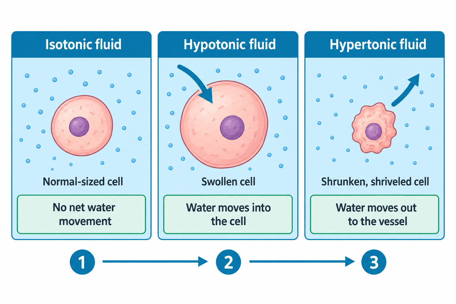
IV fluids are classified by tonicity; their concentration compared to plasma:

Type Examples Use
Isotonic 0.9% NS, LR, D5W (initially isotonic in the bag) Hydration, hypotension, post-op fluids
Hypotonic 0.45% NS, 0.33% NS Hypernatremia and cellular dehydration (use with caution)
Hypertonic 3% NS, D5NS, D5 0.45% NS, D10W Selected indications (e.g., severe hyponatremia, cerebral edema)
alt_text
//////Caption: IV Fluid Tonicity and Fluid Shifts
Type: Three-panel comparison diagram///////
Definitions
Isotonic fluids
Similar osmolarity to plasma; expands intravascular volume without significant fluid shifts
Hypotonic fluids
Lower osmolarity than plasma causes water to shift into cells
Hypertonic fluids
Higher osmolarity; pulls fluid from cells into the intravascular space

Peripheral IVs

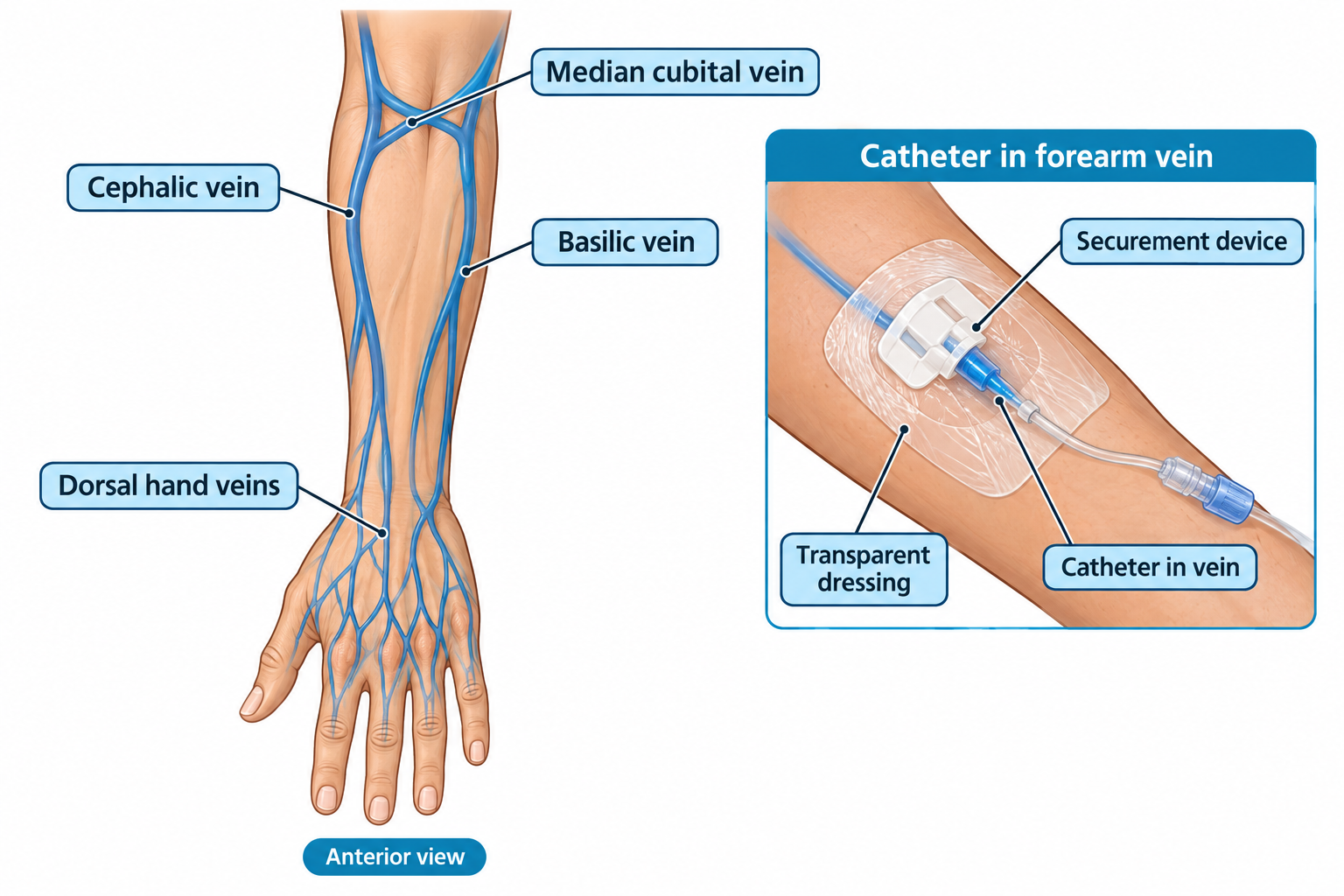
Peripheral IV lines are placed in the arm or hand for short-term access.

Nursing interventions:

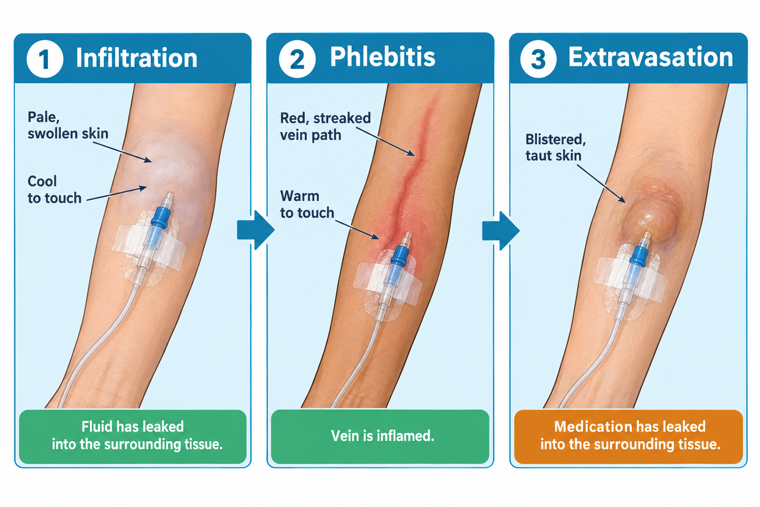
  • Inspect for signs of infiltration (swelling, cool skin), phlebitis (redness, warmth), or infection.
  • Replace peripheral IV catheters based on clinical indication and institutional policy.
  • Secure tubing and label lines.
  • Flush per protocol to prevent occlusion.
  • Avoid using the same arm as an arteriovenous fistula, significant injury, or affected arm after breast surgery (per institutional policy).
alt_text
//////Caption: Common Peripheral IV Insertion Sites
Type: Labeled anatomical illustration
Description: diagram of peripheral IV with Preferred upper-extremity veins and labeled key nursing considerations for peripheral IV placement.
Illustration note: Dorsal hand veins, Cephalic vein, Basilic vein, Median cubital vein, Catheter, Securement device, Dressing///////

Central venous access devices (CVADs)

Used for long-term therapy, vesicant drugs, TPN, or poor peripheral access.

Type Examples Nursing notes
PICC (Peripherally Inserted Central Catheter) Inserted in arm, tip ends in SVC Use sterile technique for dressing changes. Monitor the external catheter length for migration.
Central line (CVC) Subclavian, internal jugular, femoral High infection risk. Verify tip placement before use per institutional policy.
Implanted port Under skin (e.g., for chemo) Accessed with a Huber needle. Flush when not in use (typically every 4 to 12 weeks per institutional policy).
Definitions
Vesicant drugs
Medications (e.g., chemo) that cause tissue damage if extravasated
SVC
Superior vena cava: ideal tip placement for central catheters
Huber needle
Non-coring needle used to access implanted ports

IV complications and nursing interventions

Complication Signs Nursing action
Infiltration Cool, swollen, pale skin Stop infusion, elevate the limb, and apply warm or cold compresses as indicated
Phlebitis Red, warm, tender vein Discontinue IV, apply warm compress, document
Infection Redness, pus, fever Notify provider, send cultures, start antibiotics
Air embolism Chest pain, dyspnea, cyanosis, ↓BP Clamp the line, administer oxygen, position the client appropriately, and activate the rapid response team
Fluid overload Crackles, edema, ↑BP, JVD Reduce or stop the infusion as indicated, elevate the head of the bed, notify the provider, and administer diuretics as ordered
Extravasation Burning, stinging, blistering Stop infusion, aspirate the drug if appropriate, and follow the institution’s extravasation protocol.
alt_text
//////Caption: Recognizing Common IV Complications
Type: Comparison infographic
Showing: infiltration vs. phlebitis vs. extravasation///////

Nursing documentation and safety

  • Verify orders: correct fluid, rate, additives, duration.
  • Label bags, tubing, and sites.
  • Use electronic pumps for accuracy.
  • Document start time, site, fluid type, rate, and client tolerance.
  • Change tubing and dressings per institutional protocol.
alt_text
//////Caption: Essential IV Pump Safety Checks
Type: Labeled illustration of an electronic infusion pump and IV setup///////

Clinical vignette: A nurse is caring for a client receiving D5 0.45% NS at 125 mL/hr via a PICC. During a flush, the client reports burning. The nurse stops the flush, checks for blood return (none), and notes swelling at the site. Suspecting catheter migration with possible extravasation, she halts the infusion, elevates the arm, applies a warm compress, and notifies the provider. The PICC is removed and replaced after confirmation of tip migration.

Key actions: Recognized a complication, stopped the infusion, initiated appropriate care, and documented thoroughly.

Common pitfalls on the NCLEX

  • Starting IV fluids without verifying the correct rate or drug
  • Ignoring signs of fluid overload in cardiac or renal patients
  • Failing to check for blood return before administering chemo via a central line
  • Not using aseptic technique when accessing CVADs
  • Forgetting to clamp the catheter during removal, risking air embolism
  • IV therapy requires knowledge of fluids, access types, and complications
  • Always use sterile technique for central line care
  • Watch for fluid overload, especially in older or cardiac clients
  • Flush per protocol and assess site regularly
  • Infiltration ≠ phlebitis ≠ infection — know the difference

Sign up for free to take 6 quiz questions on this topic

Previous
Next  | 4.2.2 Medication administration principles
All rights reserved ©2016 - 2026 Achievable, Inc.

Intravenous therapy

Introduction

Intravenous (IV) therapy is a vital route for administering fluids, medications, blood products, and nutrition, especially when rapid absorption is required. For nurses, proficiency in IV care goes beyond just inserting a line: it includes fluid selection, complication prevention, monitoring for infection, and recognizing when central access is needed.

The NCLEX frequently tests on IV site assessments, complication management, and types of access devices.

NGN insight:
IV therapy is more than just “getting the line.” It’s about maintaining vascular access, preventing harm, and ensuring what enters the vein heals, not harms.

Learning objectives

By the end of this section, you should be able to:

  • Identify types of IV fluids and their clinical indications
  • Differentiate between peripheral and central lines, including PICCs and ports
  • Recognize and manage IV-related complications
  • Follow sterile and aseptic protocols when handling IV access
  • Document and monitor IV fluid administration accurately

Types of IV fluids

IV fluids are classified by tonicity; their concentration compared to plasma:

Type Examples Use
Isotonic 0.9% NS, LR, D5W (initially isotonic in the bag) Hydration, hypotension, post-op fluids
Hypotonic 0.45% NS, 0.33% NS Hypernatremia and cellular dehydration (use with caution)
Hypertonic 3% NS, D5NS, D5 0.45% NS, D10W Selected indications (e.g., severe hyponatremia, cerebral edema)
alt_text
//////Caption: IV Fluid Tonicity and Fluid Shifts
Type: Three-panel comparison diagram///////
Definitions
Isotonic fluids
Similar osmolarity to plasma; expands intravascular volume without significant fluid shifts
Hypotonic fluids
Lower osmolarity than plasma causes water to shift into cells
Hypertonic fluids
Higher osmolarity; pulls fluid from cells into the intravascular space

Peripheral IVs

Peripheral IV lines are placed in the arm or hand for short-term access.

Nursing interventions:

  • Inspect for signs of infiltration (swelling, cool skin), phlebitis (redness, warmth), or infection.
  • Replace peripheral IV catheters based on clinical indication and institutional policy.
  • Secure tubing and label lines.
  • Flush per protocol to prevent occlusion.
  • Avoid using the same arm as an arteriovenous fistula, significant injury, or affected arm after breast surgery (per institutional policy).
alt_text
//////Caption: Common Peripheral IV Insertion Sites
Type: Labeled anatomical illustration
Description: diagram of peripheral IV with Preferred upper-extremity veins and labeled key nursing considerations for peripheral IV placement.
Illustration note: Dorsal hand veins, Cephalic vein, Basilic vein, Median cubital vein, Catheter, Securement device, Dressing///////

Central venous access devices (CVADs)

Used for long-term therapy, vesicant drugs, TPN, or poor peripheral access.

Type Examples Nursing notes
PICC (Peripherally Inserted Central Catheter) Inserted in arm, tip ends in SVC Use sterile technique for dressing changes. Monitor the external catheter length for migration.
Central line (CVC) Subclavian, internal jugular, femoral High infection risk. Verify tip placement before use per institutional policy.
Implanted port Under skin (e.g., for chemo) Accessed with a Huber needle. Flush when not in use (typically every 4 to 12 weeks per institutional policy).
Definitions
Vesicant drugs
Medications (e.g., chemo) that cause tissue damage if extravasated
SVC
Superior vena cava: ideal tip placement for central catheters
Huber needle
Non-coring needle used to access implanted ports

IV complications and nursing interventions

Complication Signs Nursing action
Infiltration Cool, swollen, pale skin Stop infusion, elevate the limb, and apply warm or cold compresses as indicated
Phlebitis Red, warm, tender vein Discontinue IV, apply warm compress, document
Infection Redness, pus, fever Notify provider, send cultures, start antibiotics
Air embolism Chest pain, dyspnea, cyanosis, ↓BP Clamp the line, administer oxygen, position the client appropriately, and activate the rapid response team
Fluid overload Crackles, edema, ↑BP, JVD Reduce or stop the infusion as indicated, elevate the head of the bed, notify the provider, and administer diuretics as ordered
Extravasation Burning, stinging, blistering Stop infusion, aspirate the drug if appropriate, and follow the institution’s extravasation protocol.
alt_text
//////Caption: Recognizing Common IV Complications
Type: Comparison infographic
Showing: infiltration vs. phlebitis vs. extravasation///////

Nursing documentation and safety

  • Verify orders: correct fluid, rate, additives, duration.
  • Label bags, tubing, and sites.
  • Use electronic pumps for accuracy.
  • Document start time, site, fluid type, rate, and client tolerance.
  • Change tubing and dressings per institutional protocol.
alt_text
//////Caption: Essential IV Pump Safety Checks
Type: Labeled illustration of an electronic infusion pump and IV setup///////

Clinical vignette: A nurse is caring for a client receiving D5 0.45% NS at 125 mL/hr via a PICC. During a flush, the client reports burning. The nurse stops the flush, checks for blood return (none), and notes swelling at the site. Suspecting catheter migration with possible extravasation, she halts the infusion, elevates the arm, applies a warm compress, and notifies the provider. The PICC is removed and replaced after confirmation of tip migration.

Key actions: Recognized a complication, stopped the infusion, initiated appropriate care, and documented thoroughly.

Common pitfalls on the NCLEX

  • Starting IV fluids without verifying the correct rate or drug
  • Ignoring signs of fluid overload in cardiac or renal patients
  • Failing to check for blood return before administering chemo via a central line
  • Not using aseptic technique when accessing CVADs
  • Forgetting to clamp the catheter during removal, risking air embolism
Key points
  • IV therapy requires knowledge of fluids, access types, and complications
  • Always use sterile technique for central line care
  • Watch for fluid overload, especially in older or cardiac clients
  • Flush per protocol and assess site regularly
  • Infiltration ≠ phlebitis ≠ infection — know the difference

More from Pharmacological and parenteral therapies

  • Medication administration principles
  • Blood and blood product administration
  • Pharmacodynamics and side effects
  • High-risk medications
  • Adverse effects, contraindications, and interactions