


Achievable logoAchievable logo
NCLEX
Sign in
Sign up
Purchase
Textbook
Practice exams
Support
How it works
Exam catalog
Mountain with a flag at the peak
Textbook
Introduction
1. Safe and effective care environment
2. Health promotion and maintenance
2.1 Growth and development across the lifespan
2.2 Antepartum, intrapartum and postpartum
2.2.1 Antepartum care
2.2.2 Intrapartum care
2.2.3 Postpartum care
2.3 Newborn care and developmental milestones
2.4 Health screenings and preventive care (vision, cancer, immunizations)
2.5 Lifestyle counseling and high-risk behaviors
2.6 Aging, transitions, and end-of-life considerations
3. Psychosocial Integrity
4. Physiological Integrity
Wrapping up
Achievable logoAchievable logo
2.2.2 Intrapartum care
Achievable NCLEX
2. Health promotion and maintenance
2.2. Antepartum, intrapartum and postpartum
Our NCLEX course is currently in development and is a work-in-progress.

Intrapartum care

12 min read
Font
Discuss
Share
Feedback

Introduction

Intrapartum care is the high-stakes center of maternity nursing, where every contraction, every fetal heart tracing, every assessment carries the weight of two lives. Labor is a physiologic process, but it can shift into an emergency within seconds, requiring nurses to respond with precision, clarity, and calm.

On the NCLEX, intrapartum questions test your ability to prioritize maternal safety, protect fetal oxygenation, and recognize early warning signs that require immediate intervention.

Learning objectives

By the end of this section, you should be able to:

  • Describe the stages and phases of labor.
  • Interpret fetal heart rate patterns and identify Category I, II, III tracings.
  • Recognize obstetric emergencies and apply correct interventions.
  • Manage maternal comfort, positioning, and pharmacologic pain relief.
  • Provide evidence-based support for physiologic labor and safe delivery.
  • Distinguish normal from abnormal labor patterns.
  • Apply NCLEX reasoning to “first,” “best,” and “priority” nursing actions.
Definitions
McRoberts maneuver
Hyperflexing maternal legs to open the pelvic outlet.
Station
Fetal head position relative to the ischial spines.
Uterine atony
Failure of the uterine muscles to contract effectively after childbirth, which prevents the compression of placental blood vessels. The leading cause of postpartum hemorrhage.
Fetal heart rate monitoring
The continuous or intermittent assessment of the fetal pulse during labor to evaluate oxygenation and identify patterns of distress that may require clinical intervention.
Fundal massage
A manual technique where a nurse applies firm, repetitive circular pressure to the top of the uterus to stimulate contractions and stop postpartum bleeding.
Left lateral positioning
Placing the mother on her left side to relieve pressure on the inferior vena cava, thereby optimizing cardiac output and placental blood flow.
Tachysystole
More than 5 contractions in 10 minutes, averaged over 30 minutes → stop oxytocin.
Bloody show
Mucus plug discharge indicating cervical dilation.

Stages and phases of labor

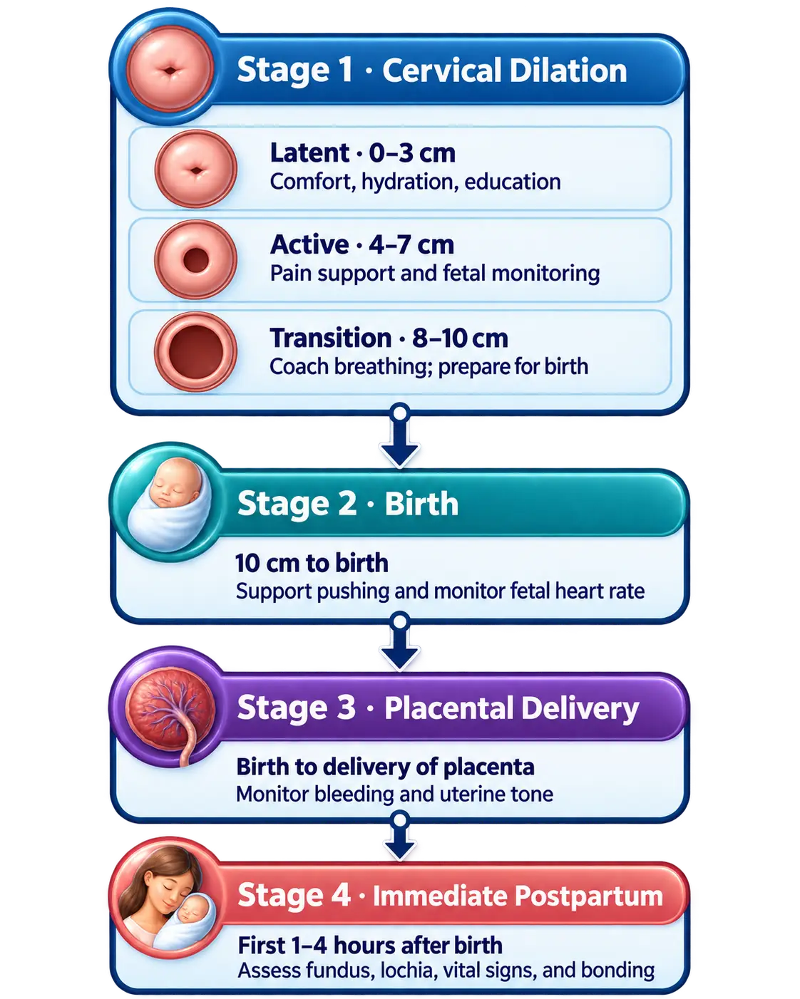
Labor occurs in four stages, each with distinct nursing priorities.

alt_text
//////Caption: The four stages of labor
Illustration type: Timeline infographic
Illustration note: Horizontal timeline showing Stage 1 (latent, active, transition), Stage 2 (birth), Stage 3 (placental delivery), and Stage 4 (immediate postpartum), highlighting cervical dilation and key nursing priorities at each stage.///////

Stage 1: Dilation (onset of labor → 10 cm)

Includes three phases:

1. Latent phase (0–5 cm or early labor)

  • Contractions: mild, irregular → moderate, every 5–10 min
  • Mother alert, talkative; may be anxious or excited
  • Best time for teaching, support, ambulation

Nursing priorities:

  • Encourage ambulation, hydration
  • Breathing techniques
  • Nonpharmacologic pain management
  • Assess fetal heart rate (FHR) regularly

2. Active phase (6–7 cm)

  • Contractions: moderate to strong, q3–5 min
  • Cervical change more rapid
  • Increased discomfort; decreased talking

Nursing priorities:

  • Continuous monitoring of maternal–fetal status
  • Pain management (epidural often placed here)
  • Encourage frequent position changes
  • Observe for signs of progression

3. Transition phase (8–10 cm)

  • Contractions: intense, q2–3 min
  • Nausea/vomiting, shaking, irritability
  • Urge to push often begins

Nursing priorities:

  • Provide reassurance and coaching
  • Slow breathing to prevent premature pushing
  • Prepare room for delivery
  • Monitor FHR closely for distress
alt_text
//////Caption: Phases of Stage 1 labor
Illustration type: Comparison infographic
Illustration note: Side-by-side comparison of latent, active, and transition phases showing cervical dilation, contraction pattern, maternal behaviors, and priority nursing interventions.///////

Stage 2: Pushing and birth (10 cm → birth)

  • Strong urge to push
  • Contractions q2–3 min
  • Fetal descent through pelvis
  • Duration highly variable (minutes → hours)

Nursing priorities:

  • Encourage effective pushing techniques
  • Maintain open glottis breathing
  • Support perineum
  • Monitor FHR q5–15 min
  • Assist provider with delivery
  • Document time of birth, Apgar at 1 and 5 minutes

Stage 3: Delivery of placenta

Usually occurs 5–30 minutes after birth.

Signs of placental separation:

  • Gush of blood
  • Lengthening of the cord
  • Firm, globular uterus rising in abdomen

Nursing priorities:

  • Inspect placenta (intact vs missing lobes)
  • Administer oxytocin to contract uterus
  • Monitor for hemorrhage
  • Fundal massage as needed

Stage 4: Immediate postpartum (first 1–2 hours)

Critical period for maternal stability.

Nursing priorities:

  • Assess fundus, lochia, vitals q15 min
  • Observe for hemorrhage
  • Support bonding, breastfeeding initiation
  • Manage perineal pain
  • Warm blankets, hydration, safety

Fetal heart rate monitoring (NCLEX priority)

Monitoring fetal oxygenation is one of the most heavily tested areas for intrapartum care. This is often estimated through certain adjacent measures, as listed below.

Fetal heart rate characteristics

Intrapartum fetal heart rate monitoring allows nurses to assess fetal oxygenation and identify specific patterns, such as late or variable decelerations, that indicate physiological stress. By recognizing these patterns early, nurses can implement timely interventions, like maternal repositioning or oxygen administration, to mitigate distress and ensure a safe delivery for both mother and child.

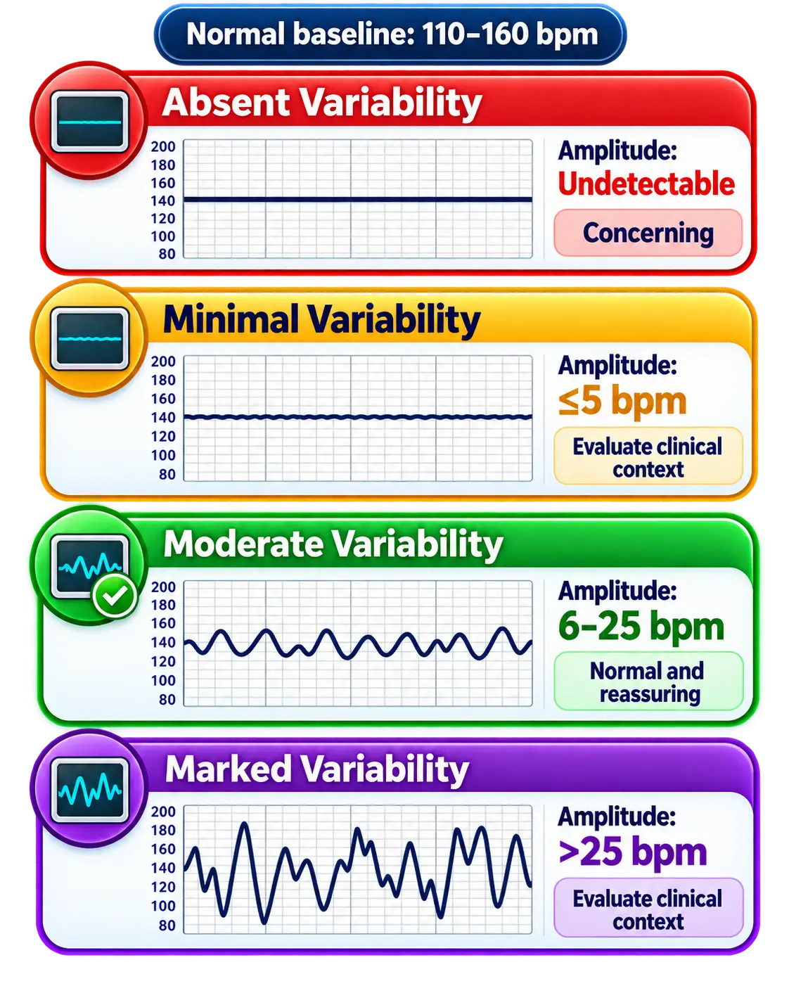
Baseline fetal heart rate: 110–160 bpm

Variability: variation of the heart rate above or below the baseline heart rate. The most important indicator of fetal oxygenation

  • Absent: none (ominous)- sign of lack of oxygenation
  • Minimal: < 5 bpm
  • Moderate: 6–25 bpm (normal)
  • Marked: > 25 bpm
Different variations of fetal heart rates
Fetal heart rate baseline characteristics

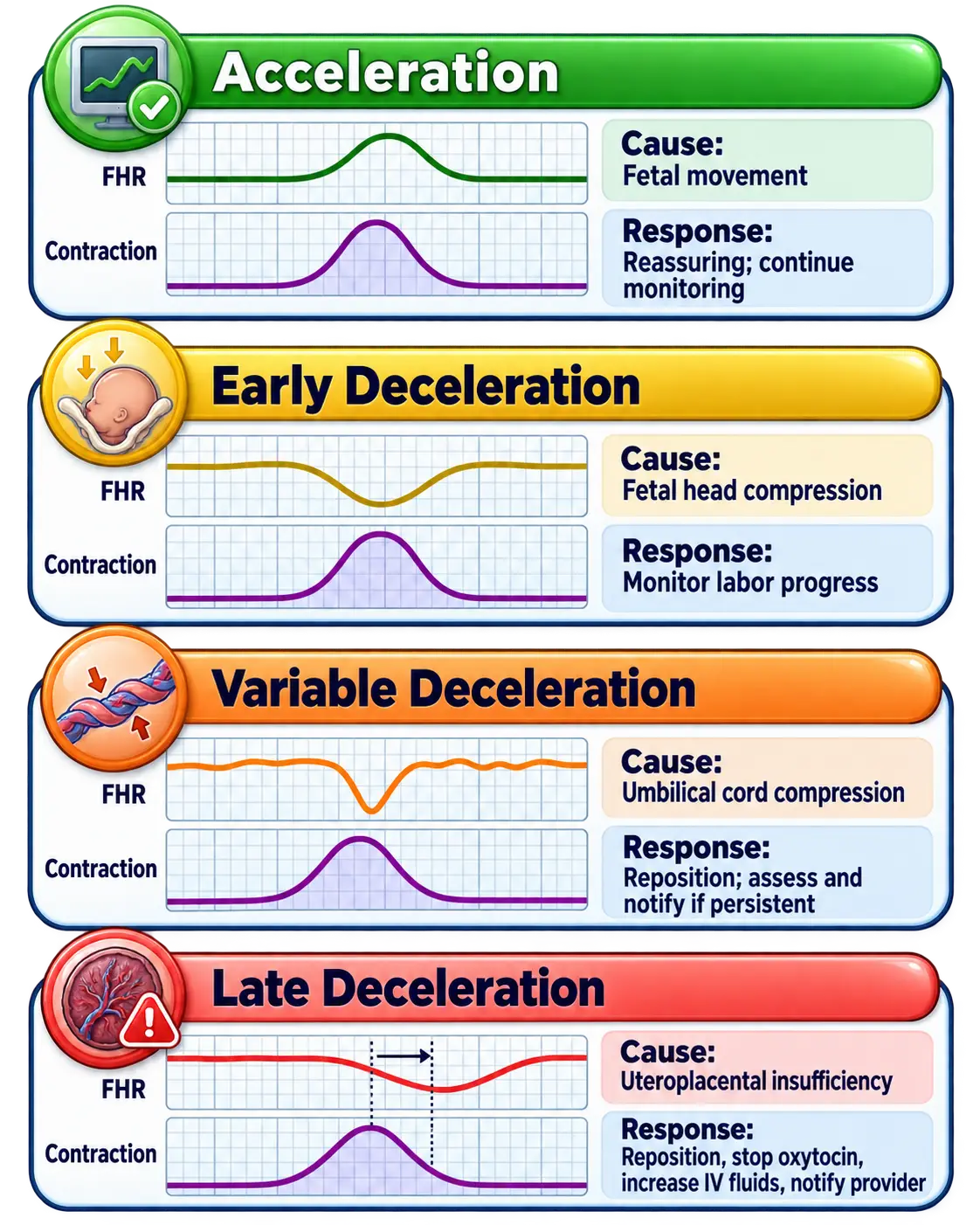
Accelerations:

  • Increase of 15 bpm x 15 seconds
  • Reassuring → fetal well-being

Early decelerations

  • Mirror contractions
  • Caused by head compression
  • Benign

Variable decelerations (V-shaped)

  • Cord compression
  • Interventions:
    • Change maternal position
    • Discontinue oxytocin
    • Administer oxygen

Late decelerations

  • Occur after the contraction peak
  • Sign of uteroplacental insufficiency
  • Interventions (in order):
    1. Reposition to the left side
    2. Stop oxytocin
    3. Oxygen at 8–10 L/min
    4. IV fluids
    5. Notify provider
Acceleration and deceleration of fetal heart rates and implication
Fetal heart rate correlations with intrapartum complications
  • Late decelerations = placental problem.
  • Variable decelerations = cord problem.
  • Early decelerations = head problem (benign).

Category I, II, III tracings

Category I (normal)

  • Baseline 110–160
  • Moderate variability
  • May have early decels or accelerations

Category II (indeterminate)

  • Minimal or marked variability
  • Recurrent variables
  • Prolonged decels
  • Requires close monitoring + interventions

Category III (abnormal)

  • Absent variability with:
    • Recurrent late decels
    • Recurrent variables
    • Bradycardia (< 110)
  • Requires immediate intervention → possible emergency birth

Pain management in labor

This section details nonpharmacologic pain relief methods (like breathing and movement) and pharmacologic options (including epidural anesthesia, systemic opioids, and nitrous oxide) used during labor.

Nonpharmacologic options

  • Breathing, relaxation, massage
  • Warm baths or showers
  • Movement and position changes
  • Counterpressure for back labor

Pharmacologic options

Pharmacological interventions during the intrapartum period require nurses to balance effective pain management and labor progression with the continuous monitoring of maternal and fetal safety. This section explores the clinical indications, administration protocols, and critical nursing considerations for common agents such as oxytocin for induction and systemic opioids or epidurals for analgesia.

Epidural anesthesia

  • This is the most common route of administering a combination of local anesthetics (lidocaine) and opioid analgesics (fentanyl). Regional anesthetics eliminate pain below the waist level while leaving the mother conscious; it is placed during active labor.

  • Watch for hypotension as a complication of the opioid, as the reduced blood pressure can lead to fetal distress.

    Hypotension Interventions:

  • Preload with fluids

  • Left lateral position

  • Monitor BP & FHR regularly

  • Treat hypotension with fluids + ephedrine if ordered

Systemic opioids

Rather than regional anesthesia with epidurals, intravenous opioids allow systemic analgesia and pain relief. Avoid within 1 hour of delivery as these can cause neonatal respiratory depression

Nitrous oxide

Nitrous oxide is administered as a self-inhaled mixture of 50% nitrous oxide and 50% oxygen via a mask or mouthpiece, allowing the mother to control the timing of the dose in coordination with her contractions. This “laughing gas” provides rapid-onset analgesia by increasing endorphin levels and reducing anxiety, while being quickly eliminated from the system to ensure there are no long-term sedative effects on the newborn. It is safe for both the mother and the baby.

Obstetric emergencies (NCLEX heavy)

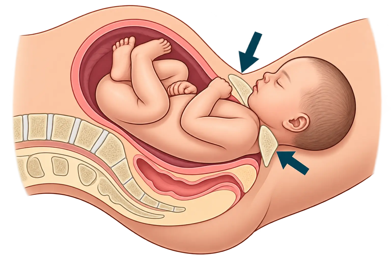
1. Shoulder dystocia

A delivery emergency where the fetal head is born but the anterior shoulder becomes wedged behind the pelvic bone ( look for turtle sign), requiring specific maneuvers like McRoberts to dislodge it.

Interventions:

  • McRoberts maneuver involves sharply flexing the mother’s thighs toward her abdomen to rotate the pelvis and release an impacted fetal shoulder
  • Suprapubic pressure- is applied to the maternal pubic bone to dislodge the fetal anterior shoulder.
  • Do NOT apply fundal pressure as this can further wedge the shoulder against the pubic symphysis and increase the risk of uterine rupture or brachial plexus injury.
Brith complication where fetal obstructs delivery
Shoulder dystocia

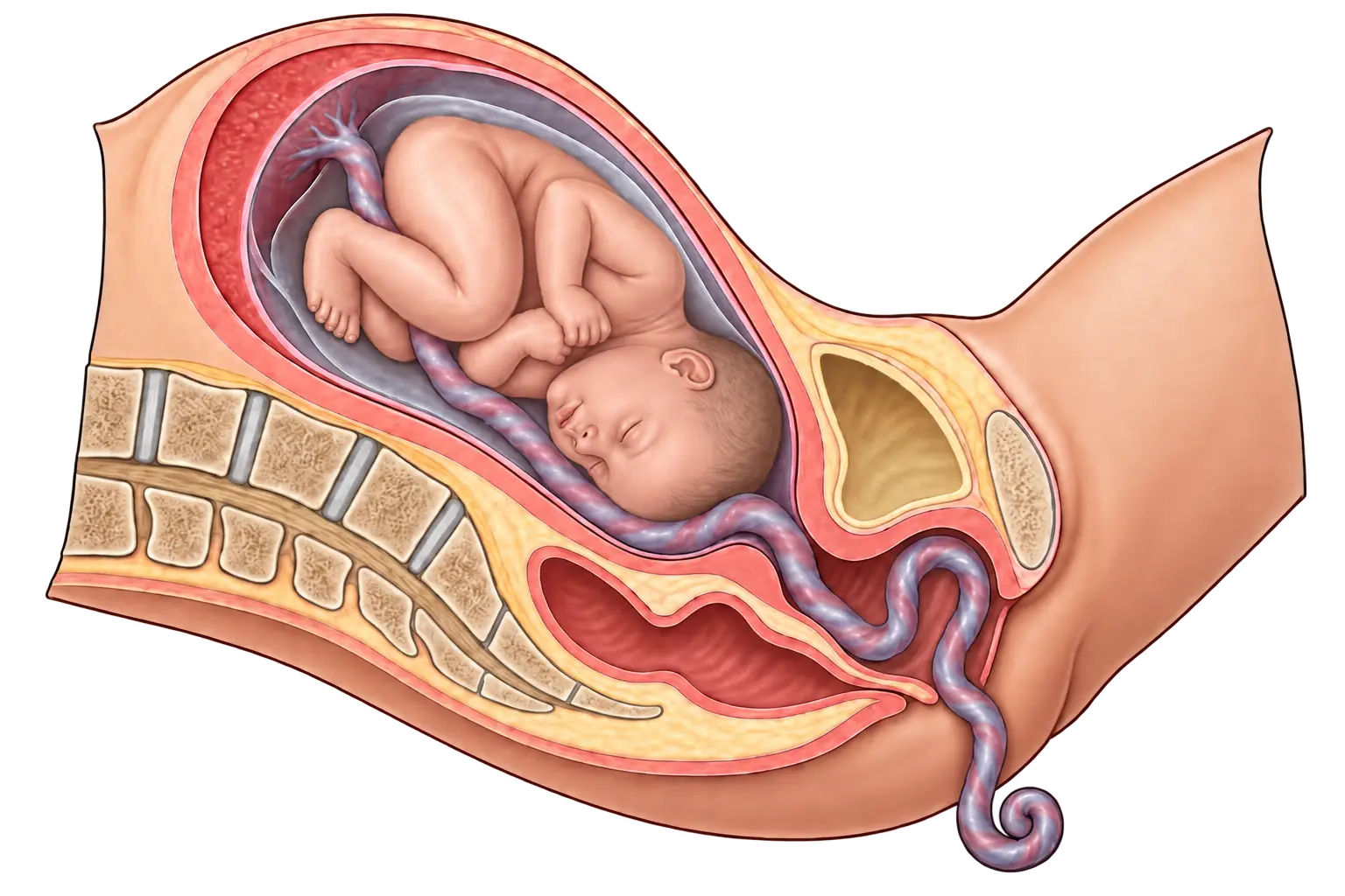
2. Umbilical cord prolapse

A critical complication where the umbilical cord slips through the cervix ahead of the baby, causing cord compression and fetal hypoxia that necessitates immediate elevation of the presenting part to relieve pressure on the umbilicus and an emergency C-section.

Interventions:

  • Call for help
  • Elevate the presenting part ( e.g. fetal head) with the gloved hand
  • Knee-chest or Trendelenburg position
  • Emergency C-section
Birth complication where umbilical cord is delivered first and interrupt fetal blood supply
Umbilical cord prolapse

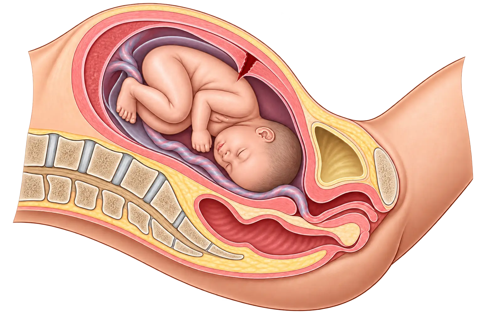
3. Uterine rupture

A catastrophic tearing of the uterine wall, often at the site of a previous scar(usually a C-section performed during a previous pregnancy), resulting in severe maternal hemorrhage and fetal distress that requires immediate surgical intervention.

Risk: Vaginal birth after C-section(VBAC), high parity, obstructed labor.

Signs: sudden pain, loss of fetal station, fetal distress.

Birth complicaiton where the uterus tears leading to heavy bleeding and shock
Uterine rupture

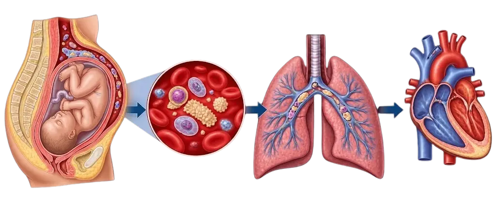
4. Amniotic fluid embolism

A rare but life-threatening condition where amniotic fluid or fetal debris enters the maternal bloodstream, triggering a rapid systemic inflammatory response, cardiovascular collapse, and coagulopathy.

Symptoms: sudden dyspnea → cardiac collapse.

Management: oxygen, CPR, emergency response.

Birth complication where fetal tissue cause massive immue respons in the mother
Amniotic fluid embolism

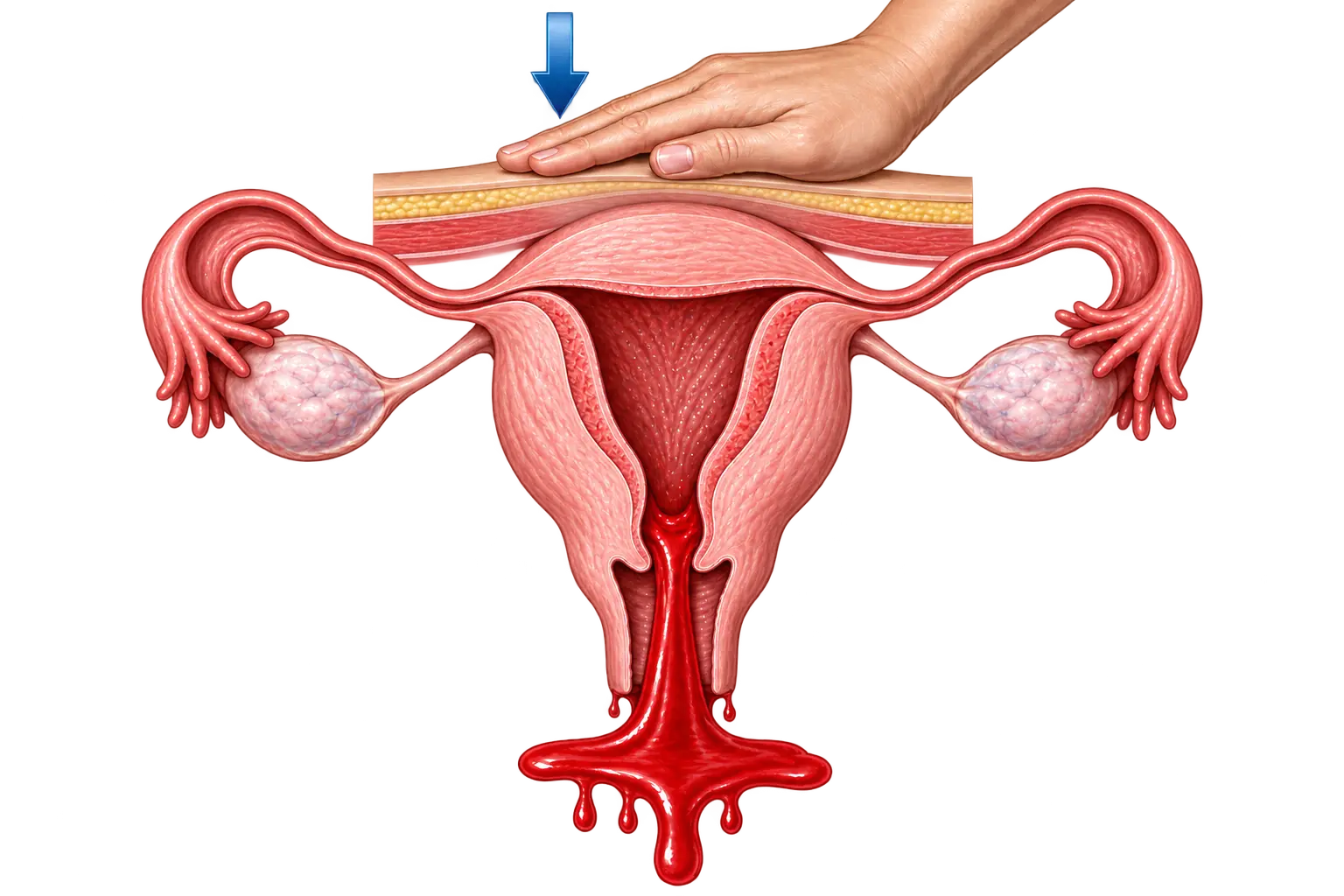
5. Postpartum hemorrhage during stage 3–4

Defined as cumulative blood loss ≥1000 mL, or blood loss accompanied by signs/symptoms of hypovolemia, within 24 hours of birth, regardless of delivery route. This is most commonly caused by uterine atony and managed with fundal massage and uterotonic medications.

Early recognition saves lives.

Interventions include:

  • Fundal massage: stimulates the uterus to contract.
  • Oxytocin, methylergonovine, carboprost: these increase the tone of the uterus.
  • Treat shock from massive blood loss.
  • Locate cause (boggy uterus vs laceration).

NOTE: Older sources define PPH as >500 mL for vaginal and >1000 mL for cesarean delivery. The current ACOG standard (≥1000 mL with hypovolemia signs, any delivery type) is the definition used for current clinical practice and NCLEX.

Birth complication characterised by excessive bleeding during partution
Postpartum hemorrhage

Nursing interventions during labor

  • Monitor FHR continuously
  • Assess the cervix only when indicated
  • Maintain maternal hydration
  • Encourage movement and upright positioning
  • Provide emotional support and labor coaching
  • Ensure safe environment and equipment readiness
  • Manage pain (nonpharmacologic + pharmacologic)
  • Identify early signs of distress or complications

Clinical instance: During active labor, the fetal heart monitor shows recurrent late decelerations with minimal variability. The nurse immediately turns the client on her left side, stops the oxytocin infusion, applies oxygen, boluses IV fluids, and notifies the provider. The fetal tracing improves, preventing an emergent delivery.

Client education

  • What to expect during each stage of labor
  • When to come to the hospital (contractions q5 min, ROM, bleeding, decreased movement)
  • Pain management options
  • Importance of repositioning
  • What fetal monitoring shows
  • Breastfeeding initiation and skin-to-skin after birth

Common pitfalls on the NCLEX

  • Confusing early and late decelerations.
  • Continuing oxytocin during fetal distress.
  • Performing vaginal exams with bleeding of unknown origin.
  • Using fundal pressure during shoulder dystocia.
  • Delaying treatment of epidural-induced hypotension.
  • Underestimating maternal reports of decreased fetal movement.
  • Late decels = placenta; variable decels = cord. Epidurals require fluid preload.
  • McRoberts + suprapubic pressure treat shoulder dystocia.
  • Cord prolapse = manual elevation + emergency C-section.
  • Stage 4 is the most dangerous period for maternal hemorrhage.

Sign up for free to take 5 quiz questions on this topic

Previous
Next  | 2.2.3 Postpartum care
All rights reserved ©2016 - 2026 Achievable, Inc.

Intrapartum care

Introduction

Intrapartum care is the high-stakes center of maternity nursing, where every contraction, every fetal heart tracing, every assessment carries the weight of two lives. Labor is a physiologic process, but it can shift into an emergency within seconds, requiring nurses to respond with precision, clarity, and calm.

On the NCLEX, intrapartum questions test your ability to prioritize maternal safety, protect fetal oxygenation, and recognize early warning signs that require immediate intervention.

Learning objectives

By the end of this section, you should be able to:

  • Describe the stages and phases of labor.
  • Interpret fetal heart rate patterns and identify Category I, II, III tracings.
  • Recognize obstetric emergencies and apply correct interventions.
  • Manage maternal comfort, positioning, and pharmacologic pain relief.
  • Provide evidence-based support for physiologic labor and safe delivery.
  • Distinguish normal from abnormal labor patterns.
  • Apply NCLEX reasoning to “first,” “best,” and “priority” nursing actions.
Definitions
McRoberts maneuver
Hyperflexing maternal legs to open the pelvic outlet.
Station
Fetal head position relative to the ischial spines.
Uterine atony
Failure of the uterine muscles to contract effectively after childbirth, which prevents the compression of placental blood vessels. The leading cause of postpartum hemorrhage.
Fetal heart rate monitoring
The continuous or intermittent assessment of the fetal pulse during labor to evaluate oxygenation and identify patterns of distress that may require clinical intervention.
Fundal massage
A manual technique where a nurse applies firm, repetitive circular pressure to the top of the uterus to stimulate contractions and stop postpartum bleeding.
Left lateral positioning
Placing the mother on her left side to relieve pressure on the inferior vena cava, thereby optimizing cardiac output and placental blood flow.
Tachysystole
More than 5 contractions in 10 minutes, averaged over 30 minutes → stop oxytocin.
Bloody show
Mucus plug discharge indicating cervical dilation.

Stages and phases of labor

Labor occurs in four stages, each with distinct nursing priorities.

alt_text
//////Caption: The four stages of labor
Illustration type: Timeline infographic
Illustration note: Horizontal timeline showing Stage 1 (latent, active, transition), Stage 2 (birth), Stage 3 (placental delivery), and Stage 4 (immediate postpartum), highlighting cervical dilation and key nursing priorities at each stage.///////

Stage 1: Dilation (onset of labor → 10 cm)

Includes three phases:

1. Latent phase (0–5 cm or early labor)

  • Contractions: mild, irregular → moderate, every 5–10 min
  • Mother alert, talkative; may be anxious or excited
  • Best time for teaching, support, ambulation

Nursing priorities:

  • Encourage ambulation, hydration
  • Breathing techniques
  • Nonpharmacologic pain management
  • Assess fetal heart rate (FHR) regularly

2. Active phase (6–7 cm)

  • Contractions: moderate to strong, q3–5 min
  • Cervical change more rapid
  • Increased discomfort; decreased talking

Nursing priorities:

  • Continuous monitoring of maternal–fetal status
  • Pain management (epidural often placed here)
  • Encourage frequent position changes
  • Observe for signs of progression

3. Transition phase (8–10 cm)

  • Contractions: intense, q2–3 min
  • Nausea/vomiting, shaking, irritability
  • Urge to push often begins

Nursing priorities:

  • Provide reassurance and coaching
  • Slow breathing to prevent premature pushing
  • Prepare room for delivery
  • Monitor FHR closely for distress
alt_text
//////Caption: Phases of Stage 1 labor
Illustration type: Comparison infographic
Illustration note: Side-by-side comparison of latent, active, and transition phases showing cervical dilation, contraction pattern, maternal behaviors, and priority nursing interventions.///////

Stage 2: Pushing and birth (10 cm → birth)

  • Strong urge to push
  • Contractions q2–3 min
  • Fetal descent through pelvis
  • Duration highly variable (minutes → hours)

Nursing priorities:

  • Encourage effective pushing techniques
  • Maintain open glottis breathing
  • Support perineum
  • Monitor FHR q5–15 min
  • Assist provider with delivery
  • Document time of birth, Apgar at 1 and 5 minutes

Stage 3: Delivery of placenta

Usually occurs 5–30 minutes after birth.

Signs of placental separation:

  • Gush of blood
  • Lengthening of the cord
  • Firm, globular uterus rising in abdomen

Nursing priorities:

  • Inspect placenta (intact vs missing lobes)
  • Administer oxytocin to contract uterus
  • Monitor for hemorrhage
  • Fundal massage as needed

Stage 4: Immediate postpartum (first 1–2 hours)

Critical period for maternal stability.

Nursing priorities:

  • Assess fundus, lochia, vitals q15 min
  • Observe for hemorrhage
  • Support bonding, breastfeeding initiation
  • Manage perineal pain
  • Warm blankets, hydration, safety

Fetal heart rate monitoring (NCLEX priority)

Monitoring fetal oxygenation is one of the most heavily tested areas for intrapartum care. This is often estimated through certain adjacent measures, as listed below.

Fetal heart rate characteristics

Intrapartum fetal heart rate monitoring allows nurses to assess fetal oxygenation and identify specific patterns, such as late or variable decelerations, that indicate physiological stress. By recognizing these patterns early, nurses can implement timely interventions, like maternal repositioning or oxygen administration, to mitigate distress and ensure a safe delivery for both mother and child.

Baseline fetal heart rate: 110–160 bpm

Variability: variation of the heart rate above or below the baseline heart rate. The most important indicator of fetal oxygenation

  • Absent: none (ominous)- sign of lack of oxygenation
  • Minimal: < 5 bpm
  • Moderate: 6–25 bpm (normal)
  • Marked: > 25 bpm

Accelerations:

  • Increase of 15 bpm x 15 seconds
  • Reassuring → fetal well-being

Early decelerations

  • Mirror contractions
  • Caused by head compression
  • Benign

Variable decelerations (V-shaped)

  • Cord compression
  • Interventions:
    • Change maternal position
    • Discontinue oxytocin
    • Administer oxygen

Late decelerations

  • Occur after the contraction peak
  • Sign of uteroplacental insufficiency
  • Interventions (in order):
    1. Reposition to the left side
    2. Stop oxytocin
    3. Oxygen at 8–10 L/min
    4. IV fluids
    5. Notify provider
  • Late decelerations = placental problem.
  • Variable decelerations = cord problem.
  • Early decelerations = head problem (benign).

Category I, II, III tracings

Category I (normal)

  • Baseline 110–160
  • Moderate variability
  • May have early decels or accelerations

Category II (indeterminate)

  • Minimal or marked variability
  • Recurrent variables
  • Prolonged decels
  • Requires close monitoring + interventions

Category III (abnormal)

  • Absent variability with:
    • Recurrent late decels
    • Recurrent variables
    • Bradycardia (< 110)
  • Requires immediate intervention → possible emergency birth

Pain management in labor

This section details nonpharmacologic pain relief methods (like breathing and movement) and pharmacologic options (including epidural anesthesia, systemic opioids, and nitrous oxide) used during labor.

Nonpharmacologic options

  • Breathing, relaxation, massage
  • Warm baths or showers
  • Movement and position changes
  • Counterpressure for back labor

Pharmacologic options

Pharmacological interventions during the intrapartum period require nurses to balance effective pain management and labor progression with the continuous monitoring of maternal and fetal safety. This section explores the clinical indications, administration protocols, and critical nursing considerations for common agents such as oxytocin for induction and systemic opioids or epidurals for analgesia.

Epidural anesthesia

  • This is the most common route of administering a combination of local anesthetics (lidocaine) and opioid analgesics (fentanyl). Regional anesthetics eliminate pain below the waist level while leaving the mother conscious; it is placed during active labor.

  • Watch for hypotension as a complication of the opioid, as the reduced blood pressure can lead to fetal distress.

    Hypotension Interventions:

  • Preload with fluids

  • Left lateral position

  • Monitor BP & FHR regularly

  • Treat hypotension with fluids + ephedrine if ordered

Systemic opioids

Rather than regional anesthesia with epidurals, intravenous opioids allow systemic analgesia and pain relief. Avoid within 1 hour of delivery as these can cause neonatal respiratory depression

Nitrous oxide

Nitrous oxide is administered as a self-inhaled mixture of 50% nitrous oxide and 50% oxygen via a mask or mouthpiece, allowing the mother to control the timing of the dose in coordination with her contractions. This “laughing gas” provides rapid-onset analgesia by increasing endorphin levels and reducing anxiety, while being quickly eliminated from the system to ensure there are no long-term sedative effects on the newborn. It is safe for both the mother and the baby.

Obstetric emergencies (NCLEX heavy)

1. Shoulder dystocia

A delivery emergency where the fetal head is born but the anterior shoulder becomes wedged behind the pelvic bone ( look for turtle sign), requiring specific maneuvers like McRoberts to dislodge it.

Interventions:

  • McRoberts maneuver involves sharply flexing the mother’s thighs toward her abdomen to rotate the pelvis and release an impacted fetal shoulder
  • Suprapubic pressure- is applied to the maternal pubic bone to dislodge the fetal anterior shoulder.
  • Do NOT apply fundal pressure as this can further wedge the shoulder against the pubic symphysis and increase the risk of uterine rupture or brachial plexus injury.

2. Umbilical cord prolapse

A critical complication where the umbilical cord slips through the cervix ahead of the baby, causing cord compression and fetal hypoxia that necessitates immediate elevation of the presenting part to relieve pressure on the umbilicus and an emergency C-section.

Interventions:

  • Call for help
  • Elevate the presenting part ( e.g. fetal head) with the gloved hand
  • Knee-chest or Trendelenburg position
  • Emergency C-section

3. Uterine rupture

A catastrophic tearing of the uterine wall, often at the site of a previous scar(usually a C-section performed during a previous pregnancy), resulting in severe maternal hemorrhage and fetal distress that requires immediate surgical intervention.

Risk: Vaginal birth after C-section(VBAC), high parity, obstructed labor.

Signs: sudden pain, loss of fetal station, fetal distress.

4. Amniotic fluid embolism

A rare but life-threatening condition where amniotic fluid or fetal debris enters the maternal bloodstream, triggering a rapid systemic inflammatory response, cardiovascular collapse, and coagulopathy.

Symptoms: sudden dyspnea → cardiac collapse.

Management: oxygen, CPR, emergency response.

5. Postpartum hemorrhage during stage 3–4

Defined as cumulative blood loss ≥1000 mL, or blood loss accompanied by signs/symptoms of hypovolemia, within 24 hours of birth, regardless of delivery route. This is most commonly caused by uterine atony and managed with fundal massage and uterotonic medications.

Early recognition saves lives.

Interventions include:

  • Fundal massage: stimulates the uterus to contract.
  • Oxytocin, methylergonovine, carboprost: these increase the tone of the uterus.
  • Treat shock from massive blood loss.
  • Locate cause (boggy uterus vs laceration).

NOTE: Older sources define PPH as >500 mL for vaginal and >1000 mL for cesarean delivery. The current ACOG standard (≥1000 mL with hypovolemia signs, any delivery type) is the definition used for current clinical practice and NCLEX.

Nursing interventions during labor

  • Monitor FHR continuously
  • Assess the cervix only when indicated
  • Maintain maternal hydration
  • Encourage movement and upright positioning
  • Provide emotional support and labor coaching
  • Ensure safe environment and equipment readiness
  • Manage pain (nonpharmacologic + pharmacologic)
  • Identify early signs of distress or complications

Clinical instance: During active labor, the fetal heart monitor shows recurrent late decelerations with minimal variability. The nurse immediately turns the client on her left side, stops the oxytocin infusion, applies oxygen, boluses IV fluids, and notifies the provider. The fetal tracing improves, preventing an emergent delivery.

Client education

  • What to expect during each stage of labor
  • When to come to the hospital (contractions q5 min, ROM, bleeding, decreased movement)
  • Pain management options
  • Importance of repositioning
  • What fetal monitoring shows
  • Breastfeeding initiation and skin-to-skin after birth

Common pitfalls on the NCLEX

  • Confusing early and late decelerations.
  • Continuing oxytocin during fetal distress.
  • Performing vaginal exams with bleeding of unknown origin.
  • Using fundal pressure during shoulder dystocia.
  • Delaying treatment of epidural-induced hypotension.
  • Underestimating maternal reports of decreased fetal movement.
Key points
  • Late decels = placenta; variable decels = cord. Epidurals require fluid preload.
  • McRoberts + suprapubic pressure treat shoulder dystocia.
  • Cord prolapse = manual elevation + emergency C-section.
  • Stage 4 is the most dangerous period for maternal hemorrhage.

More from Antepartum, intrapartum and postpartum

  • Antepartum care
  • Postpartum care