Respiratory system
General function/physiology
The gas exchange process in the lungs depends on diffusion: oxygen moves into the bloodstream, and carbon dioxide moves out of it. This movement is driven by Henry’s law, which states that a gas will dissolve in a liquid until it reaches an equilibrium concentration.
Blood arriving at the lungs has relatively low oxygen because body tissues have used it, so oxygen diffuses from the air in the lungs into the blood. At the same time, this blood carries relatively high carbon dioxide, so carbon dioxide diffuses from the blood into the air spaces of the lungs to be exhaled.
Thermoregulation is also influenced by breathing, because exhaling warm, moist air contributes to body heat loss.
Protection against disease
The respiratory system uses several protective mechanisms against pathogens and particulate matter:
Nostril hair acts as an initial filter, catching larger particles before they travel deeper into the airways.
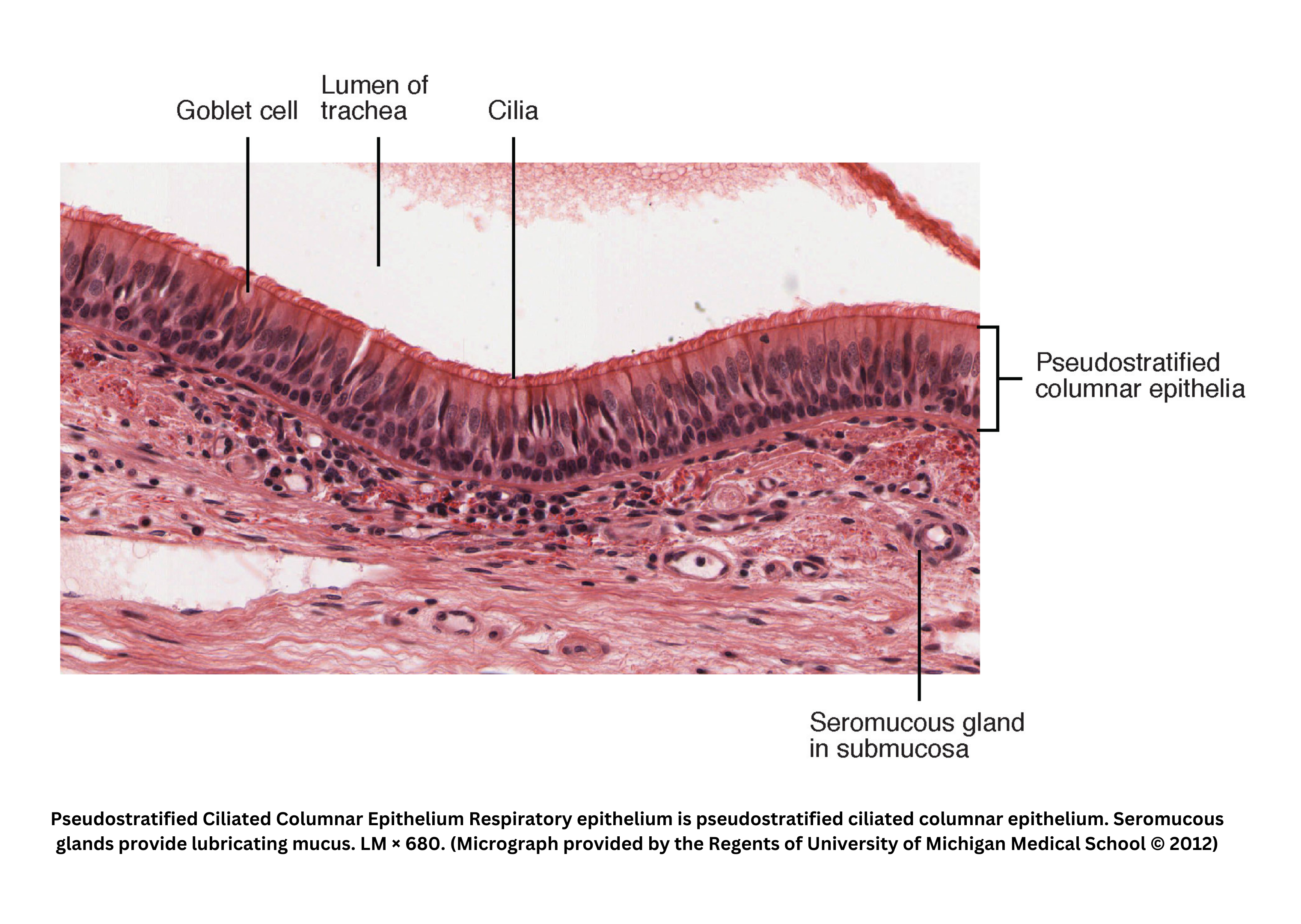
Once inhaled, mucus produced by Goblet cells - specialized, columnar epithelial cell lining the respiratory tract - traps pathogens and finer particles. The trapped material is then moved out by cilia, tiny hair-like structures that sweep contaminants toward the throat, where they are either expelled or swallowed and neutralized by stomach acid.
In the alveoli, macrophages provide a final line of defense by engulfing and destroying foreign substances that escape the cilia and reach these delicate air sacs.
Structure of lungs and alveoli
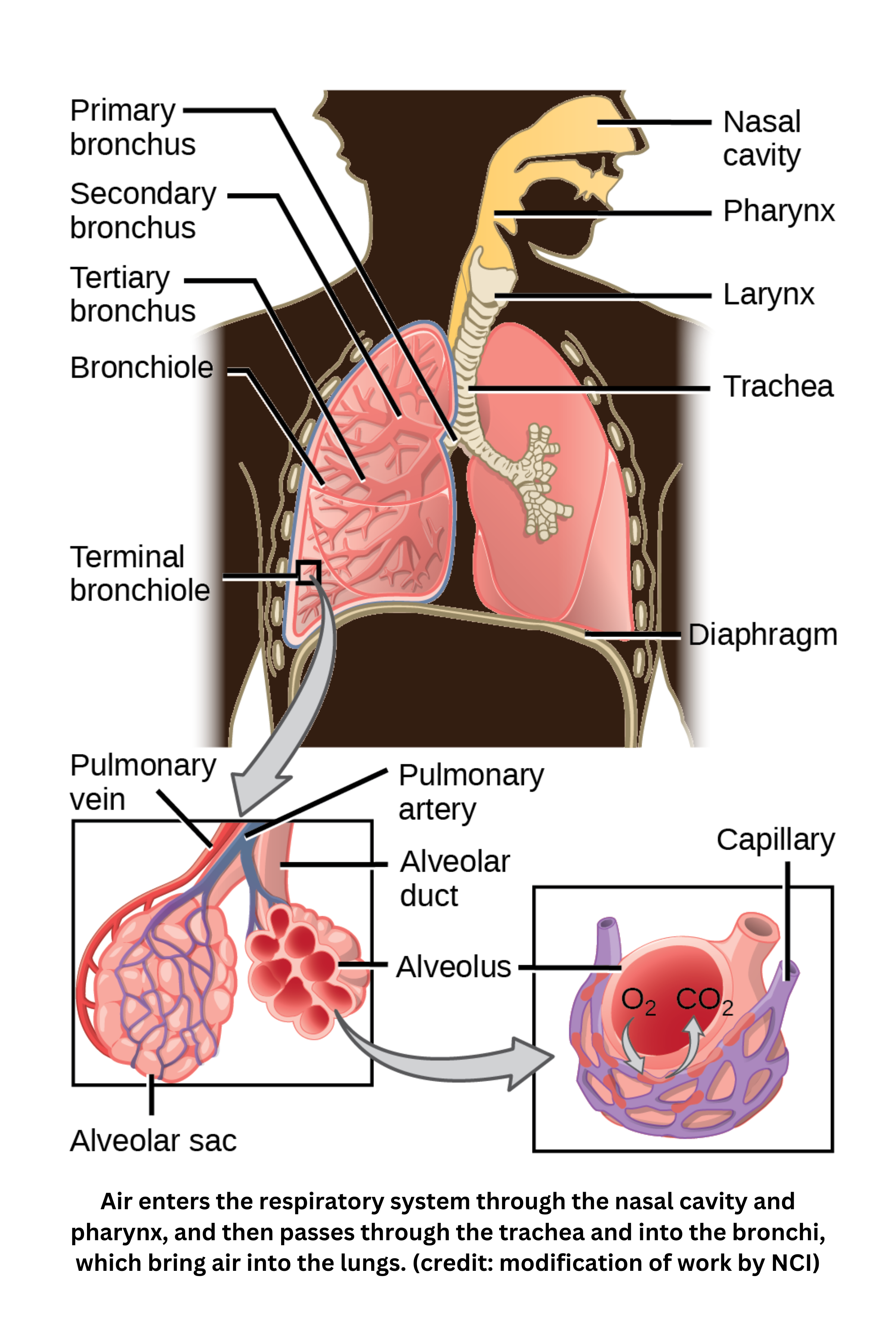
Air enters through the nasal cavity, where it is warmed, humidified, and filtered by hair, mucus, and cilia. It then passes through the pharynx and the larynx into the trachea, a tube about 10-12 cm long. The trachea is supported by hyaline cartilage rings and smooth muscle that can contract during forced exhalation (e.g., coughing).
The trachea splits into two primary bronchi, each entering a lung. Within the lungs, these bronchi branch into secondary and tertiary bronchi, then into smaller bronchioles, which rely on air pressure (rather than cartilage) to stay open.
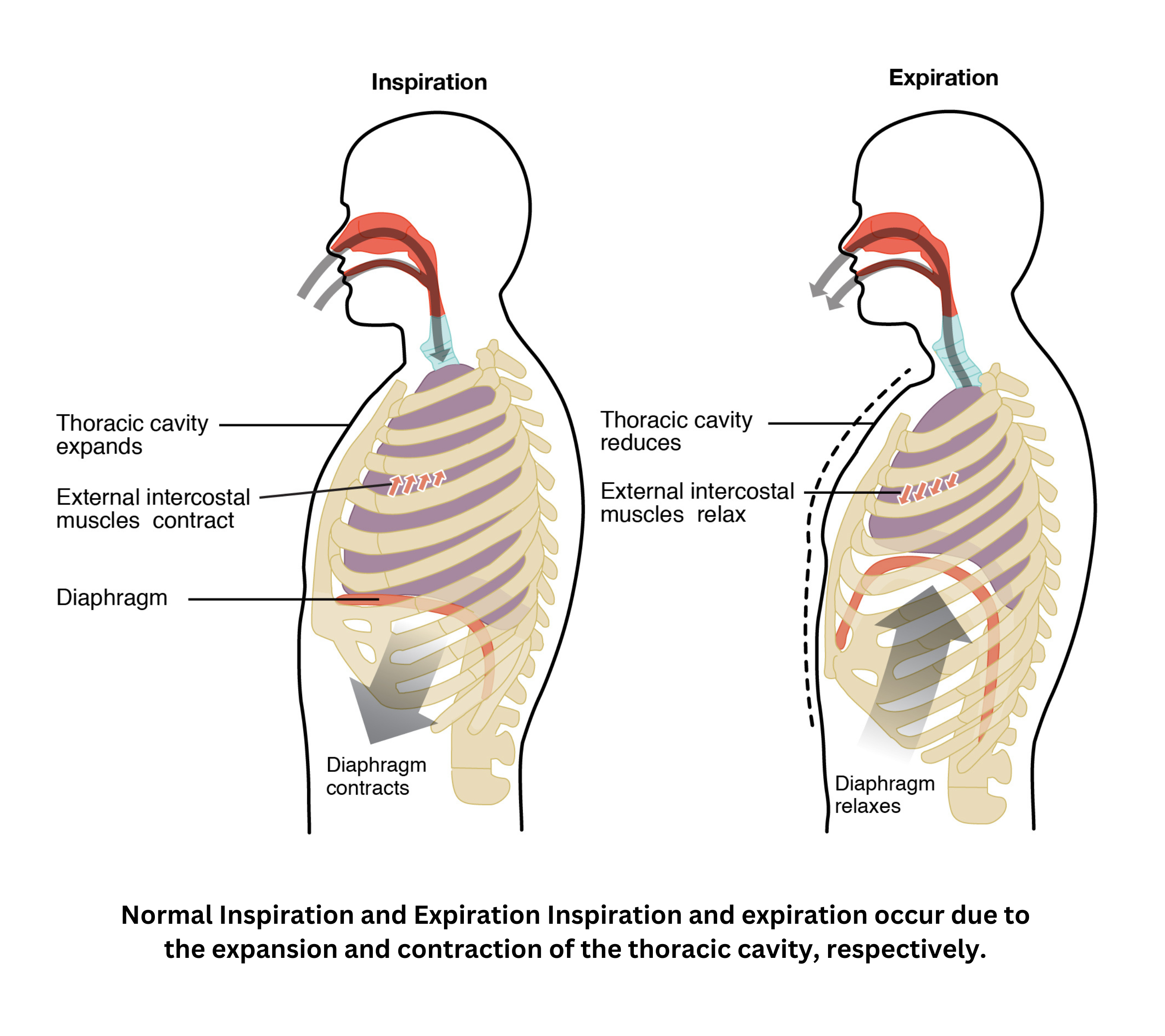
The lungs differ in size: the right lung has three lobes, whereas the left lung has two. Below them lies the diaphragm, a muscular boundary at the base of the thoracic cavity. When it contracts, it lowers and expands the chest volume, helping drive inhalation.
At the end of each bronchiole, alveolar ducts lead to clusters of alveoli, which resemble tiny bubbles arranged in alveolar sacs. Each alveolus is surrounded by capillaries; both are one cell thick, allowing gas exchange (oxygen in, carbon dioxide out) across their thin walls. With around 300 million alveoli per lung creating a total surface area of ~75 m², this structure maximizes oxygen delivery to the bloodstream and removal of carbon dioxide from it, giving the lungs their soft, sponge-like texture.
Breathing mechanisms
Diaphragm, rib cage, differential pressure
The diaphragm is a dome-shaped muscle. When it contracts, it moves downward, increasing chest volume and creating negative pressure that draws air into the lungs. At the same time, the rib cage expands outward - supported by intercostal muscles - and helps maintain lung volume even at rest.
Airflow is driven by differential pressure. The pressure inside the lungs (intrapulmonary) moves toward atmospheric levels, while the intrapleural pressure remains lower. This lower intrapleural pressure helps pull the lungs open and prevents collapse.
Resiliency and surface tension effects
A key feature of lung resiliency is elastic recoil. Without the outward support of the rib cage, the lungs would collapse further after exhalation.
Surface tension in the alveoli also promotes collapse. To counter this, the alveoli produce surfactants that reduce surface tension and help keep the air sacs open.
Alveolar gas exchange and pH control
In alveolar gas exchange, oxygen diffuses into blood and carbon dioxide diffuses out. This is guided by differences in partial pressures and can be described quantitatively by Henry’s Law.
Gas movement also connects to pH control, because elevated in the blood can lower pH.
Nervous system control
The body’s nervous control responds by adjusting breathing rate, reflecting its sensitivity.
Together, these coordinated mechanisms support oxygen delivery and carbon dioxide removal while also helping regulate acid-base balance.