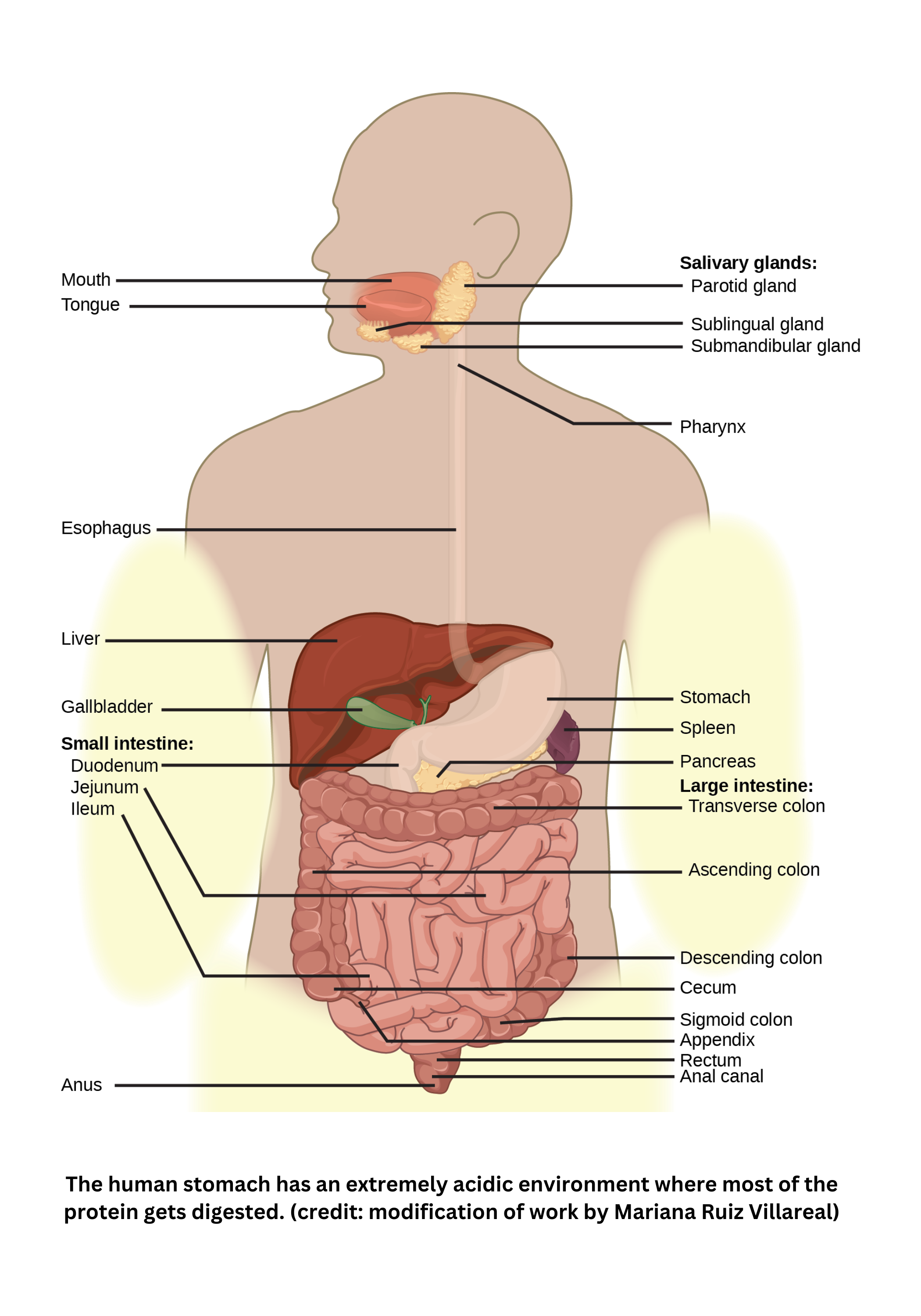
Digestive system
Ingestion
The process of ingestion begins in the mouth. Saliva dissolves food and contains:
- Mucin for lubrication
- Amylase to begin breaking down polysaccharides
- Antimicrobial components such as antibodies and lysozyme
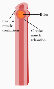
As you swallow, the epiglottis closes off the airway to help prevent choking. The pharynx then directs the food bolus into the esophagus, a muscular tube that moves food toward the stomach by peristalsis (rhythmic muscle contractions).
Once food reaches the stomach, digestion continues through both mechanical mixing and chemical breakdown. The stomach’s acidic environment is produced by parietal cells, which secrete hydrochloric acid. The low pH activates pepsin, a protease that breaks down proteins. At the same time, goblet cells secrete a protective mucus layer that helps prevent the stomach lining from being digested.
The stomach is an elastic, banana-shaped organ that can stretch considerably. It’s sealed by the cardiac sphincter at the top (where the esophagus enters) and the pyloric sphincter at the bottom (where contents exit into the small intestine).
The liver supports digestion and metabolism in several ways. It synthesizes bile from cholesterol. Bile is stored in the gall bladder and released to emulsify fats into smaller droplets called micelles, which increases surface area for lipase to act. The liver also makes and stores glycogen, carries out gluconeogenesis, and performs detoxification (including ammonia removal). In addition, it stores certain vitamins and iron.
Finally, bile may be held in the gall bladder until it’s needed. When released, it enters the duodenum to assist fat digestion.
Major digestive enzymes
| Enzyme | Produced in | Site of release | pH level |
|---|---|---|---|
| Carbohydrate digestion: | |||
| Salivary amylase | Salivary glands | Mouth | Neutral |
| Pancreatic amylase | Pancreas | Small intestine | Basic |
| Maltase | Small intestine | Small intestine | Basic |
| Protein digestion: | |||
| Pepsin | Gastric glands | Stomach | Acidic |
| Trypsin | Pancreas | Small intestine | Basic |
| Peptidases | Small intestine | Small intestine | Basic |
| Nucleic acid digestion: | |||
| Nuclease | Pancreas | Small intestine | Basic |
| Nucleosidases | Pancreas | Small intestine | Basic |
| Fat digestion: | |||
| Lipase | Pancreas | Small intestine | Basic |
Table adapted from Wikimedia
Pancreas
The pancreas is a tadpole-shaped gland that produces a wide range of digestive enzymes and bicarbonate. Its enzymes include Amylase (starch digestion), various Proteases (protein digestion), Lipase (fat digestion), and Ribonuclease (nucleic acid digestion).
The pancreas also synthesizes , which neutralizes acidic chyme arriving from the stomach. This helps create an optimal pH for digestive enzymes in the small intestine. Because this secretion is exocrine, pancreatic enzymes and bicarbonate travel through a duct into the duodenum, the first part of the small intestine.
Small intestine
The small intestine is the main site of digestion and absorption. It has three sections: the duodenum, jejunum, and ileum. Its inner lining contains folds, villi, and microvilli, which greatly increase surface area and improve nutrient uptake.
Inside each villus:
- Blood capillaries absorb most nutrients.
- Lacteals (specialized lymphatic capillaries) absorb digested fats.
Enterocytes (intestinal absorptive cells) move molecules into circulation using active transport or facilitated diffusion, depending on concentration gradients.
Most digestive enzymes come from the pancreas, but the small intestine also produces additional enzymes (for example, certain proteases and amylases). Together, pancreatic secretions, intestinal enzymes, and bicarbonate’s neutralizing effect allow nutrients to be efficiently broken down and absorbed.
Large intestine
The large intestine is divided into several regions:
- The cecum, which is a blind pouch containing the appendix
- The ascending colon, transverse colon, descending colon, and sigmoid colon
- The rectum, which stores feces.
Unlike the small intestine, the large intestine has no folds or villi. Its primary function is to absorb any remaining water that wasn’t absorbed in the small intestine.
Within the large intestine, bacterial flora ferment undigested nutrients (producing gas) and synthesize vitamin K, which is necessary for blood clotting. The rectum connects to the outside through the anal sphincter, which opens during defecation to release waste.
Muscular control
Muscular control in the digestive tract depends on valves (sphincters) and coordinated movement. The cardiac sphincter (gastroesophageal sphincter) at the junction of the esophagus and stomach helps prevent food from moving backward. The pyloric sphincter at the stomach’s exit controls the flow of stomach contents into the small intestine.
Peristalsis - rhythmic contractions of smooth muscle - moves food through each segment of the gastrointestinal tract so digestion and absorption can proceed.
Endocrine control
The endocrine system regulates digestion by releasing hormones that act on specific target tissues in the gastrointestinal tract:
- gastrin is secreted by G cells in the stomach and stimulates parietal cells to produce gastric acid, supporting digestion.
- When acidic chyme enters the small intestine, secretin is released from duodenal S cells. Secretin prompts the pancreas and liver to secrete bicarbonate and bile, respectively.
- cholecystokinin (CCK) is produced by I cells in the small intestine. It causes the gallbladder to contract and stimulates the pancreas to release digestive enzymes.
- Hormones such as glucagon-like peptide-1 (GLP-1) and peptide YY (PYY) help regulate satiety and slow gastric emptying, fine-tuning the overall digestive process.
Nervous control
The enteric nervous system (ENS) provides autonomous control over digestion by coordinating local reflexes. This network of neurons, embedded in the walls of the gastrointestinal tract, regulates motility, secretion, and blood flow.
Although the ENS can function independently, it communicates with the central nervous system through the vagus nerve, allowing digestive activity to adjust in response to food intake and other stimuli.