


This browser is too old to run Achievable.

Update it, or open this page in a current version of Chrome, Safari, Firefox or Edge. On an iPhone or iPad, update iOS: every browser there runs on the version of Safari that comes with it.

Achievable logoAchievable logo
NPTE-PTA
Sign in
Sign up
Purchase
Textbook
Practice exams
Support
How it works
Resources
Exam catalog
Mountain with a flag at the peak
Textbook
Introduction
1. Cardiovascular system
2. Pulmonary system
3. Neuromuscular system
4. Pediatrics
4.1 Pediatric foundations
4.2 Congenital disorders
4.3 Developmental and acquired disorders
4.3.1 Hip and knee conditions
4.3.2 Scoliosis, autism, and birth injuries
5. Musculoskeletal system
6. Other systems
7. Non-systems
Wrapping up
Achievable logoAchievable logo
4.3.1 Hip and knee conditions
Achievable NPTE-PTA
4. Pediatrics
4.3. Developmental and acquired disorders
Our NPTE-PTA course is now in "early access" - get 50% off for a limited time.

Hip and knee conditions

9 min read
Font
Discuss
Share
Feedback
Definitions
Acquired pediatric conditions
Conditions that develop after birth and do not have a link to genetic abnormalities.

Unlike congenital disorders, which are present at birth due to genetic or developmental causes, acquired conditions develop afterward from causes such as trauma, growth-related stress, or infection. This chapter covers common acquired conditions affecting the hip and knee, along with the physical therapy interventions used to manage each one.

PTA role reminder: For each condition below, the PTA carries out the plan of care established by the supervising PT - applying prescribed bracing, orthotics, or equipment; collecting data on the patient’s response; and communicating any changes to the PT - rather than evaluating, prescribing, or modifying the plan.

Legg-Calve-Perthes disease

Definitions
Legg-Calve-Perthes disease
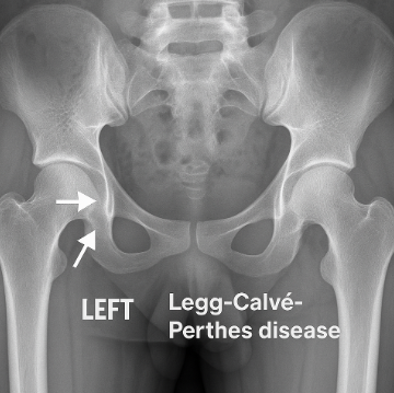
A condition that occurs when there is a temporary interruption in blood flow to the femoral head, causing the femoral head to die and eventually collapse. The cause of the interruption is idiopathic (unknown); repetitive minor trauma and blood-clotting abnormalities are proposed contributing factors.

Medical confirmation is performed through physical examination, MRI, and X-ray. The condition is most common in children under 10 years old.

Phases of Legg-Calve-Perthes disease

  1. Necrosis: part of the femoral head has died
  2. Fragmentation: the body attempts to clean up the broken pieces of bone from the area
  3. Re-ossification: blood flow is restored to the femoral head, and the bone begins to regrow
  4. Remodeling: new bone is now in place

Each phase can last months to years; the re-ossification and remodeling phases show the femoral head can heal once blood flow returns.

Physical therapy interventions for Legg-Calve-Perthes disease

  • Activity modifications
  • Bracing/splinting to keep the femoral head in contact with the acetabulum
  • Strengthening and range of motion activities of the hip
  • Report any increased pain, new limp, or skin breakdown under the brace to the supervising PT

Medical management of Legg-Calve-Perthes disease

  • Use of anti-inflammatory medications
  • Surgical containment (femoral or pelvic osteotomy) to keep the femoral head seated in the acetabulum - generally considered for children older than about 8 at diagnosis, or when more than half of the femoral head is involved

Transient synovitis is a self-limiting cause of hip pain and limited hip motion in children; most children have symptoms for less than two weeks, a viral cause is suspected, and children are followed to avoid missing Legg-Calve-Perthes disease.

Radiograph of Legg-Calvé-Perthes disease
Achievable

Slipped capital femoral epiphysis (SCFE)

Definitions
Slipped capital femoral epiphysis (SCFE)
A condition in which the femoral head (capital epiphysis) slips off the femoral neck at the growth plate (physis), usually posteriorly and inferiorly.

Etiology can be due to hormonal changes during puberty, a growth spurt, obesity, weakening of the growth plate, and family history. The most common age of injury is between 10-16 years of age.

Medical management for SCFE

Surgery is indicated once confirmed through physical examination and X-ray imaging. Delay in intervention can cause serious damage such as avascular necrosis, arthritis, or deformity of the hip joint. Surgery (in situ pinning) places a metal screw across the growth plate to fix the femoral head to the femoral neck and prevent further slipping.

Physical therapy interventions for SCFE

Physical therapy interventions are dependent upon the surgeon’s protocol post-op and will gauge the progression of activities until return to full weight bearing. Increased pain, inability to tolerate the next weight-bearing stage, or new limping during progression should be reported to the supervising PT rather than advanced independently.

The following table compares Legg-Calve-Perthes disease and slipped capital femoral epiphysis by age, typical child, what happens in the hip, findings, and management.


Feature Legg-Calve-Perthes disease Slipped capital femoral epiphysis
Typical age Under 10 years, most often 4-8 10-16 years, around the pubertal growth spurt
Typical child More often boys; often small for their age More often boys; obesity is a major risk factor
What happens Blood flow to the femoral head is interrupted, so part of the head dies and collapses The femoral head slips at the growth plate
Findings Groin or thigh pain, antalgic gait, leg length discrepancy, limited abduction, flexion, and internal rotation Similar pain, limp, and ROM limits, plus obligatory external rotation when the hip is flexed and frequent referred knee pain; an unstable slip may prevent weight bearing
Management Containment: activity modification, bracing, ROM and strengthening; surgical containment in older children Urgent: non-weight-bearing and surgical screw fixation; PT follows the surgeon’s post-op protocol

Osgood-Schlatter disease

Definitions
Osgood-Schlatter disease
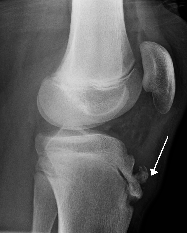
A condition in which an individual experiences knee pain and swelling at the tibial tuberosity due to increased pulling by the quadriceps on the patellar tendon.

Common causes are repetitive movements, excessive growth during puberty, and participation in sports that require increased running and jumping. Onset is typically during the adolescent growth spurt, around ages 12-15 in boys and 8-13 in girls. Confirmation of diagnosis is through physical examination and X-ray. Sinding-Larsen-Johansson syndrome is a related overuse condition of active adolescents in which the patellar tendon’s pull irritates the inferior pole of the patella, so pain and tenderness are at the bottom of the kneecap rather than at the tibial tuberosity.

Osgood Schlatter
Osgood Schlatter
James Heilman, MD
/
Wikimedia Commons
/
CC BY-SA 3.0

Symptoms

  • Palpable swelling at the tibial tuberosity
  • Pain at anterior knee
  • Increased pain during running and jumping activities, with relief of pain during rest

Physical therapy interventions for Osgood-Schlatter disease

  • Conservative care: activity modification (reducing high-impact running/jumping until pain improves), ice therapy, and patient education on physician-directed use of over-the-counter pain relievers
  • Strengthening of lower extremity muscles to create an even pull of muscles
  • Stretching of the quadriceps and hamstrings to reduce tensile pull on the tibial tuberosity
  • Dynamic balance and landing-control activities that simulate sport demands
  • Postural re-education when performing running, jumping activities
  • Report any worsening pain, new swelling, or lack of improvement to the supervising PT

Blount’s disease

Definitions
Blount’s disease (tibia vara)
A growth disturbance of the medial (inner) part of the proximal tibial growth plate that causes progressive bowing (genu varum), often with internal rotation of the tibia.

Bowing is normal in infants and should straighten by about 18 to 20 months. Bowing that persists or worsens after that age, centered at the proximal tibia, is the warning sign. Obesity, early walking and large stature are risk factors, and body weight loading the medial growth plate drives progression.

  • Infantile form: noticed before about age 4; about half of cases affect both legs, and the knee may thrust laterally in stance
  • Adolescent form: diagnosed after age 10; usually one leg, usually in a child who is overweight

Medical management of Blount’s disease

Bracing with a knee-ankle-foot orthosis (KAFO) is the first-line treatment for a child about 3 or younger. The brace unloads the medial growth plate and is worn during all waking hours, locked in extension for standing. It works best in a young child with one leg involved and early disease, and tends to fail with obesity, a lateral thrust, or a start after age 3. Surgery (an osteotomy of the proximal tibia, or guided growth) is used for deformity not corrected by about age 4, for advanced disease, and for the adolescent form.

Physical therapy interventions for Blount’s disease

  • Teach caregivers to put on the brace and follow its wear schedule
  • Check the skin under the brace
  • Report increasing bowing, a new lateral thrust, skin breakdown or pain to the supervising PT

Osteochondritis dissecans (OCD)

Definitions
Osteochondritis dissecans (OCD)
A disorder of the subchondral bone and its overlying cartilage in which a fragment of bone and cartilage can loosen from the joint surface.

The knee is the most common site, classically the lateral aspect of the medial femoral condyle. OCD is rare before age 6 and peaks between 13 and 17. It is two to four times more common in males and is linked to sport; repetitive microtrauma is a leading theory.

Symptoms

  • Poorly localized, activity-related knee pain, sometimes with swelling
  • Catching or locking in later stages, when the fragment becomes unstable or loose
  • Ligament tests are normal; imaging shows the defect in the femoral condyle, which tells OCD apart from a meniscal tear

Medical management of OCD

Most lesions in children and adolescents are stable (the fragment is still attached) and are treated without surgery for at least 3 to 6 months: no running or jumping sport, often with a brace or crutch-protected partial weight bearing for the first 4 to 6 weeks. About half to two-thirds heal in 6 to 12 months, and healing is better while the growth plates are open. Unstable lesions (a loose, hinged or detached fragment), and stable lesions that do not heal, are treated surgically.

Physical therapy interventions for OCD

  • Keep to the weight-bearing restriction in the plan of care
  • Keep the athlete out of impact and pivoting sport until cleared
  • Report new locking, giving way or joint swelling to the supervising PT: the fragment may have become unstable

Overview

  • Acquired pediatric conditions develop after birth (trauma, growth stress, infection) vs. congenital conditions present at birth
  • PTA role: implement PT’s plan of care (bracing, equipment, exercises), collect data, report changes — never evaluate or modify plan

Legg-Calve-Perthes disease

  • Idiopathic interruption of blood flow to femoral head → death/collapse; most common under age 10
  • Four phases: necrosis → fragmentation → re-ossification → remodeling (each can last months-years)
  • Management: activity modification, bracing to maintain femoral head containment, ROM/strengthening; surgical osteotomy considered if >age 8 or >50% head involvement
  • Report increased pain, new limp, or skin breakdown under brace

Slipped capital femoral epiphysis (SCFE)

  • Femoral head slips off femoral neck at growth plate, usually posteriorly/inferiorly; linked to puberty, obesity, growth spurts; typical age 10-16
  • Urgent surgical fixation (in situ pinning) to prevent progression; delay risks avascular necrosis/arthritis
  • PT follows surgeon’s post-op weight-bearing protocol; report pain, inability to progress, or new limp

Perthes vs. SCFE comparison

  • Perthes: younger (4-8), often small stature, blood flow disruption, managed conservatively/bracing
  • SCFE: adolescent (10-16), often obese, growth plate slippage, managed surgically/urgently
  • Both show groin/thigh pain, antalgic gait, limited hip ROM; SCFE adds obligatory external rotation with flexion and referred knee pain

Osgood-Schlatter disease

  • Traction injury at tibial tuberosity from quadriceps pull via patellar tendon; linked to growth spurts and running/jumping sports (ages ~8-15)
  • Symptoms: swelling/pain at tibial tuberosity, worse with activity, relieved by rest
  • PT: activity modification, ice, quad/hamstring stretching, lower extremity strengthening, landing-control training; report worsening pain or swelling

Blount’s disease (tibia vara)

  • Growth disturbance of medial proximal tibial growth plate → progressive bowing (genu varum); differentiate from normal infant bowing (resolves by 18-20 months)
  • Infantile form (before age 4, often bilateral) vs. adolescent form (after age 10, usually unilateral, linked to obesity)
  • Management: KAFO bracing (best under age 3, worn during waking hours) first-line; surgery (osteotomy/guided growth) if uncorrected by age 4 or in adolescent form
  • PT: caregiver brace education, skin checks, report worsening bowing or skin issues

Osteochondritis dissecans (OCD)

  • Bone/cartilage fragment loosens from joint surface, most common at lateral aspect of medial femoral condyle; peaks ages 13-17, more common in males, linked to repetitive microtrauma
  • Symptoms: activity-related knee pain, swelling, catching/locking if unstable; ligament tests normal; imaging showing the femoral condyle defect distinguishes it from a meniscal tear
  • Stable lesions: nonsurgical management (activity restriction, bracing/partial weight bearing) for 3-6 months; unstable or non-healing lesions require surgery
  • PT: enforce weight-bearing restrictions, restrict impact/pivoting sports, report new locking/giving way/swelling

Sign up for free to take 17 quiz questions on this topic

Previous
Next  | 4.3.2 Scoliosis, autism, and birth injuries
All rights reserved ©2016 - 2026 Achievable, Inc.

Hip and knee conditions

Definitions
Acquired pediatric conditions
Conditions that develop after birth and do not have a link to genetic abnormalities.

Unlike congenital disorders, which are present at birth due to genetic or developmental causes, acquired conditions develop afterward from causes such as trauma, growth-related stress, or infection. This chapter covers common acquired conditions affecting the hip and knee, along with the physical therapy interventions used to manage each one.

PTA role reminder: For each condition below, the PTA carries out the plan of care established by the supervising PT - applying prescribed bracing, orthotics, or equipment; collecting data on the patient’s response; and communicating any changes to the PT - rather than evaluating, prescribing, or modifying the plan.

Legg-Calve-Perthes disease

Definitions
Legg-Calve-Perthes disease
A condition that occurs when there is a temporary interruption in blood flow to the femoral head, causing the femoral head to die and eventually collapse. The cause of the interruption is idiopathic (unknown); repetitive minor trauma and blood-clotting abnormalities are proposed contributing factors.

Medical confirmation is performed through physical examination, MRI, and X-ray. The condition is most common in children under 10 years old.

Phases of Legg-Calve-Perthes disease

  1. Necrosis: part of the femoral head has died
  2. Fragmentation: the body attempts to clean up the broken pieces of bone from the area
  3. Re-ossification: blood flow is restored to the femoral head, and the bone begins to regrow
  4. Remodeling: new bone is now in place

Each phase can last months to years; the re-ossification and remodeling phases show the femoral head can heal once blood flow returns.

Physical therapy interventions for Legg-Calve-Perthes disease

  • Activity modifications
  • Bracing/splinting to keep the femoral head in contact with the acetabulum
  • Strengthening and range of motion activities of the hip
  • Report any increased pain, new limp, or skin breakdown under the brace to the supervising PT

Medical management of Legg-Calve-Perthes disease

  • Use of anti-inflammatory medications
  • Surgical containment (femoral or pelvic osteotomy) to keep the femoral head seated in the acetabulum - generally considered for children older than about 8 at diagnosis, or when more than half of the femoral head is involved

Transient synovitis is a self-limiting cause of hip pain and limited hip motion in children; most children have symptoms for less than two weeks, a viral cause is suspected, and children are followed to avoid missing Legg-Calve-Perthes disease.

Slipped capital femoral epiphysis (SCFE)

Definitions
Slipped capital femoral epiphysis (SCFE)
A condition in which the femoral head (capital epiphysis) slips off the femoral neck at the growth plate (physis), usually posteriorly and inferiorly.

Etiology can be due to hormonal changes during puberty, a growth spurt, obesity, weakening of the growth plate, and family history. The most common age of injury is between 10-16 years of age.

Medical management for SCFE

Surgery is indicated once confirmed through physical examination and X-ray imaging. Delay in intervention can cause serious damage such as avascular necrosis, arthritis, or deformity of the hip joint. Surgery (in situ pinning) places a metal screw across the growth plate to fix the femoral head to the femoral neck and prevent further slipping.

Physical therapy interventions for SCFE

Physical therapy interventions are dependent upon the surgeon’s protocol post-op and will gauge the progression of activities until return to full weight bearing. Increased pain, inability to tolerate the next weight-bearing stage, or new limping during progression should be reported to the supervising PT rather than advanced independently.

The following table compares Legg-Calve-Perthes disease and slipped capital femoral epiphysis by age, typical child, what happens in the hip, findings, and management.


Feature Legg-Calve-Perthes disease Slipped capital femoral epiphysis
Typical age Under 10 years, most often 4-8 10-16 years, around the pubertal growth spurt
Typical child More often boys; often small for their age More often boys; obesity is a major risk factor
What happens Blood flow to the femoral head is interrupted, so part of the head dies and collapses The femoral head slips at the growth plate
Findings Groin or thigh pain, antalgic gait, leg length discrepancy, limited abduction, flexion, and internal rotation Similar pain, limp, and ROM limits, plus obligatory external rotation when the hip is flexed and frequent referred knee pain; an unstable slip may prevent weight bearing
Management Containment: activity modification, bracing, ROM and strengthening; surgical containment in older children Urgent: non-weight-bearing and surgical screw fixation; PT follows the surgeon’s post-op protocol

Osgood-Schlatter disease

Definitions
Osgood-Schlatter disease
A condition in which an individual experiences knee pain and swelling at the tibial tuberosity due to increased pulling by the quadriceps on the patellar tendon.

Common causes are repetitive movements, excessive growth during puberty, and participation in sports that require increased running and jumping. Onset is typically during the adolescent growth spurt, around ages 12-15 in boys and 8-13 in girls. Confirmation of diagnosis is through physical examination and X-ray. Sinding-Larsen-Johansson syndrome is a related overuse condition of active adolescents in which the patellar tendon’s pull irritates the inferior pole of the patella, so pain and tenderness are at the bottom of the kneecap rather than at the tibial tuberosity.

Symptoms

  • Palpable swelling at the tibial tuberosity
  • Pain at anterior knee
  • Increased pain during running and jumping activities, with relief of pain during rest

Physical therapy interventions for Osgood-Schlatter disease

  • Conservative care: activity modification (reducing high-impact running/jumping until pain improves), ice therapy, and patient education on physician-directed use of over-the-counter pain relievers
  • Strengthening of lower extremity muscles to create an even pull of muscles
  • Stretching of the quadriceps and hamstrings to reduce tensile pull on the tibial tuberosity
  • Dynamic balance and landing-control activities that simulate sport demands
  • Postural re-education when performing running, jumping activities
  • Report any worsening pain, new swelling, or lack of improvement to the supervising PT

Blount’s disease

Definitions
Blount’s disease (tibia vara)
A growth disturbance of the medial (inner) part of the proximal tibial growth plate that causes progressive bowing (genu varum), often with internal rotation of the tibia.

Bowing is normal in infants and should straighten by about 18 to 20 months. Bowing that persists or worsens after that age, centered at the proximal tibia, is the warning sign. Obesity, early walking and large stature are risk factors, and body weight loading the medial growth plate drives progression.

  • Infantile form: noticed before about age 4; about half of cases affect both legs, and the knee may thrust laterally in stance
  • Adolescent form: diagnosed after age 10; usually one leg, usually in a child who is overweight

Medical management of Blount’s disease

Bracing with a knee-ankle-foot orthosis (KAFO) is the first-line treatment for a child about 3 or younger. The brace unloads the medial growth plate and is worn during all waking hours, locked in extension for standing. It works best in a young child with one leg involved and early disease, and tends to fail with obesity, a lateral thrust, or a start after age 3. Surgery (an osteotomy of the proximal tibia, or guided growth) is used for deformity not corrected by about age 4, for advanced disease, and for the adolescent form.

Physical therapy interventions for Blount’s disease

  • Teach caregivers to put on the brace and follow its wear schedule
  • Check the skin under the brace
  • Report increasing bowing, a new lateral thrust, skin breakdown or pain to the supervising PT

Osteochondritis dissecans (OCD)

Definitions
Osteochondritis dissecans (OCD)
A disorder of the subchondral bone and its overlying cartilage in which a fragment of bone and cartilage can loosen from the joint surface.

The knee is the most common site, classically the lateral aspect of the medial femoral condyle. OCD is rare before age 6 and peaks between 13 and 17. It is two to four times more common in males and is linked to sport; repetitive microtrauma is a leading theory.

Symptoms

  • Poorly localized, activity-related knee pain, sometimes with swelling
  • Catching or locking in later stages, when the fragment becomes unstable or loose
  • Ligament tests are normal; imaging shows the defect in the femoral condyle, which tells OCD apart from a meniscal tear

Medical management of OCD

Most lesions in children and adolescents are stable (the fragment is still attached) and are treated without surgery for at least 3 to 6 months: no running or jumping sport, often with a brace or crutch-protected partial weight bearing for the first 4 to 6 weeks. About half to two-thirds heal in 6 to 12 months, and healing is better while the growth plates are open. Unstable lesions (a loose, hinged or detached fragment), and stable lesions that do not heal, are treated surgically.

Physical therapy interventions for OCD

  • Keep to the weight-bearing restriction in the plan of care
  • Keep the athlete out of impact and pivoting sport until cleared
  • Report new locking, giving way or joint swelling to the supervising PT: the fragment may have become unstable
Key points

Overview

  • Acquired pediatric conditions develop after birth (trauma, growth stress, infection) vs. congenital conditions present at birth
  • PTA role: implement PT’s plan of care (bracing, equipment, exercises), collect data, report changes — never evaluate or modify plan

Legg-Calve-Perthes disease

  • Idiopathic interruption of blood flow to femoral head → death/collapse; most common under age 10
  • Four phases: necrosis → fragmentation → re-ossification → remodeling (each can last months-years)
  • Management: activity modification, bracing to maintain femoral head containment, ROM/strengthening; surgical osteotomy considered if >age 8 or >50% head involvement
  • Report increased pain, new limp, or skin breakdown under brace

Slipped capital femoral epiphysis (SCFE)

  • Femoral head slips off femoral neck at growth plate, usually posteriorly/inferiorly; linked to puberty, obesity, growth spurts; typical age 10-16
  • Urgent surgical fixation (in situ pinning) to prevent progression; delay risks avascular necrosis/arthritis
  • PT follows surgeon’s post-op weight-bearing protocol; report pain, inability to progress, or new limp

Perthes vs. SCFE comparison

  • Perthes: younger (4-8), often small stature, blood flow disruption, managed conservatively/bracing
  • SCFE: adolescent (10-16), often obese, growth plate slippage, managed surgically/urgently
  • Both show groin/thigh pain, antalgic gait, limited hip ROM; SCFE adds obligatory external rotation with flexion and referred knee pain

Osgood-Schlatter disease

  • Traction injury at tibial tuberosity from quadriceps pull via patellar tendon; linked to growth spurts and running/jumping sports (ages ~8-15)
  • Symptoms: swelling/pain at tibial tuberosity, worse with activity, relieved by rest
  • PT: activity modification, ice, quad/hamstring stretching, lower extremity strengthening, landing-control training; report worsening pain or swelling

Blount’s disease (tibia vara)

  • Growth disturbance of medial proximal tibial growth plate → progressive bowing (genu varum); differentiate from normal infant bowing (resolves by 18-20 months)
  • Infantile form (before age 4, often bilateral) vs. adolescent form (after age 10, usually unilateral, linked to obesity)
  • Management: KAFO bracing (best under age 3, worn during waking hours) first-line; surgery (osteotomy/guided growth) if uncorrected by age 4 or in adolescent form
  • PT: caregiver brace education, skin checks, report worsening bowing or skin issues

Osteochondritis dissecans (OCD)

  • Bone/cartilage fragment loosens from joint surface, most common at lateral aspect of medial femoral condyle; peaks ages 13-17, more common in males, linked to repetitive microtrauma
  • Symptoms: activity-related knee pain, swelling, catching/locking if unstable; ligament tests normal; imaging showing the femoral condyle defect distinguishes it from a meniscal tear
  • Stable lesions: nonsurgical management (activity restriction, bracing/partial weight bearing) for 3-6 months; unstable or non-healing lesions require surgery
  • PT: enforce weight-bearing restrictions, restrict impact/pivoting sports, report new locking/giving way/swelling

More from Developmental and acquired disorders

  • Scoliosis, autism, and birth injuries