



Congenital musculoskeletal disorders
The following congenital disorders primarily affect the bones, joints, and connective tissue.
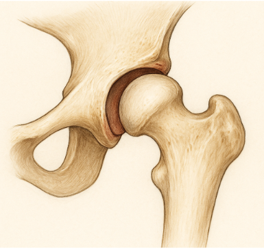
Developmental dysplasia of the hip (DDH)
Factors that contribute to the development of DDH include first pregnancy, breech birth, swaddling too tightly with the legs extended, increased infant size, female sex, and family history.
Common symptoms
- Leg length discrepancy (affected leg is shorter than the unaffected)
- Increased folds in the skin of the thigh/buttocks of the affected extremity
- Limited hip abduction on the affected side (the main sign after about 3 months of age, once Ortolani/Barlow are no longer positive)
- Increased popping noted when moving the affected leg
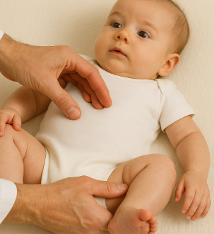
- Ortolani test
- Abduction of the flexed hip reduces a dislocated femoral head back into the acetabulum, producing a palpable clunk
- Barlow test
- Adduction of the flexed hip provokes (dislocates) an unstable hip, producing a palpable clunk
- Ortolani test
Physical therapy interventions for DDH
- Education provided to the family for the appropriate positioning of the infant to reduce hip dislocation occurrence
- Gentle range of motion activities that do not cause dislocation
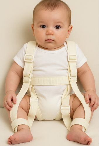
- Wearing of Pavlik harness
- The Pavlik harness is a soft splint used to allow for consistent contact between the acetabulum and the femoral head
- Positioned in abduction of legs, hip flexion, and knee flexion through a series of straps to keep the infant in this position
- Used for infants 6 months or younger, with a goal of 24-hour wear for the first 6 weeks, progressing to nighttime-only wear for the following 6 weeks
- The parents must learn how to care for the baby fully during the period of continuous wear
- Change from wearing 24 hours a day to only at night is determined by an orthopedic physician via x-ray and ultrasound
- Developmental delay may occur in an infant due to constant positioning, as mentioned above, with decreased ability to move out of position
- The Pavlik harness is a soft splint used to allow for consistent contact between the acetabulum and the femoral head
Example: Pavlik harness adherence
During the 24-hour wear phase, a parent asks the PTA to remove the harness for bathing.
Answer: The PTA reinforces the current wear schedule. Changing it is a physician decision based on follow-up imaging, not something the PTA or family adjusts on their own.
Clubfoot (talipes equinovarus)
Phases of intervention
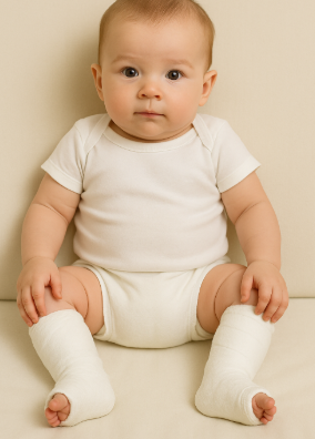
- Phase 1: serial casting
- Clubfoot is stretched/manipulated to improve the range of motion, and then a hard cast is set in place over the affected extremity
- The hard cast is removed, and stretching occurs again, with a new cast put in place with the new range of motion that has been achieved
- The goal is to wear the hard cast until normal alignment is achieved - can take 4-10 weeks to achieve normal alignment
- Most infants then undergo a percutaneous Achilles tenotomy to correct the remaining equinus before the final cast
- Clubfoot is stretched/manipulated to improve the range of motion, and then a hard cast is set in place over the affected extremity
- Phase 2: bracing
- Once serial casting is complete (normal alignment is achieved), bracing is put in place to aid in maintaining the normal alignment of the foot
- A foot-abduction brace (boots on a bar) is worn about 23 hours a day for roughly 3 months, then during nighttime sleep and naps until about age 4
- Non-adherence to bracing is the leading cause of relapse
- Once serial casting is complete (normal alignment is achieved), bracing is put in place to aid in maintaining the normal alignment of the foot
Surgical intervention may be necessary depending on the severity of the deformity and the inability to change Achilles tendon length through conservative measures.
Metatarsus adductus is a common foot deformity usually noted at birth in which the forefoot turns inward.
Physical therapy interventions for clubfoot
Within the PT’s plan of care, the PTA may assist with stretching between casts, teach families how to put on the brace and check the skin, monitor motor milestones, and report skin breakdown, poor brace tolerance, or loss of correction to the supervising PT. Choosing or changing the brace is a PT or physician decision.
Osteogenesis imperfecta
Symptoms of osteogenesis imperfecta
- Short stature
- Joint laxity
- Frequent fractures
- Bone deformities
- Muscle weakness
- Hearing loss
- Dental problems
- Blue or gray-blue sclerae (a classic sign)
There are varying degrees of severity of osteogenesis imperfecta, ranging from mild symptoms that persist throughout life to severe symptoms in which the newborn dies within weeks of birth.
Rickets, which is usually not genetic (inherited forms such as X-linked hypophosphatemic rickets are rare), also causes soft, weak bones in children, usually because they do not get enough vitamin D, which helps growing bones absorb calcium and phosphorus. Symptoms include bone pain or tenderness, impaired growth, and deformities of the bones and teeth.
Physical therapy interventions for osteogenesis imperfecta
- Support the trunk and limbs with open hands; never lift by the ankles or pull on a limb
- Avoid twisting/torsional forces; use active or gentle assisted ROM only, never forceful stretching
- Aquatic therapy allows safe strengthening with reduced joint stress
- Education on swaddling, positioning, handling, and fall-prevention strategies as the child progresses through developmental milestones
Arthrogryposis multiplex congenita (AMC)
The condition is not a single disease but a clinical finding that may have several underlying causes, primarily involving the neuromuscular system. When a fetus does not move enough in utero, the joints do not develop normally and soft tissues such as muscles and tendons can become contracted.
Clinical presentation
- Multiple joint contractures at birth (e.g., clubfoot, extended knees, flexed wrists, thumb-in-palm deformity)
- Muscle hypoplasia or replacement of muscle tissue with fibrous or fatty tissue
- Thin, atrophic limbs
- No progressive neurological decline
- Normal cognitive function in most cases
Medical and physical therapy management
- Physical therapy: early passive range of motion and daily stretching to prevent worsening contractures, positioning and serial casting to correct deformities, strengthening of available muscle groups, and mobility/gait training with assistive devices (walkers, wheelchairs, KAFOs) as needed
- Orthotic management: AFOs or KAFOs for ambulation, hand splints to improve function, and spinal orthoses if scoliosis develops
- Surgical management: surgery (soft-tissue releases, tendon transfers, osteotomies) may follow if conservative care fails; post-op PT follows the surgeon’s protocol
AMC-related clubfoot is stiffer, more prone to relapse, and more often needs surgery than idiopathic clubfoot.
| Condition | Core problem | Key sign | Main precaution |
|---|---|---|---|
| DDH | Shallow acetabulum, hip instability | Palpable clunk or limited abduction | Avoid dislocating positions; follow Pavlik schedule |
| Idiopathic clubfoot | Soft-tissue contracture | Foot fixed down and inward at birth | Maintain casting/bracing schedule |
| Osteogenesis imperfecta | Defective bone collagen | Frequent fractures, blue sclerae | Gentle handling; no forceful ROM |
| AMC | Limited fetal movement | Multiple fixed contractures at birth | Early, consistent stretching |