


This browser is too old to run Achievable.

Update it, or open this page in a current version of Chrome, Safari, Firefox or Edge. On an iPhone or iPad, update iOS: every browser there runs on the version of Safari that comes with it.

Achievable logoAchievable logo
NPTE-PTA
Sign in
Sign up
Purchase
Textbook
Practice exams
Support
How it works
Resources
Exam catalog
Mountain with a flag at the peak
Textbook
Introduction
1. Cardiovascular system
2. Pulmonary system
3. Neuromuscular system
4. Pediatrics
4.1 Pediatric foundations
4.2 Congenital disorders
4.2.1 Congenital neurological and genetic disorders
4.2.2 Congenital musculoskeletal disorders
4.3 Developmental and acquired disorders
5. Musculoskeletal system
6. Other systems
7. Non-systems
Wrapping up
Achievable logoAchievable logo
4.2.1 Congenital neurological and genetic disorders
Achievable NPTE-PTA
4. Pediatrics
4.2. Congenital disorders
Our NPTE-PTA course is now in "early access" - get 50% off for a limited time.

Congenital neurological and genetic disorders

11 min read
Font
Discuss
Share
Feedback
Definitions
Congenital disorders
Conditions that develop in an individual in utero, during birth, or shortly after birth, due to gene mutations, deoxygenation to the brain or heart, infections, or micronutrient deficiencies.

Cerebral palsy

Definitions
Cerebral palsy (CP)
A group of disorders causing deficits in movement and coordination, caused by abnormal development or damage to the brain.

Damage can occur before, during, or shortly after birth, and may result from intracranial hemorrhage, lack of oxygen to the brain, abnormal brain development, or damage to the brain’s white matter.

Risk factors for CP include multiple births, toxin exposure, maternal or infant infection, low birth weight, seizures, complicated birth or labor, jaundice, and breech (buttocks- or feet-first) delivery.

Common comorbidities among individuals with cerebral palsy include intellectual disabilities, visual impairments, speech and language deficits, seizures, hearing impairments, and various orthopedic conditions.

Types of cerebral palsy

  • Spastic cerebral palsy (hypertonic cerebral palsy)
    • Type of cerebral palsy in which the individual exhibits hypertonia, causing stiff and jerky movements
      • Spastic hemiplegia
        • Hypertonia is located on one side of the body - the ipsilateral arm and leg
        • The affected side is typically shorter and thinner, scoliosis may be present, and intellect is often normal
      • Spastic diplegia
        • Hypertonia is located primarily in the lower extremities
        • Hyperreflexia can be present, along with scissoring of gait; intellect is often normal
      • Spastic quadriplegia
        • Hypertonia is located throughout the entire body
        • The most severe type of spastic cerebral palsy, due to widespread damage to the entire brain
        • Individuals with this type of CP rarely walk, demonstrate difficulty speaking, and often have moderate-to-severe intellectual disability and frequent seizures
  • Hypotonic cerebral palsy
    • Characterized by low tone and floppiness of extremities and axial skeleton
    • Individuals may demonstrate poor muscle tone, excessive range of motion, increased weight gain, impaired speech due to poor oral motor control, and a wide base of support with gait
  • Dyskinetic cerebral palsy (athetoid cerebral palsy)
    • Caused by damage to the basal ganglia; characterized by slow and uncontrollable writhing or jerky movements of the hands, feet, arms, and legs
    • Individuals may demonstrate postural deficits, hearing problems, and breathing difficulties; intellect is often preserved, though intellectual disability is common
  • Ataxic cerebral palsy
    • Characterized by poor coordination, balance impairments, and impairments in depth perception
    • Individuals may demonstrate a wide base of support, dysmetria, and difficulty with precision of movement
  • Mixed types
    • Characterized by a mix of hypertonia and hypotonia with varied symptom presentation

Classifications of cerebral palsy (Gross Motor Function Classification System, GMFCS)

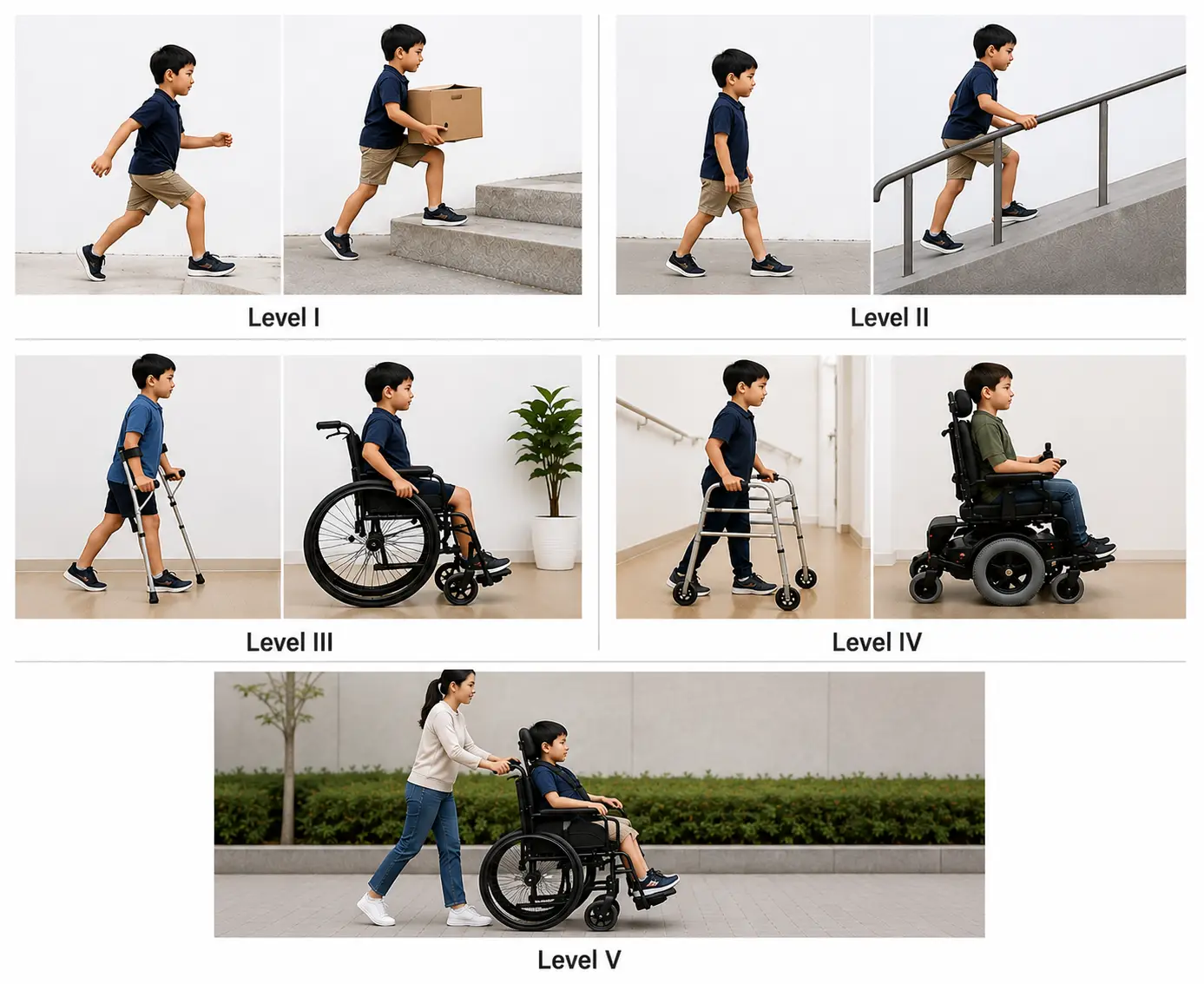
  • Level I: Walks independently without limitations. Climbs stairs without using a railing and can run and jump, although speed, balance, and coordination may be limited.
  • Level II: Walks independently but uses a railing for stairs. May have difficulty with long distances, uneven ground, or crowded spaces and has limited ability to run or jump.
  • Level III: Walks using a hand-held mobility device, such as crutches or a walker. Typically, self-propels a wheelchair for longer distances.
  • Level IV: Has limited self-mobility and usually relies on a wheelchair or powered mobility for most movement. May stand or take a few steps using a supportive walker.
  • Level V: Has severe limitations in mobility and posture, with difficulty controlling head, trunk, arm, and leg movements. Depends on others for transfers and mobility and is typically transported in a wheelchair
Illustration of the five GMFCS levels, showing increasing mobility limitations and use of assistive devices.
Gross Motor Function Classification System (GMFCS)
Achievable

Physical therapy interventions for cerebral palsy

  • Positioning is key to promote improvements in mobility, ADL participation, gait and balance improvements, and interaction with the environment
    • Symmetrical posture
    • Alignment of trunk, pelvis, and extremities
    • Head in midline
    • Hips and knees positioned at 90 degrees while sitting
    • Checking fit and skin integrity of orthoses prescribed by the PT, and training don/doff
    • Optimizing functional motor skills
  • Treatment of orthopedic conditions such as
    • Scoliosis
    • Joint contractures
    • Kyphosis
    • Clubfoot
    • Hip or shoulder dislocation

Medical management for cerebral palsy

  • Anti-seizure medication
  • Spasticity medication
    • Botox injections for local hypertonic muscles
    • Baclofen taken orally or by implantation for multiple areas of hypertonia
  • Surgical interventions
    • Dorsal rhizotomy
      • Selected dorsal (sensory) nerve rootlets - those driving the spasticity - are cut, leaving the rest intact, to decrease spasticity and improve overall function
    • Z-plasty
      • Release of muscle or tendons to release contractures

Down syndrome

Definitions
Down syndrome
A condition resulting from a chromosomal abnormality on chromosome 21 - instead of the usual pair, three copies of chromosome 21 are present (trisomy 21).

Characteristics of Down syndrome

  • Small ears and a protruding tongue
  • Microcephaly with flattened occiput
  • Short stature
  • Hypotonia and hypermobility
  • Congenital heart defects
  • Speech deficits
  • Developmental delays
  • Vertebral instability at the atlanto-axial joint (C1-C2)
  • Intellectual disabilities

Physical therapy interventions for Down syndrome

  • Promote gross motor development
  • Increase motor control and postural awareness
  • Improve oral-motor development
  • Train in use of DME selected by the PT; report fit or functional changes to the PT
  • Patient and family education

Special considerations with Down syndrome

  • Avoidance of diving, tumbling, headstands, and contact sports, due to an increased risk of hyperflexion injury from atlanto-axial instability

PTA role: Data collection and communication - not changing the plan of care - are within the PTA’s scope. If a session reveals new neurological signs (neck pain, numbness, weakness, or changes in coordination) that suggest atlanto-axial instability, stop the activity and report the finding to the supervising PT.

Duchenne muscular dystrophy

Definitions
Duchenne muscular dystrophy
A progressive disease process in which the protein dystrophin is not produced, causing an increased breakdown of muscle tissue over time. It most often affects boys; with modern care (steroids, ventilatory and cardiac support), life expectancy commonly extends into the late 20s or 30s.

Common symptoms of Duchenne muscular dystrophy

  • Progressive muscle weakness
  • Pseudohypertrophic muscles appear hypertrophied, but the muscle has been replaced by fat and connective tissue in calves, deltoids, quadriceps, and tongue
  • Contracture
  • Cardiac myopathy
  • Gowers’ sign
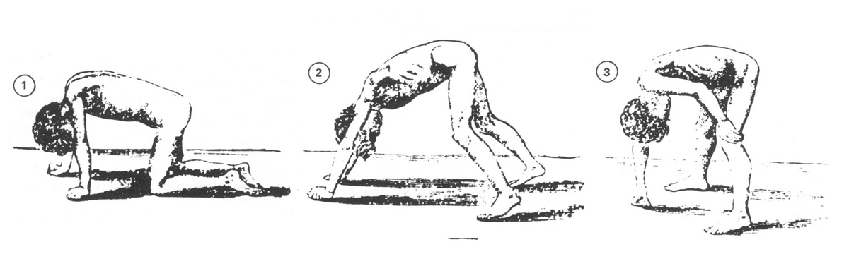
    • The child pushes up from the floor with their hands, walking their hands up their legs to stand - this is due to weak hip and knee extensors; typically noticeable by ages 3-5
  • Waddling gait
  • Increased risk of falls
Gowers' sign
Gowers' sign
William Richard Gowers (1845-1915), Gowers W. R. Clinical lecture on pseudohypertrophic muscular paralysis. Lancet 1879; ii, 73-5.
/
Wikimedia Commons
/
Public domain

Progression of Duchenne muscular dystrophy

  • Early childhood (age 3-5): weakness, tripping, and Gowers’ sign appear
  • Later childhood (age 9-14): loss of ambulation as gait deviations, poor endurance, and lower extremity contractures progress; use of a manual wheelchair begins
  • Adolescence and beyond (age 15+): increased respiratory and cardiac compromise and total dependence for ADLs and mobility; with modern cardiopulmonary care, many individuals now live into their late 20s or 30s

Physical therapy interventions for Duchenne muscular dystrophy

  • Maintain range of motion
  • Collect data on mobility and functional status to help the PT maintain the current level of function
  • Use submaximal, low-fatigue activity and avoid eccentric and high-resistance exercise; stop and report muscle pain or weakness lasting more than 24 hours, or dark urine
  • Train in use of DME selected by the PT; report fit or need changes to the PT
  • Parent and patient education

Example: Fatigue during gait training

A child with Duchenne muscular dystrophy begins to breathe harder and lean more heavily on the walker partway through a gait-training session.

Answer: The PTA stops the activity, lets the child rest, and documents the change in endurance and effort, then reports the finding to the supervising PT. The PTA does not push through the fatigue or increase resistance, since overworking already-weakened muscle fibers in DMD can accelerate muscle breakdown.

Medical management for Duchenne muscular dystrophy

  • Corticosteroids (e.g., prednisone, deflazacort) to slow strength loss; cardiac and respiratory management as needed
  • Treatment of orthopedic conditions through injections or surgery

Spinal muscular atrophy (SMA)

Definitions
Spinal muscular atrophy (SMA)
A group of congenital disorders in which the motor neurons within the spinal cord are dysfunctional, caused by mutations in the survival motor neuron 1 (SMN1) gene, leading to loss of anterior horn cell motor neurons.

Common symptoms associated with SMA include muscle contractures, muscle weakness, scoliosis, difficulty with functional mobility, difficulty swallowing, and respiratory impairment. Symptoms are progressive throughout the individual’s lifespan.

Five types of SMA

  • Type 0 (zero)
    • Presents at birth; muscle atrophy and severe muscle weakness present; leads to life-threatening respiratory compromise
  • Type 1 (infantile)
    • Presentation within the first 6 months of life; muscle weakness specifically impacting feeding, crawling, and sitting
  • Type 2 (intermediate)
    • Presentation between 6-18 months of life; progressive muscle weakness in the hips, legs, and trunk
  • Type 3 (juvenile)
    • Presentation between 18 months and 18 years of age; muscle weakness in the back, legs, and feet
  • Type 4 (adult)
    • Presentation after 18 years of age; mild symptoms that present later in life

Physical therapy interventions for SMA

Physical therapy interventions are based upon the level of impairment associated with SMA and can include progression to meet developmental milestones, improvements in range of motion, improvements in balance, improvements in posture, strengthening muscles, monitoring respiratory status (stop the activity and report any decline in respiratory tolerance to the supervising PT), and initiation of energy conservation strategies.

Comparing cerebral palsy, SMA, and Duchenne muscular dystrophy

These three conditions can all present as a weak or floppy child, but the lesion site and course differ, which changes what the PTA should expect and watch for.

Condition Lesion site Tone and reflexes Course Management emphasis
Cerebral palsy Upper motor neuron (brain) Hypertonia (or hypotonia in the hypotonic type), reflexes often increased Non-progressive, but presentation changes with growth Positioning, orthotics, spasticity management
Spinal muscular atrophy Lower motor neuron (anterior horn cell) Hypotonia, reflexes decreased or absent Progressive Energy conservation, respiratory monitoring, equipment
Duchenne muscular dystrophy Muscle fiber (myopathy) Hypotonia; reflexes diminish as muscle is lost (knee jerks lost early, ankle jerks kept until late); proximal weakness, pseudohypertrophy, Gowers’ sign Progressive Maintain ROM and function, submaximal activity, avoid overwork

Spina bifida

Definitions
Spina bifida
A neural tube defect resulting in vertebral and/or spinal cord malformation. The cause is multifactorial, with maternal folic acid deficiency as a major modifiable risk factor. Elevated maternal serum and amniotic alpha-fetoprotein is a screening marker of an open neural tube defect in utero - it is not the cause of the defect.

Common symptoms associated with spina bifida

  • Flaccid or spastic paralysis
  • Bladder incontinence
  • Musculoskeletal deformities (scoliosis, hip dysplasia, hip dislocation, clubfoot, hip/knee contracture)
  • Hydrocephalus, commonly with Arnold-Chiari type II malformation (especially with myelomeningocele)
Definitions
Hydrocephalus
Increased cerebrospinal fluid in the ventricles of the brain; the fluid accumulation causes increased pressure on other brain structures.
Arnold-Chiari malformation
A structural defect in which the cerebellum pushes down into the spinal canal; often associated with hydrocephalus.

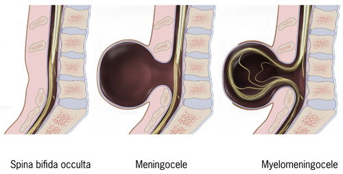
Three types of spina bifida

Type Cord involvement Skin/presentation Disability PT implications
Occulta None Depression or dimple, dark hair patch, fatty deposit, or port-wine nevus over an intact lower back Minimal, if any Routine monitoring; PT usually not needed
Meningocele None (sac contains meninges and CSF only) Sac protrudes through the vertebral defect, usually skin-covered Usually mild Monitor for associated clubfoot or hip dysplasia; promote typical motor development
Myelomeningocele Spinal cord and nerve roots involved Sac protrudes through the skin, often open Severe; paralysis, hydrocephalus, and Arnold-Chiari II malformation are common ROM, positioning, mobility training, skin and bowel/bladder management
Types of spina bifida
Courtesy CDC
/
CDC
/
Public domain

Physical therapy and spina bifida interventions

  • Joint ROM
  • Axial and trunk strengthening and engagement
  • Positioning and handling (specifically for infants)
  • Mobility and balance
  • Use latex-free equipment and inspect insensate skin regularly
  • Stop and report signs of shunt malfunction (headache, vomiting, irritability, lethargy, bulging fontanelle) to the PT

Cerebral palsy (CP) overview

  • Caused by abnormal brain development/damage before, during, or shortly after birth (hemorrhage, hypoxia, white matter damage)
  • Risk factors: multiple births, infection, low birth weight, complicated/breech delivery, jaundice
  • Common comorbidities: intellectual disability, seizures, visual/hearing/speech deficits, orthopedic conditions

Types of CP

  • Spastic (hypertonic): hemiplegia (one side), diplegia (legs, scissoring gait), quadriplegia (whole body, most severe)
  • Hypotonic: low tone, floppy, wide base of support
  • Dyskinetic (athetoid): writhing/jerky involuntary movements, intellect intact
  • Ataxic: poor coordination/balance, dysmetria
  • Mixed: combination of hyper/hypotonia

GMFCS levels (I-V)

  • I: independent, no limitations
  • II: independent, needs railing for stairs
  • III: uses hand-held device (crutches/walker)
  • IV: limited self-mobility, relies on wheelchair
  • V: severe limitations, dependent for mobility/posture

CP management

  • PT focus: symmetrical positioning, orthotic fit/training, functional motor skills, orthopedic condition management (scoliosis, contractures)
  • Medical: anti-seizure meds, Botox/baclofen for spasticity, surgery (dorsal rhizotomy, Z-plasty)

Down syndrome

  • Trisomy 21; features: hypotonia, short stature, microcephaly, congenital heart defects, intellectual disability
  • Key risk: atlanto-axial instability (C1-C2) — avoid diving, tumbling, headstands, contact sports
  • PTA role: report new neuro signs (neck pain, weakness, numbness) immediately; don’t alter plan of care

Duchenne muscular dystrophy (DMD)

  • X-linked, lack of dystrophin → progressive muscle breakdown; mainly affects boys; life expectancy now often into late 20s-30s with modern care
  • Signs: pseudohypertrophy (calves), Gowers’ sign (ages 3-5), waddling gait, cardiac myopathy
  • Progression: early weakness → loss of ambulation (9-14) → respiratory/cardiac decline, full dependence (15+)
  • PT: maintain ROM, submaximal/low-fatigue exercise, avoid eccentric/high-resistance work; stop/report prolonged muscle pain, dark urine
  • Medical: corticosteroids, cardiac/respiratory management, orthopedic treatment

Spinal muscular atrophy (SMA)

  • SMN1 gene mutation → anterior horn cell motor neuron loss; progressive
  • Types 0-4 based on onset age (birth → adulthood) and severity (Type 0 most severe, Type 4 mildest)
  • PT: support developmental milestones, ROM, posture, strength, respiratory monitoring, energy conservation
  • PTA: report respiratory decline immediately (applies to both DMD and SMA)

Comparing CP, SMA, DMD

  • CP: upper motor neuron, hypertonia, non-progressive but changes with growth
  • SMA: lower motor neuron, hypotonia/decreased reflexes, progressive
  • DMD: muscle fiber disorder, proximal weakness/pseudohypertrophy, progressive
  • Each requires different management emphasis (positioning vs. energy conservation vs. submaximal activity)

Spina bifida

  • Neural tube defect; linked to maternal folic acid deficiency; elevated AFP is a screening marker, not a cause
  • Symptoms: paralysis, incontinence, musculoskeletal deformities, hydrocephalus (often with Arnold-Chiari II malformation)
  • Three types: Occulta (mildest, no cord involvement), Meningocele (sac, no cord involvement), Myelomeningocele (severe, cord/nerve involvement, sac often open)
  • PT: ROM, trunk strengthening, positioning, mobility/balance training
  • Use latex-free equipment, monitor insensate skin, report shunt malfunction signs (headache, vomiting, lethargy, bulging fontanelle) immediately

Sign up for free to take 25 quiz questions on this topic

Previous
Next  | 4.2.2 Congenital musculoskeletal disorders
All rights reserved ©2016 - 2026 Achievable, Inc.

Congenital neurological and genetic disorders

Definitions
Congenital disorders
Conditions that develop in an individual in utero, during birth, or shortly after birth, due to gene mutations, deoxygenation to the brain or heart, infections, or micronutrient deficiencies.

Cerebral palsy

Definitions
Cerebral palsy (CP)
A group of disorders causing deficits in movement and coordination, caused by abnormal development or damage to the brain.

Damage can occur before, during, or shortly after birth, and may result from intracranial hemorrhage, lack of oxygen to the brain, abnormal brain development, or damage to the brain’s white matter.

Risk factors for CP include multiple births, toxin exposure, maternal or infant infection, low birth weight, seizures, complicated birth or labor, jaundice, and breech (buttocks- or feet-first) delivery.

Common comorbidities among individuals with cerebral palsy include intellectual disabilities, visual impairments, speech and language deficits, seizures, hearing impairments, and various orthopedic conditions.

Types of cerebral palsy

  • Spastic cerebral palsy (hypertonic cerebral palsy)
    • Type of cerebral palsy in which the individual exhibits hypertonia, causing stiff and jerky movements
      • Spastic hemiplegia
        • Hypertonia is located on one side of the body - the ipsilateral arm and leg
        • The affected side is typically shorter and thinner, scoliosis may be present, and intellect is often normal
      • Spastic diplegia
        • Hypertonia is located primarily in the lower extremities
        • Hyperreflexia can be present, along with scissoring of gait; intellect is often normal
      • Spastic quadriplegia
        • Hypertonia is located throughout the entire body
        • The most severe type of spastic cerebral palsy, due to widespread damage to the entire brain
        • Individuals with this type of CP rarely walk, demonstrate difficulty speaking, and often have moderate-to-severe intellectual disability and frequent seizures
  • Hypotonic cerebral palsy
    • Characterized by low tone and floppiness of extremities and axial skeleton
    • Individuals may demonstrate poor muscle tone, excessive range of motion, increased weight gain, impaired speech due to poor oral motor control, and a wide base of support with gait
  • Dyskinetic cerebral palsy (athetoid cerebral palsy)
    • Caused by damage to the basal ganglia; characterized by slow and uncontrollable writhing or jerky movements of the hands, feet, arms, and legs
    • Individuals may demonstrate postural deficits, hearing problems, and breathing difficulties; intellect is often preserved, though intellectual disability is common
  • Ataxic cerebral palsy
    • Characterized by poor coordination, balance impairments, and impairments in depth perception
    • Individuals may demonstrate a wide base of support, dysmetria, and difficulty with precision of movement
  • Mixed types
    • Characterized by a mix of hypertonia and hypotonia with varied symptom presentation

Classifications of cerebral palsy (Gross Motor Function Classification System, GMFCS)

  • Level I: Walks independently without limitations. Climbs stairs without using a railing and can run and jump, although speed, balance, and coordination may be limited.
  • Level II: Walks independently but uses a railing for stairs. May have difficulty with long distances, uneven ground, or crowded spaces and has limited ability to run or jump.
  • Level III: Walks using a hand-held mobility device, such as crutches or a walker. Typically, self-propels a wheelchair for longer distances.
  • Level IV: Has limited self-mobility and usually relies on a wheelchair or powered mobility for most movement. May stand or take a few steps using a supportive walker.
  • Level V: Has severe limitations in mobility and posture, with difficulty controlling head, trunk, arm, and leg movements. Depends on others for transfers and mobility and is typically transported in a wheelchair

Physical therapy interventions for cerebral palsy

  • Positioning is key to promote improvements in mobility, ADL participation, gait and balance improvements, and interaction with the environment
    • Symmetrical posture
    • Alignment of trunk, pelvis, and extremities
    • Head in midline
    • Hips and knees positioned at 90 degrees while sitting
    • Checking fit and skin integrity of orthoses prescribed by the PT, and training don/doff
    • Optimizing functional motor skills
  • Treatment of orthopedic conditions such as
    • Scoliosis
    • Joint contractures
    • Kyphosis
    • Clubfoot
    • Hip or shoulder dislocation

Medical management for cerebral palsy

  • Anti-seizure medication
  • Spasticity medication
    • Botox injections for local hypertonic muscles
    • Baclofen taken orally or by implantation for multiple areas of hypertonia
  • Surgical interventions
    • Dorsal rhizotomy
      • Selected dorsal (sensory) nerve rootlets - those driving the spasticity - are cut, leaving the rest intact, to decrease spasticity and improve overall function
    • Z-plasty
      • Release of muscle or tendons to release contractures

Down syndrome

Definitions
Down syndrome
A condition resulting from a chromosomal abnormality on chromosome 21 - instead of the usual pair, three copies of chromosome 21 are present (trisomy 21).

Characteristics of Down syndrome

  • Small ears and a protruding tongue
  • Microcephaly with flattened occiput
  • Short stature
  • Hypotonia and hypermobility
  • Congenital heart defects
  • Speech deficits
  • Developmental delays
  • Vertebral instability at the atlanto-axial joint (C1-C2)
  • Intellectual disabilities

Physical therapy interventions for Down syndrome

  • Promote gross motor development
  • Increase motor control and postural awareness
  • Improve oral-motor development
  • Train in use of DME selected by the PT; report fit or functional changes to the PT
  • Patient and family education

Special considerations with Down syndrome

  • Avoidance of diving, tumbling, headstands, and contact sports, due to an increased risk of hyperflexion injury from atlanto-axial instability

PTA role: Data collection and communication - not changing the plan of care - are within the PTA’s scope. If a session reveals new neurological signs (neck pain, numbness, weakness, or changes in coordination) that suggest atlanto-axial instability, stop the activity and report the finding to the supervising PT.

Duchenne muscular dystrophy

Definitions
Duchenne muscular dystrophy
A progressive disease process in which the protein dystrophin is not produced, causing an increased breakdown of muscle tissue over time. It most often affects boys; with modern care (steroids, ventilatory and cardiac support), life expectancy commonly extends into the late 20s or 30s.

Common symptoms of Duchenne muscular dystrophy

  • Progressive muscle weakness
  • Pseudohypertrophic muscles appear hypertrophied, but the muscle has been replaced by fat and connective tissue in calves, deltoids, quadriceps, and tongue
  • Contracture
  • Cardiac myopathy
  • Gowers’ sign
    • The child pushes up from the floor with their hands, walking their hands up their legs to stand - this is due to weak hip and knee extensors; typically noticeable by ages 3-5
  • Waddling gait
  • Increased risk of falls

Progression of Duchenne muscular dystrophy

  • Early childhood (age 3-5): weakness, tripping, and Gowers’ sign appear
  • Later childhood (age 9-14): loss of ambulation as gait deviations, poor endurance, and lower extremity contractures progress; use of a manual wheelchair begins
  • Adolescence and beyond (age 15+): increased respiratory and cardiac compromise and total dependence for ADLs and mobility; with modern cardiopulmonary care, many individuals now live into their late 20s or 30s

Physical therapy interventions for Duchenne muscular dystrophy

  • Maintain range of motion
  • Collect data on mobility and functional status to help the PT maintain the current level of function
  • Use submaximal, low-fatigue activity and avoid eccentric and high-resistance exercise; stop and report muscle pain or weakness lasting more than 24 hours, or dark urine
  • Train in use of DME selected by the PT; report fit or need changes to the PT
  • Parent and patient education

Example: Fatigue during gait training

A child with Duchenne muscular dystrophy begins to breathe harder and lean more heavily on the walker partway through a gait-training session.

Answer: The PTA stops the activity, lets the child rest, and documents the change in endurance and effort, then reports the finding to the supervising PT. The PTA does not push through the fatigue or increase resistance, since overworking already-weakened muscle fibers in DMD can accelerate muscle breakdown.

Medical management for Duchenne muscular dystrophy

  • Corticosteroids (e.g., prednisone, deflazacort) to slow strength loss; cardiac and respiratory management as needed
  • Treatment of orthopedic conditions through injections or surgery

Spinal muscular atrophy (SMA)

Definitions
Spinal muscular atrophy (SMA)
A group of congenital disorders in which the motor neurons within the spinal cord are dysfunctional, caused by mutations in the survival motor neuron 1 (SMN1) gene, leading to loss of anterior horn cell motor neurons.

Common symptoms associated with SMA include muscle contractures, muscle weakness, scoliosis, difficulty with functional mobility, difficulty swallowing, and respiratory impairment. Symptoms are progressive throughout the individual’s lifespan.

Five types of SMA

  • Type 0 (zero)
    • Presents at birth; muscle atrophy and severe muscle weakness present; leads to life-threatening respiratory compromise
  • Type 1 (infantile)
    • Presentation within the first 6 months of life; muscle weakness specifically impacting feeding, crawling, and sitting
  • Type 2 (intermediate)
    • Presentation between 6-18 months of life; progressive muscle weakness in the hips, legs, and trunk
  • Type 3 (juvenile)
    • Presentation between 18 months and 18 years of age; muscle weakness in the back, legs, and feet
  • Type 4 (adult)
    • Presentation after 18 years of age; mild symptoms that present later in life

Physical therapy interventions for SMA

Physical therapy interventions are based upon the level of impairment associated with SMA and can include progression to meet developmental milestones, improvements in range of motion, improvements in balance, improvements in posture, strengthening muscles, monitoring respiratory status (stop the activity and report any decline in respiratory tolerance to the supervising PT), and initiation of energy conservation strategies.

Comparing cerebral palsy, SMA, and Duchenne muscular dystrophy

These three conditions can all present as a weak or floppy child, but the lesion site and course differ, which changes what the PTA should expect and watch for.

Condition Lesion site Tone and reflexes Course Management emphasis
Cerebral palsy Upper motor neuron (brain) Hypertonia (or hypotonia in the hypotonic type), reflexes often increased Non-progressive, but presentation changes with growth Positioning, orthotics, spasticity management
Spinal muscular atrophy Lower motor neuron (anterior horn cell) Hypotonia, reflexes decreased or absent Progressive Energy conservation, respiratory monitoring, equipment
Duchenne muscular dystrophy Muscle fiber (myopathy) Hypotonia; reflexes diminish as muscle is lost (knee jerks lost early, ankle jerks kept until late); proximal weakness, pseudohypertrophy, Gowers’ sign Progressive Maintain ROM and function, submaximal activity, avoid overwork

Spina bifida

Definitions
Spina bifida
A neural tube defect resulting in vertebral and/or spinal cord malformation. The cause is multifactorial, with maternal folic acid deficiency as a major modifiable risk factor. Elevated maternal serum and amniotic alpha-fetoprotein is a screening marker of an open neural tube defect in utero - it is not the cause of the defect.

Common symptoms associated with spina bifida

  • Flaccid or spastic paralysis
  • Bladder incontinence
  • Musculoskeletal deformities (scoliosis, hip dysplasia, hip dislocation, clubfoot, hip/knee contracture)
  • Hydrocephalus, commonly with Arnold-Chiari type II malformation (especially with myelomeningocele)
Definitions
Hydrocephalus
Increased cerebrospinal fluid in the ventricles of the brain; the fluid accumulation causes increased pressure on other brain structures.
Arnold-Chiari malformation
A structural defect in which the cerebellum pushes down into the spinal canal; often associated with hydrocephalus.

Three types of spina bifida

Type Cord involvement Skin/presentation Disability PT implications
Occulta None Depression or dimple, dark hair patch, fatty deposit, or port-wine nevus over an intact lower back Minimal, if any Routine monitoring; PT usually not needed
Meningocele None (sac contains meninges and CSF only) Sac protrudes through the vertebral defect, usually skin-covered Usually mild Monitor for associated clubfoot or hip dysplasia; promote typical motor development
Myelomeningocele Spinal cord and nerve roots involved Sac protrudes through the skin, often open Severe; paralysis, hydrocephalus, and Arnold-Chiari II malformation are common ROM, positioning, mobility training, skin and bowel/bladder management

Physical therapy and spina bifida interventions

  • Joint ROM
  • Axial and trunk strengthening and engagement
  • Positioning and handling (specifically for infants)
  • Mobility and balance
  • Use latex-free equipment and inspect insensate skin regularly
  • Stop and report signs of shunt malfunction (headache, vomiting, irritability, lethargy, bulging fontanelle) to the PT
Key points

Cerebral palsy (CP) overview

  • Caused by abnormal brain development/damage before, during, or shortly after birth (hemorrhage, hypoxia, white matter damage)
  • Risk factors: multiple births, infection, low birth weight, complicated/breech delivery, jaundice
  • Common comorbidities: intellectual disability, seizures, visual/hearing/speech deficits, orthopedic conditions

Types of CP

  • Spastic (hypertonic): hemiplegia (one side), diplegia (legs, scissoring gait), quadriplegia (whole body, most severe)
  • Hypotonic: low tone, floppy, wide base of support
  • Dyskinetic (athetoid): writhing/jerky involuntary movements, intellect intact
  • Ataxic: poor coordination/balance, dysmetria
  • Mixed: combination of hyper/hypotonia

GMFCS levels (I-V)

  • I: independent, no limitations
  • II: independent, needs railing for stairs
  • III: uses hand-held device (crutches/walker)
  • IV: limited self-mobility, relies on wheelchair
  • V: severe limitations, dependent for mobility/posture

CP management

  • PT focus: symmetrical positioning, orthotic fit/training, functional motor skills, orthopedic condition management (scoliosis, contractures)
  • Medical: anti-seizure meds, Botox/baclofen for spasticity, surgery (dorsal rhizotomy, Z-plasty)

Down syndrome

  • Trisomy 21; features: hypotonia, short stature, microcephaly, congenital heart defects, intellectual disability
  • Key risk: atlanto-axial instability (C1-C2) — avoid diving, tumbling, headstands, contact sports
  • PTA role: report new neuro signs (neck pain, weakness, numbness) immediately; don’t alter plan of care

Duchenne muscular dystrophy (DMD)

  • X-linked, lack of dystrophin → progressive muscle breakdown; mainly affects boys; life expectancy now often into late 20s-30s with modern care
  • Signs: pseudohypertrophy (calves), Gowers’ sign (ages 3-5), waddling gait, cardiac myopathy
  • Progression: early weakness → loss of ambulation (9-14) → respiratory/cardiac decline, full dependence (15+)
  • PT: maintain ROM, submaximal/low-fatigue exercise, avoid eccentric/high-resistance work; stop/report prolonged muscle pain, dark urine
  • Medical: corticosteroids, cardiac/respiratory management, orthopedic treatment

Spinal muscular atrophy (SMA)

  • SMN1 gene mutation → anterior horn cell motor neuron loss; progressive
  • Types 0-4 based on onset age (birth → adulthood) and severity (Type 0 most severe, Type 4 mildest)
  • PT: support developmental milestones, ROM, posture, strength, respiratory monitoring, energy conservation
  • PTA: report respiratory decline immediately (applies to both DMD and SMA)

Comparing CP, SMA, DMD

  • CP: upper motor neuron, hypertonia, non-progressive but changes with growth
  • SMA: lower motor neuron, hypotonia/decreased reflexes, progressive
  • DMD: muscle fiber disorder, proximal weakness/pseudohypertrophy, progressive
  • Each requires different management emphasis (positioning vs. energy conservation vs. submaximal activity)

Spina bifida

  • Neural tube defect; linked to maternal folic acid deficiency; elevated AFP is a screening marker, not a cause
  • Symptoms: paralysis, incontinence, musculoskeletal deformities, hydrocephalus (often with Arnold-Chiari II malformation)
  • Three types: Occulta (mildest, no cord involvement), Meningocele (sac, no cord involvement), Myelomeningocele (severe, cord/nerve involvement, sac often open)
  • PT: ROM, trunk strengthening, positioning, mobility/balance training
  • Use latex-free equipment, monitor insensate skin, report shunt malfunction signs (headache, vomiting, lethargy, bulging fontanelle) immediately

More from Congenital disorders

  • Congenital musculoskeletal disorders