



Congenital neuromuscular and neural tube disorders
The contents of this chapter will cover common congenital disorders.
Cerebral palsy
Damage can occur before, during, or shortly after birth, and may result from intracranial hemorrhage, lack of oxygen to the brain, abnormal brain development, or damage to the brain’s white matter.
The risk of developing CP increases with multiple births, exposure to toxic substances, maternal or infant infections, low birth weight, seizures, complicated birth or labor, jaundice, or breech birth (feet-first delivery).
Common comorbidities among individuals with cerebral palsy include intellectual disabilities, visual impairments, speech and language deficits, seizures, hearing impairments, and various orthopedic conditions.
Types of cerebral palsy
- Spastic cerebral palsy (hypertonic cerebral palsy)
- Type of cerebral palsy in which the individual exhibits hypertonia, causing stiff and jerky movements
- Spastic hemiplegia
- Hypertonia is located on one side of the body - the ipsilateral arm and leg
- The affected side is typically shorter and thinner, scoliosis may be present, and intellect is normal
- Spastic diplegia
- Hypertonia is located primarily in the lower extremities
- Hyperreflexia can be present, along with scissoring of gait; intellect is normal
- Spastic quadriplegia
- Hypertonia is located throughout the entire body
- The most severe type of spastic cerebral palsy, due to widespread damage to the entire brain
- Individuals with this type of CP rarely walk, demonstrate difficulty speaking, and can have some intellectual difficulty
- Spastic hemiplegia
- Type of cerebral palsy in which the individual exhibits hypertonia, causing stiff and jerky movements
- Hypotonic cerebral palsy
- Characterized by low tone and floppiness of extremities and axial skeleton
- Individuals may demonstrate poor muscle tone, excessive range of motion, increased weight gain, impaired speech due to poor oral motor control, and a wide base of support with gait
- Dyskinetic cerebral palsy (athetoid cerebral palsy)
- Characterized by slow and uncontrollable writhing or jerky movements of the hands, feet, arms, and legs
- Individuals may demonstrate postural deficits, hearing problems, and breathing difficulties; intellect remains intact
- Ataxic cerebral palsy
- Characterized by poor coordination, balance impairments, and impairments in depth perception
- Individuals may demonstrate a wide base of support, dysmetria, and difficulty with precision of movement
- Mixed types
- Characterized by a mix of hypertonia and hypotonia with varied symptom presentation
Classifications of cerebral palsy (Gross Motor Function Classification System, GMFCS)
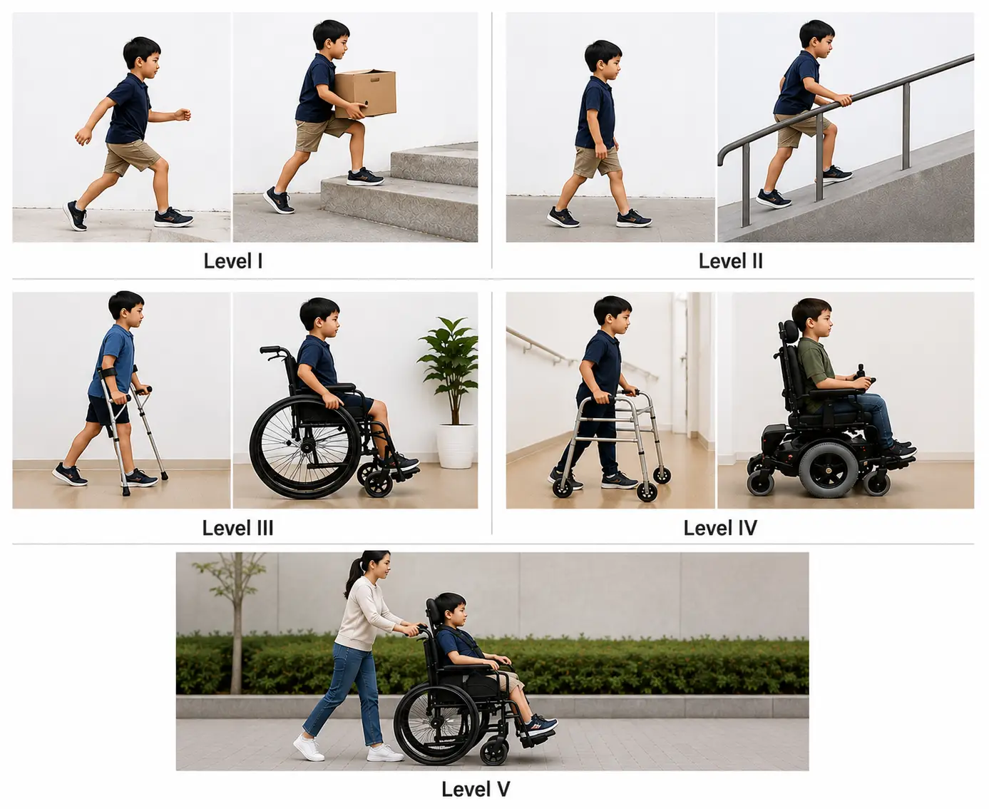
- Level I: Walks independently without limitations. Climbs stairs without using a railing and can run and jump, although speed, balance, and coordination may be limited.
- Level II: Walks independently but uses a railing for stairs. May have difficulty with long distances, uneven ground, or crowded spaces and has limited ability to run or jump.
- Level III: Walks using a hand-held mobility device, such as crutches or a walker. Typically, self-propels a wheelchair for longer distances.
- Level IV: Has limited self-mobility and usually relies on a wheelchair or powered mobility for most movement. May stand or take a few steps using a supportive walker.
- Level V: Has severe limitations in mobility and posture, with difficulty controlling head, trunk, arm, and leg movements. Depends on others for transfers and mobility and is typically transported in a wheelchair
Physical therapy interventions for cerebral palsy
- Positioning is key to promote improvements in mobility, ADL participation, gait and balance improvements, and interaction with the environment
- Symmetrical posture
- Alignment of trunk, pelvis, and extremities
- Head in midline
- Hips and knees positioned at 90 degrees while sitting
- Prescription of orthoses
- Optimizing functional motor skills
- Treatment of visual-motor and perceptual disorders
- Aids to assist with visual processing, aiding in connecting the visual system to the brain
- Treatment of orthopedic conditions such as
- Scoliosis
- Joint contractures
- Kyphosis
- Clubfoot
- Hip or shoulder dislocation
Medical management for cerebral palsy
- Anti-seizure medication
- Spasticity medication
- Botox injections for local hypertonic muscles
- Baclofen taken orally or by implantation for multiple areas of hypertonia
- Surgical interventions
- Dorsal rhizotomy
- Dorsal nerve roots are severed to aid in decreasing spasticity and improving overall function
- Z-plasty
- Release of muscle or tendons to release contractures
- Dorsal rhizotomy
Down syndrome
Characteristics of Down syndrome
- Small ears and a protruding tongue
- Microcephaly with flattened occiput
- Short stature
- Hypotonia and hypermobility
- Congenital heart defects
- Speech deficits
- Developmental delays
- Vertebral instability at the atlanto-axial joint (C1-C2)
- Intellectual disabilities
Physical therapy interventions for Down syndrome
- Promote gross motor development
- Increase motor control and postural awareness
- Improve oral-motor development
- Durable medical equipment recommendations as appropriate
- Patient and family education
Special considerations with Down syndrome
- Avoidance of diving, tumbling, headstands, and contact sports, due to an increased risk of hyperflexion injury from atlanto-axial instability
Duchenne muscular dystrophy
Common symptoms of Duchenne muscular dystrophy
- Progressive muscle weakness
- Pseudohypertrophic muscles appear hypertrophied, but the muscle has been replaced by fat and connective tissue in calves, deltoids, quadriceps, and tongue
- Contracture
- Cardiac myopathy
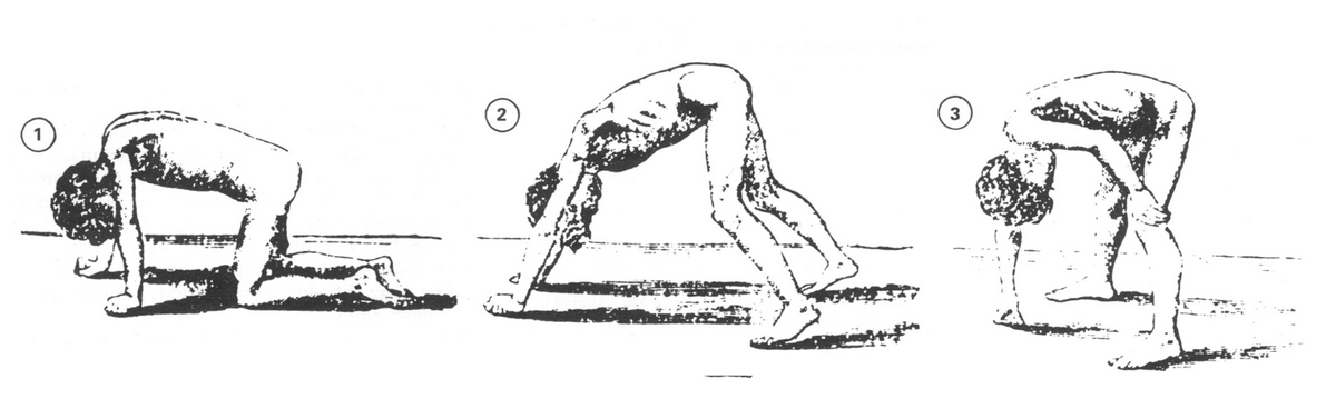
- Gower’s sign
- The child pushes up from the floor with their hands, walking their hands up their legs to stand - this is due to weak hip and knee extensors; typically begins at ages 4-7
- Waddling gait
- Increased risk of falls
Progression of Duchenne muscular dystrophy
- Early childhood (age 3-5): weakness, tripping, and Gower’s sign appear
- Later childhood (age 9-14): loss of ambulation as gait deviations, poor endurance, and lower extremity contractures progress; use of a manual wheelchair begins
- Adolescence and beyond (age 15+): increased respiratory compromise and total dependence for ADLs and mobility, with death typically occurring in early adulthood due to respiratory failure
Physical therapy interventions for Duchenne muscular dystrophy
- Maintain range of motion
- Assess mobility and attempt to maintain the current level of mobility
- Provide durable medical equipment as appropriate
- Parent and patient education
Medical management for Duchenne muscular dystrophy
- Treatment of symptoms as appropriate
- Use of medications such as steroids or antibiotics, as needed
- Treatment of orthopedic conditions through injections or surgery
Spinal muscular atrophy (SMA)
Common symptoms associated with SMA include muscle contractures, muscle weakness, scoliosis, difficulty with functional mobility, difficulty swallowing, and respiratory impairment. Symptoms are progressive throughout the individual’s lifespan.
Diagnosis is confirmed via physical examination, genetic testing, and electromyography (EMG) or nerve conduction studies.
Five types of SMA
- Type 0 (zero)
- Presents at birth; muscle atrophy and severe muscle weakness present; leads to life-threatening respiratory compromise
- Type 1 (infantile)
- Presentation within the first 6 months of life; muscle weakness specifically impacting feeding, crawling, and sitting
- Type 2 (intermediate)
- Presentation between 6-18 months of life; progressive muscle weakness in the hips, legs, and trunk
- Type 3 (juvenile)
- Presentation between 18 months and 18 years of age; muscle weakness in the back, legs, and feet
- Type 4 (adult)
- Presentation after 18 years of age; mild symptoms that present later in life
Physical therapy interventions for SMA
Physical therapy interventions are based upon the level of impairment associated with SMA and can include progression to meet developmental milestones, improvements in range of motion, improvements in balance, improvements in posture, strengthening muscles, and initiation of energy conservation strategies.
Spina bifida
Common symptoms associated with spina bifida
- Flaccid or spastic paralysis
- Bladder incontinence
- Musculoskeletal deformities (scoliosis, hip dysplasia, hip dislocation, clubfoot, hip/knee contracture)
- Hydrocephalus, along with Type I or II Arnold-Chiari malformation
Three types of spina bifida
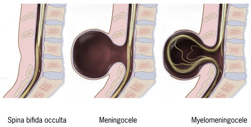
- Spina bifida occulta
- No spinal cord involvement
- Depression or dimple in the lower back
- A small patch of dark hair
- Soft fatty deposits
- Port-wine nevi (deep red-purple macular lesions)
- Minimal disability, if any
- Spina bifida meningocele
- No spinal cord involvement
- Meninges protrude through the skin
- Cerebrospinal fluid may leak
- Associated diagnoses: clubfoot, hip dysplasia, hydrocephalus
- Moderate disability
- Spina bifida myelomeningocele
- Spinal cord involvement
- Protrudes through the skin
- Severe disability will result
- Paralysis usually occurs
Physical therapy and spina bifida interventions
- Joint ROM
- Axial and trunk strengthening and engagement
- Positioning and handling (specifically for infants)
- Mobility and balance