
Achievable logoAchievable logo
NPTE-PTA
Sign in
Sign up
Purchase
Textbook
Practice exams
Support
How it works
Resources
Exam catalog
Mountain with a flag at the peak
Textbook
Introduction
1. Cardiovascular system
1.1 Physiology of cardiac system
1.2 Cardiac pathologies
1.3 Cardiac rehabilitation
1.4 Electrocardiograms
1.5 Lab values and ankle brachial index
1.6 Cardiac exercise testing
2. Pulmonary system
3. Neuromuscular system
4. Pediatrics
5. Musculoskeletal system
6. Other systems
7. Non-systems
Wrapping up
Achievable logoAchievable logo
1.1 Physiology of cardiac system
Achievable NPTE-PTA
1. Cardiovascular system
Our NPTE-PTA course is now in "early access" - get 50% off for a limited time.

Physiology of cardiac system

8 min read
Font
Discuss
Share
Feedback

Anatomy

The heart is a muscle that pumps blood throughout the body’s systems. The left side of the heart pumps oxygenated blood from the lungs to all body systems via the arterial supply. Blood then returns to the right side of the heart via the venous system, where waste products are excreted by various organs. The right side then pumps deoxygenated blood into the lungs, where carbon dioxide (CO2) is released, and oxygen is absorbed. This cycle repeats with each heartbeat.

The heart consists of:

  • External walls
  • Blood-filled chambers
  • Valves that allow blood to flow through the chambers
  • Blood vessels that carry blood to the other structures
  • Electrical signaling conduction system

The three layers of the heart wall are:

  • Endocardium; innermost layer
  • Myocardium: muscular middle layer
  • Epicardium: outermost layer

Chambers of the heart:

  • Right atrium
  • Right ventricle
  • Left atrium
  • Left ventricle

Valves of the heart:

  • Atrioventricular valves:
    • Tricuspid valve: lies between the right atrium and the right ventricle
    • Mitral valve: lies between the left atrium and left ventricle
  • Semilunar valves:
    • Aortic valve: oxygenated blood flows from the left ventricle to the aorta
    • Pulmonary valve: deoxygenated blood flows from the right ventricle to the pulmonary artery

Blood vessels:

  • Arteries: carry oxygenated blood
  • Veins: carry deoxygenated blood
  • Capillaries: smallest blood vessels, where oxygen, carbon dioxide, nutrients, and wastes are exchanged between the blood and the surrounding tissues
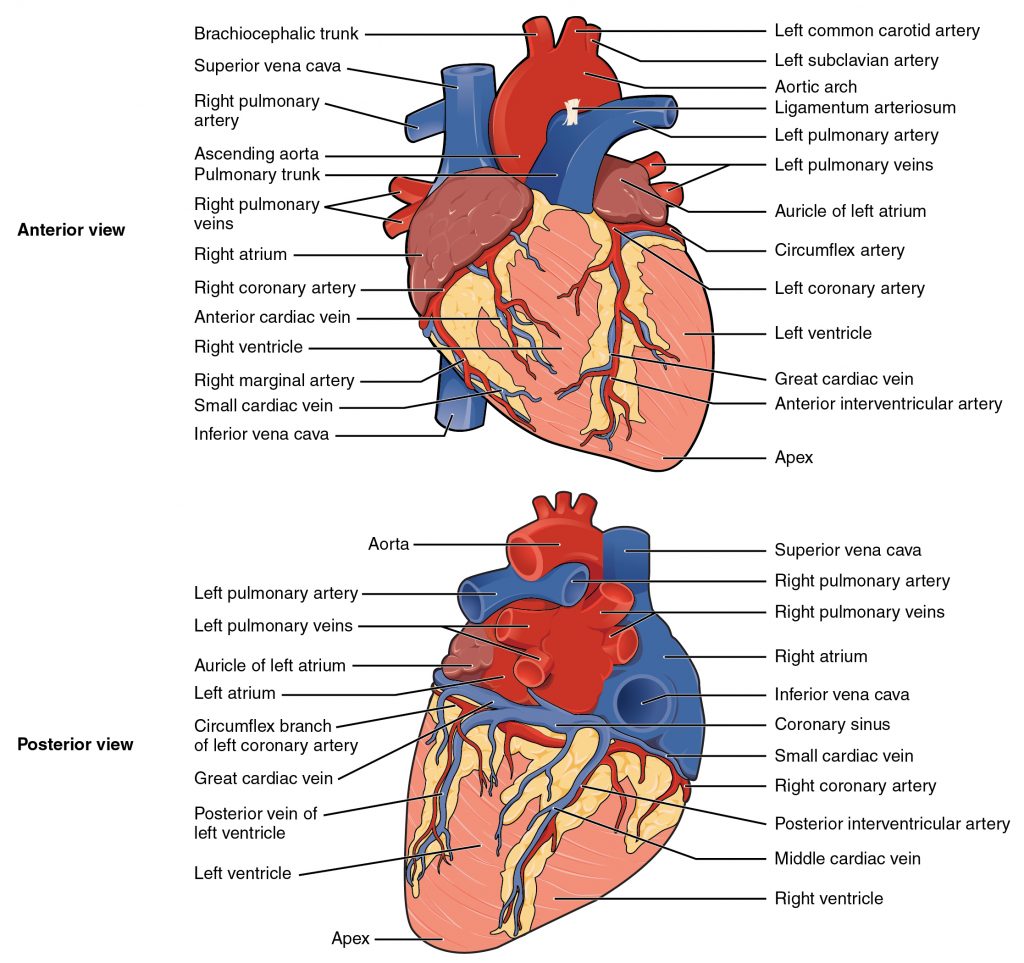
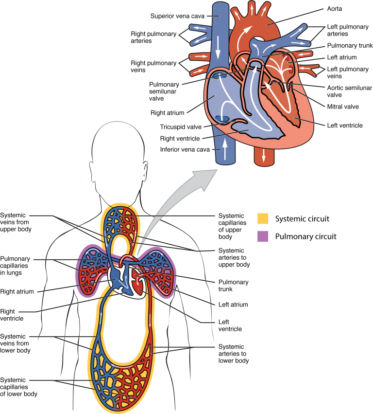
Diagram of parts of heart
Anatomy & Physiology by Lindsay M. Biga, Sierra Dawson, Amy Harwell, Robin Hopkins, Joel Kaufmann, Mike LeMaster, Philip Matern, Katie Morrison-Graham, Devon Quick & Jon Runyeon
/
Oregon State University
/
CC BY 4.0

Chambers of the heart

The heart is separated into four chambers that work together to keep blood flowing in the right direction, with the right and left sides handling deoxygenated and oxygenated blood separately, as described above.

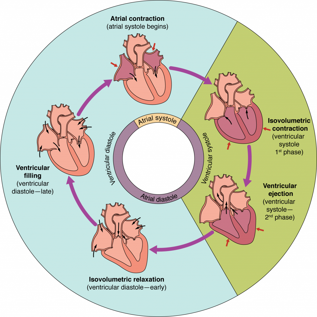
Cycle of heart
Cycle of heart
Achievable
/
Achievable
Definitions
Superior vena cava
Large vein that carries deoxygenated blood from the head, chest, arm, and neck
Inferior vena cava
Large vein that carries deoxygenated blood from the feet, legs, and abdomen
Right atrium
Receives deoxygenated blood from the body system and then pumps it to the right ventricle
Right ventricle
Receives blood from the right atrium and pumps deoxygenated blood through the pulmonary valve into the pulmonary artery to the lungs for oxygenation
Left atrium
Receives oxygenated blood from the lungs via four pulmonary veins
Left ventricle
Receives oxygenated blood from the left atrium and pumps oxygenated blood into the aorta, distributing it to the brain, organs, and tissues.
Aorta
Transports oxygenated blood from the heart to the peripheral systems

Blood flow through the heart: step-by-step

  1. Deoxygenated blood returns to the heart:
    • Blood low in oxygen returns from the body to the heart through two major veins:
      • Superior vena cava (SVC): returns blood from the upper body
      • Inferior vena cava (IVC): returns blood from the lower body
    • Both veins empty into the right atrium.
  2. Right atrium
    • Deoxygenated blood enters the right atrium, the upper right chamber of the heart.
    • Blood then flows through the tricuspid valve into the right ventricle.
  3. Right ventricle
    • The right ventricle pumps deoxygenated blood through the pulmonary semilunar valve into the pulmonary arteries.
  4. Pulmonary arteries to the lungs
    • The pulmonary arteries carry deoxygenated blood from the heart to the lungs.
      • This is an important exception because most arteries carry oxygenated blood.
  5. Lungs oxygenate the blood
    • In the lungs, carbon dioxide (CO₂) is removed and oxygen (O₂) is added to the blood through gas exchange at the alveoli.
    • The blood is now oxygenated.
  6. Left atrium
    • Oxygen-rich blood returns to the heart through the pulmonary veins and enters the left atrium, the upper left chamber of the heart.
      • This is another important exception because pulmonary veins carry oxygenated blood.
    • Blood then passes through the mitral (bicuspid) valve into the left ventricle.
  7. Left ventricle
    • The left ventricle pumps oxygenated blood through the aortic semilunar valve into the aorta.
    • The left ventricle has the thickest myocardium because it must generate enough force to pump blood throughout the entire body.
  8. Aorta to the body
    • The aorta distributes oxygen-rich blood to the systemic circulation, supplying tissues and organs throughout the body.
Blood flow of heart
Blood flow of heart
Anatomy & Physiology by Lindsay M. Biga, Sierra Dawson, Amy Harwell, Robin Hopkins, Joel Kaufmann, Mike LeMaster, Philip Matern, Katie Morrison-Graham, Devon Quick & Jon Runyeon
/
Oregon State University
/
CC BY 4.0

Electrical signals in the heart

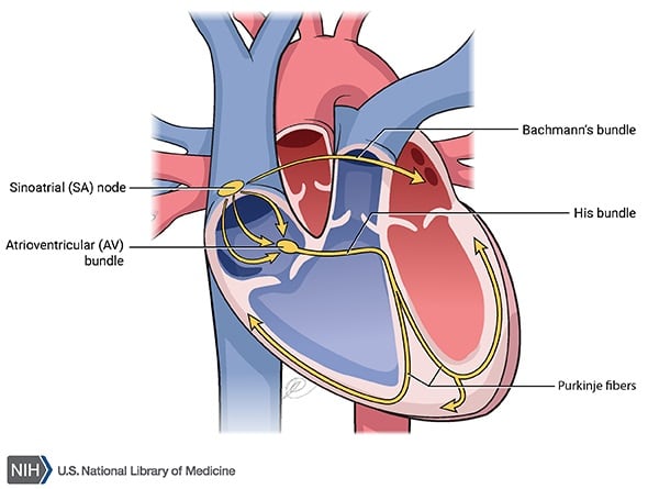
Electrical signals within the heart act as a pacemaker to the heart by assisting with controlling heart rate, coordinating the heart chambers (atria and ventricles), adapting to the changing needs of the body, and ensuring appropriate circulation. The normal conduction pathway follows this order: SA node → AV node → bundle of His → bundle branches → Purkinje fibers.

Heart electrical activity
Achievable
/
Achievable
Definitions
Sinoatrial node
The sinoatrial (SA) node is the heart’s natural pacemaker. It initiates the electrical impulse that triggers atrial contraction, setting the normal heart rate and rhythm.
Atrioventricular node
The atrioventricular (AV) node receives the impulse from the SA node and briefly delays it, allowing the ventricles to fill with blood before passing the impulse on to the bundle of His.
Bundle of His
A specialized group of conductive cardiac fibers that carries electrical impulses from the AV node to the right and left bundle branches, helping coordinate ventricular depolarization and contraction.
Purkinje fibers
Specialized conductive fibers located within the ventricular walls that rapidly transmit electrical impulses throughout the ventricles, producing coordinated contraction of the right and left ventricles.

Crucial factors of cardiac function

The amount of blood pumped with each heartbeat is crucial to maintaining the vitality of the lungs and the peripheral system. The principles that are important in this process are preload and afterload. If preload or afterload is abnormal - for example, too little blood filling the ventricles or too much resistance to ejection - stroke volume and cardiac output fall; over time, chronic volume overload (excess preload) or pressure overload (excess afterload, as in longstanding hypertension) can lead to heart failure.

Definitions
Preload
The amount of ventricular stretch at the end of diastole, primarily determined by the volume of blood filling the ventricles (end-diastolic volume)
Afterload
The resistance or pressure the ventricles must overcome to eject blood during systole.
Stroke volume
Amount of blood pumped out by the ventricle at the end of each contraction
Cardiac output
Heart rate multiplied by stroke volume to determine how much blood is pumped from the ventricles in liters per minute (normally about 4-8 L/min at rest in a healthy adult)
Diastole
Relaxation of atrium and ventricles
Systole
Contraction of the atrium and ventricle

Anatomy overview

  • Heart pumps blood: left side sends oxygenated blood to body; right side sends deoxygenated blood to lungs
  • Three wall layers: endocardium (inner), myocardium (muscular middle), epicardium (outer)
  • Four chambers: right atrium, right ventricle, left atrium, left ventricle

Valves of the heart

  • Atrioventricular valves:
    • Tricuspid: right atrium → right ventricle
    • Mitral: left atrium → left ventricle
  • Semilunar valves:
    • Aortic: left ventricle → aorta
    • Pulmonary: right ventricle → pulmonary artery

Blood vessels

  • Arteries: carry oxygenated blood (exception: pulmonary artery)
  • Veins: carry deoxygenated blood (exception: pulmonary veins)
  • Capillaries: site of gas/nutrient/waste exchange

Chambers & major vessels

  • Superior/inferior vena cava: return deoxygenated blood to right atrium
  • Right atrium → right ventricle → pulmonary artery → lungs
  • Left atrium (via pulmonary veins) → left ventricle → aorta → body
  • Aorta: distributes oxygenated blood systemically

Blood flow pathway (step-by-step)

  • Deoxygenated blood: SVC/IVC → right atrium → tricuspid valve → right ventricle → pulmonary valve → pulmonary arteries → lungs
  • Oxygenation occurs in lungs (CO2 out, O2 in)
  • Oxygenated blood: pulmonary veins → left atrium → mitral valve → left ventricle → aortic valve → aorta → body
  • Left ventricle has thickest myocardium (pumps blood to entire body)

Electrical conduction system

  • Pathway: SA node → AV node → bundle of His → bundle branches → Purkinje fibers
  • SA node: natural pacemaker, sets heart rate/rhythm
  • AV node: delays impulse, allows ventricular filling
  • Bundle of His & Purkinje fibers: coordinate ventricular depolarization/contraction

Crucial factors of cardiac function

  • Preload: ventricular stretch at end of diastole (end-diastolic volume)
  • Afterload: resistance ventricles overcome during systole
  • Stroke volume: blood ejected per contraction
  • Cardiac output = heart rate × stroke volume (~4-8 L/min at rest)
  • Abnormal preload/afterload → reduced stroke volume/cardiac output
    • Chronic excess preload or afterload (e.g., hypertension) → heart failure
  • Diastole = relaxation; Systole = contraction

Sign up for free to take 5 quiz questions on this topic

Previous
Next  | 1.2 Cardiac pathologies
All rights reserved ©2016 - 2026 Achievable, Inc.

Physiology of cardiac system

Anatomy

The heart is a muscle that pumps blood throughout the body’s systems. The left side of the heart pumps oxygenated blood from the lungs to all body systems via the arterial supply. Blood then returns to the right side of the heart via the venous system, where waste products are excreted by various organs. The right side then pumps deoxygenated blood into the lungs, where carbon dioxide (CO2) is released, and oxygen is absorbed. This cycle repeats with each heartbeat.

The heart consists of:

  • External walls
  • Blood-filled chambers
  • Valves that allow blood to flow through the chambers
  • Blood vessels that carry blood to the other structures
  • Electrical signaling conduction system

The three layers of the heart wall are:

  • Endocardium; innermost layer
  • Myocardium: muscular middle layer
  • Epicardium: outermost layer

Chambers of the heart:

  • Right atrium
  • Right ventricle
  • Left atrium
  • Left ventricle

Valves of the heart:

  • Atrioventricular valves:
    • Tricuspid valve: lies between the right atrium and the right ventricle
    • Mitral valve: lies between the left atrium and left ventricle
  • Semilunar valves:
    • Aortic valve: oxygenated blood flows from the left ventricle to the aorta
    • Pulmonary valve: deoxygenated blood flows from the right ventricle to the pulmonary artery

Blood vessels:

  • Arteries: carry oxygenated blood
  • Veins: carry deoxygenated blood
  • Capillaries: smallest blood vessels, where oxygen, carbon dioxide, nutrients, and wastes are exchanged between the blood and the surrounding tissues

Chambers of the heart

The heart is separated into four chambers that work together to keep blood flowing in the right direction, with the right and left sides handling deoxygenated and oxygenated blood separately, as described above.

Definitions
Superior vena cava
Large vein that carries deoxygenated blood from the head, chest, arm, and neck
Inferior vena cava
Large vein that carries deoxygenated blood from the feet, legs, and abdomen
Right atrium
Receives deoxygenated blood from the body system and then pumps it to the right ventricle
Right ventricle
Receives blood from the right atrium and pumps deoxygenated blood through the pulmonary valve into the pulmonary artery to the lungs for oxygenation
Left atrium
Receives oxygenated blood from the lungs via four pulmonary veins
Left ventricle
Receives oxygenated blood from the left atrium and pumps oxygenated blood into the aorta, distributing it to the brain, organs, and tissues.
Aorta
Transports oxygenated blood from the heart to the peripheral systems

Blood flow through the heart: step-by-step

  1. Deoxygenated blood returns to the heart:
    • Blood low in oxygen returns from the body to the heart through two major veins:
      • Superior vena cava (SVC): returns blood from the upper body
      • Inferior vena cava (IVC): returns blood from the lower body
    • Both veins empty into the right atrium.
  2. Right atrium
    • Deoxygenated blood enters the right atrium, the upper right chamber of the heart.
    • Blood then flows through the tricuspid valve into the right ventricle.
  3. Right ventricle
    • The right ventricle pumps deoxygenated blood through the pulmonary semilunar valve into the pulmonary arteries.
  4. Pulmonary arteries to the lungs
    • The pulmonary arteries carry deoxygenated blood from the heart to the lungs.
      • This is an important exception because most arteries carry oxygenated blood.
  5. Lungs oxygenate the blood
    • In the lungs, carbon dioxide (CO₂) is removed and oxygen (O₂) is added to the blood through gas exchange at the alveoli.
    • The blood is now oxygenated.
  6. Left atrium
    • Oxygen-rich blood returns to the heart through the pulmonary veins and enters the left atrium, the upper left chamber of the heart.
      • This is another important exception because pulmonary veins carry oxygenated blood.
    • Blood then passes through the mitral (bicuspid) valve into the left ventricle.
  7. Left ventricle
    • The left ventricle pumps oxygenated blood through the aortic semilunar valve into the aorta.
    • The left ventricle has the thickest myocardium because it must generate enough force to pump blood throughout the entire body.
  8. Aorta to the body
    • The aorta distributes oxygen-rich blood to the systemic circulation, supplying tissues and organs throughout the body.

Electrical signals in the heart

Electrical signals within the heart act as a pacemaker to the heart by assisting with controlling heart rate, coordinating the heart chambers (atria and ventricles), adapting to the changing needs of the body, and ensuring appropriate circulation. The normal conduction pathway follows this order: SA node → AV node → bundle of His → bundle branches → Purkinje fibers.

Definitions
Sinoatrial node
The sinoatrial (SA) node is the heart’s natural pacemaker. It initiates the electrical impulse that triggers atrial contraction, setting the normal heart rate and rhythm.
Atrioventricular node
The atrioventricular (AV) node receives the impulse from the SA node and briefly delays it, allowing the ventricles to fill with blood before passing the impulse on to the bundle of His.
Bundle of His
A specialized group of conductive cardiac fibers that carries electrical impulses from the AV node to the right and left bundle branches, helping coordinate ventricular depolarization and contraction.
Purkinje fibers
Specialized conductive fibers located within the ventricular walls that rapidly transmit electrical impulses throughout the ventricles, producing coordinated contraction of the right and left ventricles.

Crucial factors of cardiac function

The amount of blood pumped with each heartbeat is crucial to maintaining the vitality of the lungs and the peripheral system. The principles that are important in this process are preload and afterload. If preload or afterload is abnormal - for example, too little blood filling the ventricles or too much resistance to ejection - stroke volume and cardiac output fall; over time, chronic volume overload (excess preload) or pressure overload (excess afterload, as in longstanding hypertension) can lead to heart failure.

Definitions
Preload
The amount of ventricular stretch at the end of diastole, primarily determined by the volume of blood filling the ventricles (end-diastolic volume)
Afterload
The resistance or pressure the ventricles must overcome to eject blood during systole.
Stroke volume
Amount of blood pumped out by the ventricle at the end of each contraction
Cardiac output
Heart rate multiplied by stroke volume to determine how much blood is pumped from the ventricles in liters per minute (normally about 4-8 L/min at rest in a healthy adult)
Diastole
Relaxation of atrium and ventricles
Systole
Contraction of the atrium and ventricle
Key points

Anatomy overview

  • Heart pumps blood: left side sends oxygenated blood to body; right side sends deoxygenated blood to lungs
  • Three wall layers: endocardium (inner), myocardium (muscular middle), epicardium (outer)
  • Four chambers: right atrium, right ventricle, left atrium, left ventricle

Valves of the heart

  • Atrioventricular valves:
    • Tricuspid: right atrium → right ventricle
    • Mitral: left atrium → left ventricle
  • Semilunar valves:
    • Aortic: left ventricle → aorta
    • Pulmonary: right ventricle → pulmonary artery

Blood vessels

  • Arteries: carry oxygenated blood (exception: pulmonary artery)
  • Veins: carry deoxygenated blood (exception: pulmonary veins)
  • Capillaries: site of gas/nutrient/waste exchange

Chambers & major vessels

  • Superior/inferior vena cava: return deoxygenated blood to right atrium
  • Right atrium → right ventricle → pulmonary artery → lungs
  • Left atrium (via pulmonary veins) → left ventricle → aorta → body
  • Aorta: distributes oxygenated blood systemically

Blood flow pathway (step-by-step)

  • Deoxygenated blood: SVC/IVC → right atrium → tricuspid valve → right ventricle → pulmonary valve → pulmonary arteries → lungs
  • Oxygenation occurs in lungs (CO2 out, O2 in)
  • Oxygenated blood: pulmonary veins → left atrium → mitral valve → left ventricle → aortic valve → aorta → body
  • Left ventricle has thickest myocardium (pumps blood to entire body)

Electrical conduction system

  • Pathway: SA node → AV node → bundle of His → bundle branches → Purkinje fibers
  • SA node: natural pacemaker, sets heart rate/rhythm
  • AV node: delays impulse, allows ventricular filling
  • Bundle of His & Purkinje fibers: coordinate ventricular depolarization/contraction

Crucial factors of cardiac function

  • Preload: ventricular stretch at end of diastole (end-diastolic volume)
  • Afterload: resistance ventricles overcome during systole
  • Stroke volume: blood ejected per contraction
  • Cardiac output = heart rate × stroke volume (~4-8 L/min at rest)
  • Abnormal preload/afterload → reduced stroke volume/cardiac output
    • Chronic excess preload or afterload (e.g., hypertension) → heart failure
  • Diastole = relaxation; Systole = contraction

More from Cardiovascular system

  • Cardiac pathologies
  • Cardiac rehabilitation
  • Electrocardiograms
  • Lab values and ankle brachial index
  • Cardiac exercise testing