Olfactory, kinesthesia and vestibular
Olfaction (smell)
Like taste, the sense of olfaction (smell) responds to chemical stimuli.
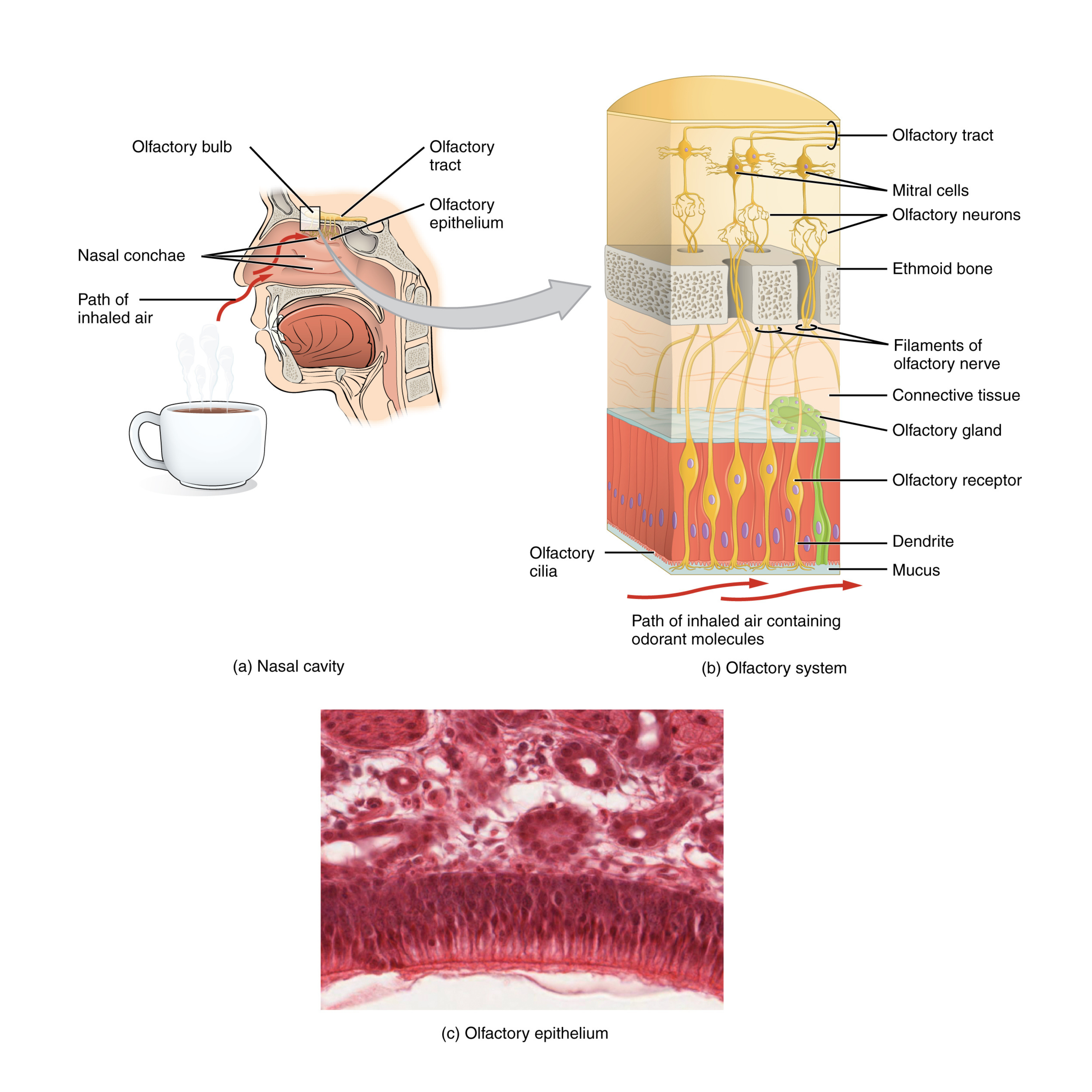
In the superior nasal cavity, a specialized region called the olfactory epithelium contains olfactory receptor neurons, which are bipolar sensory neurons. At the apical surface of this tissue, these neurons extend dendrites into the surrounding mucus.
When you inhale, odorant molecules dissolve in the mucus and bind to carrier proteins that help deliver them to the olfactory dendrites. The odorants then bind to G protein-coupled receptors on the dendritic membrane, producing a graded membrane potential.
Each olfactory neuron’s axon passes through an olfactory foramen in the cribriform plate of the ethmoid bone to reach the brain. Bundles of these axons form the olfactory tract, which connects to the olfactory bulb on the ventral side of the frontal lobe.
From the olfactory bulb, signals travel to several brain regions, including the cerebrum - specifically the primary olfactory cortex in the inferior and medial regions of the temporal lobe - as well as parts of the limbic system and hypothalamus, where smells can trigger emotional memories. Smell is unusual because it does not synapse in the thalamus before reaching the cerebral cortex, which helps explain why odors can strongly evoke emotions and memories.
The nasal epithelium, including the olfactory cells, is exposed to potentially harmful airborne chemicals. For that reason, olfactory neurons are routinely replaced. As new neurons form, their axons grow along existing pathways in the cranial nerve to reconnect with targets in the olfactory bulb.
Pheromones
Pheromones are chemical signals released by one member of a species that affect the behavior or physiology of another member of the same species.
Pheromones serve multiple functions:
- Mating and reproduction: Attracting potential mates or signaling sexual readiness.
- Territorial behavior: Marking boundaries with pheromones (common in animals like dogs or cats).
- Alarm responses: Inducing escape or defensive actions when danger is present.
- Social hierarchies: Conveying dominance or submission within a group.
Pheromone mechanism of action
- Detection: In many species, pheromones are detected by the olfactory system, often through a specialized structure called the vomeronasal organ (VNO) in the nasal cavity. Signals from the VNO travel to the accessory olfactory bulb, which then relays information to brain regions that control behavior.
- In humans: The presence and functionality of a VNO remain debated. Evidence suggests pheromones may subtly influence subconscious behaviors (e.g., mood or attraction), possibly through the regular olfactory system rather than a separate structure.
Types of pheromones
- Releaser pheromones: Trigger immediate, specific behaviors (e.g., attracting mates).
- Primer pheromones: Initiate long-term physiological changes, such as altering hormone levels.
- Signaler pheromones: Convey information about an individual’s identity or reproductive condition.
- Modulator pheromones: Influence emotional states or moods.
In humans, the potential effects of pheromones on attraction, reproduction, and mood regulation continue to be researched.
Kinesthesia
Kinesthesia is the conscious perception of body movement. It’s distinct from proprioception, which primarily detects the body’s static position.
Components: Kinesthesia depends on sensory receptors in muscles, tendons, and joints that respond to:
- Muscle stretch (via muscle spindles).
- Joint position and movement (through various mechanoreceptors).
- Tension in tendons (via Golgi tendon organs).
Distinction from proprioception:
- Kinesthesia: Awareness of movement (e.g., sensing your arm swinging).
- Proprioception: Awareness of body position (e.g., knowing the position of your hand without looking).
Neural pathways:
- Kinesthetic signals travel through the somatosensory system - via sensory neurons in muscles and joints - to the cerebellum (for coordination) and the primary somatosensory cortex (for conscious processing).
Integration with other senses:
- Kinesthesia works alongside vision and vestibular inputs (balance) to create a complete sense of body movement and coordination.
Vestibular sense and equilibrium (balance)
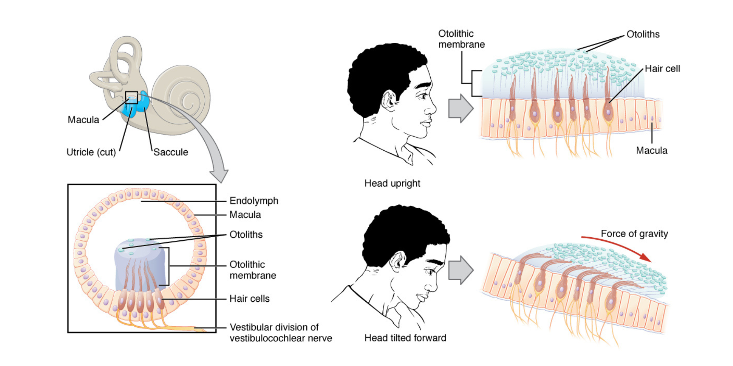
Along with hearing, the inner ear is essential for equilibrium (balance). Specialized hair cells with stereocilia detect head position, head movement, and overall motion.
These hair cells are located in the vestibule of the inner ear. The utricle and saccule detect head position, while the semicircular canals detect head rotation. Signals from the vestibular ganglion travel through the vestibulocochlear nerve to the brain stem and cerebellum.
Inside the utricle and saccule, macula tissue (plural: maculae) contains hair cells supported by surrounding cells. The stereocilia extend into the otolithic membrane, a viscous layer topped with calcium carbonate crystals called otoliths.
Because the otolithic membrane is weighted by these crystals, it shifts when the head tilts. This movement bends the stereocilia and causes differential depolarization of the hair cells. The brain interprets these signals to determine head orientation.
The semicircular canals are three looped structures extending from the vestibule: one aligned horizontally and two oriented vertically.
At the base of each canal is an enlarged region called the ampulla, which contains the hair cells that detect rotational motion (for example, turning the head side to side). The stereocilia project into the cupula, a membrane that moves when the head rotates because the fluid in the canal lags behind. As the cupula deflects, it bends the stereocilia and generates a signal that reflects the direction and speed of head movement.
By comparing input from all three semicircular canals, the vestibular system interprets movement in three-dimensional space.