Hearing
Hearing (also called audition) is the process of converting sound waves into neural signals. The ear’s structures guide sound inward and then translate mechanical vibrations into electrical activity.
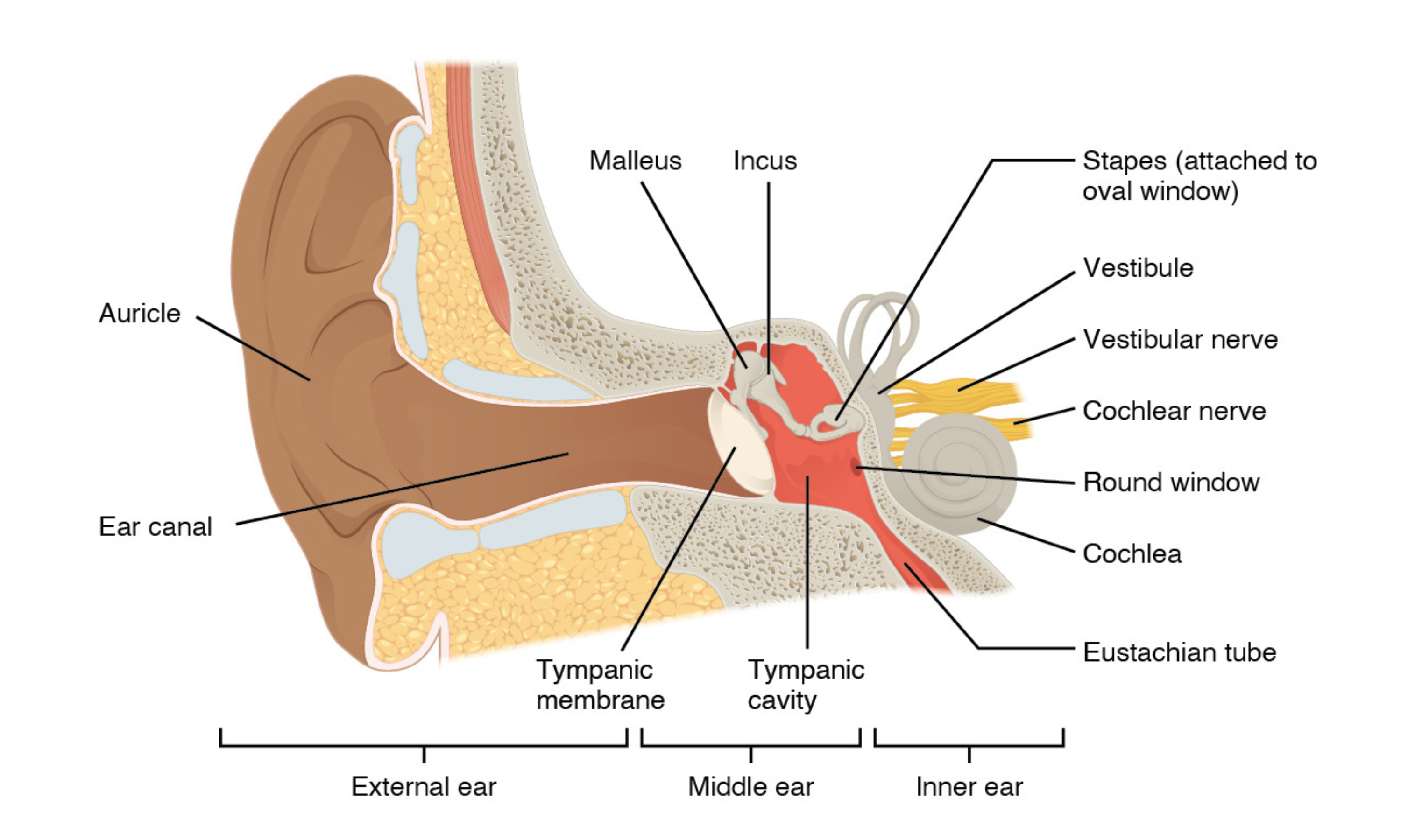
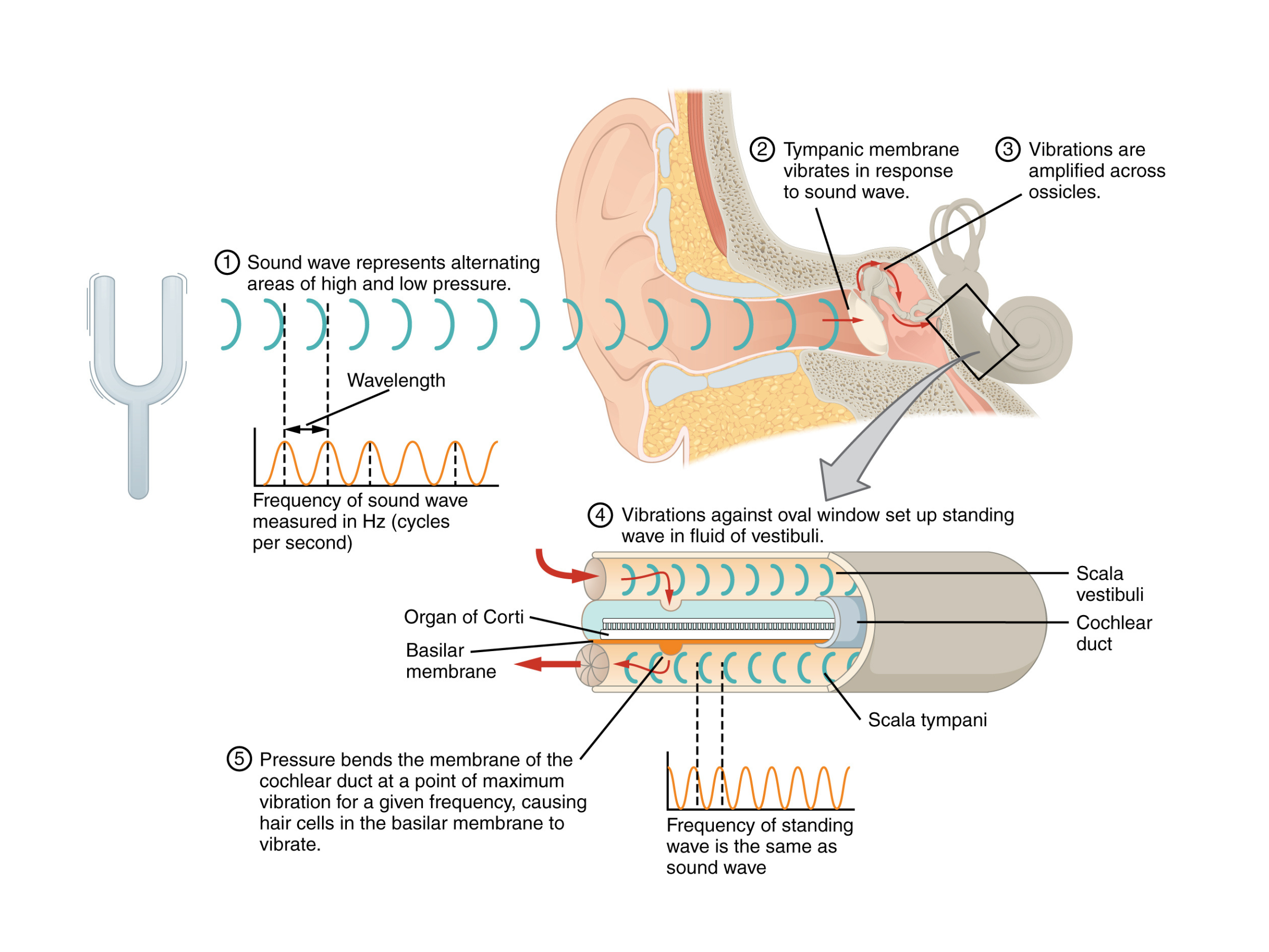
The fleshy outer part of the ear, the auricle, funnels sound waves into the auditory canal. The canal passes through the external auditory meatus of the temporal bone and ends at the tympanic membrane (eardrum). When sound waves strike the tympanic membrane, it vibrates.
External ear
- Auricle (sometimes called pinna if it’s movable, as in certain animals)
- Auditory canal
- Tympanic membrane
Middle ear
- The middle ear contains three small bones - malleus, incus, and stapes - collectively called the ossicles.
The malleus attaches to the tympanic membrane and articulates with the incus, which articulates with the stapes. Vibrations move through these bones and enter the inner ear at the oval window. - The Eustachian tube connects the middle ear to the pharynx, which helps equalize pressure on both sides of the tympanic membrane.
Inner ear
- The inner ear sits within the temporal bone and is often described as a bony labyrinth. It has two main regions:
- Cochlea - for hearing
- Vestibule - for balance
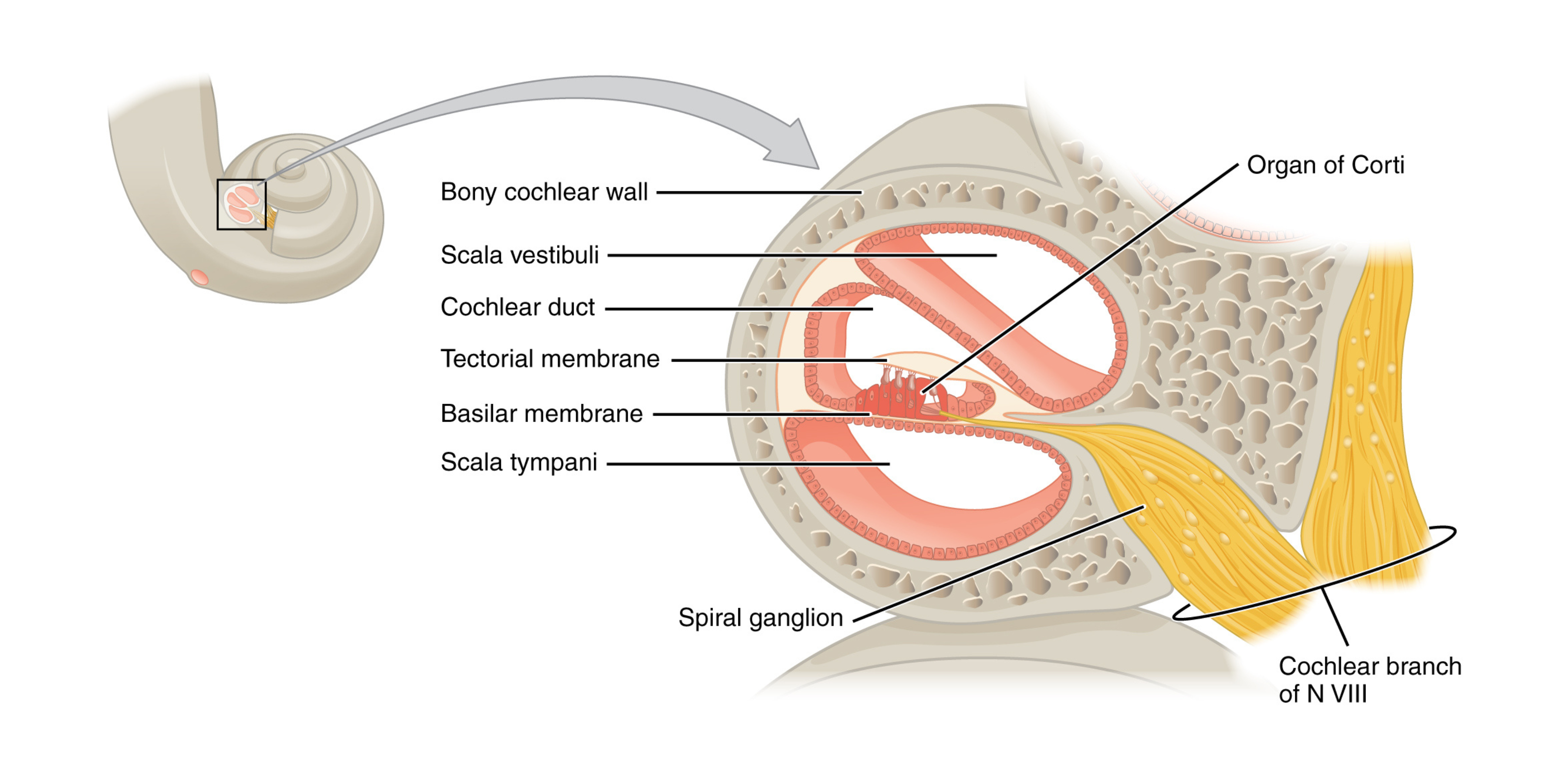
- These regions send signals along separate fiber bundles that join to form the vestibulocochlear nerve. In the cochlea, the spiral ganglia contain the neurons involved in transducing sound into neural impulses. The stapes contacts the oval window, where vibrations begin to generate fluid waves.
Scala and fluid motion
- Inside the cochlea, the scala vestibuli extends from the oval window above the cochlear duct, while the scala tympani extends below and ends at the round window.
Vibrations transmitted by the ossicles set the fluid in these scalae in motion. - High-frequency sound waves produce the greatest effect near the oval window, while low-frequency waves travel farther toward the tip of the cochlea.
- As fluid shifts within the cochlea, the membrane of the round window bulges in or out to accommodate the displacement.
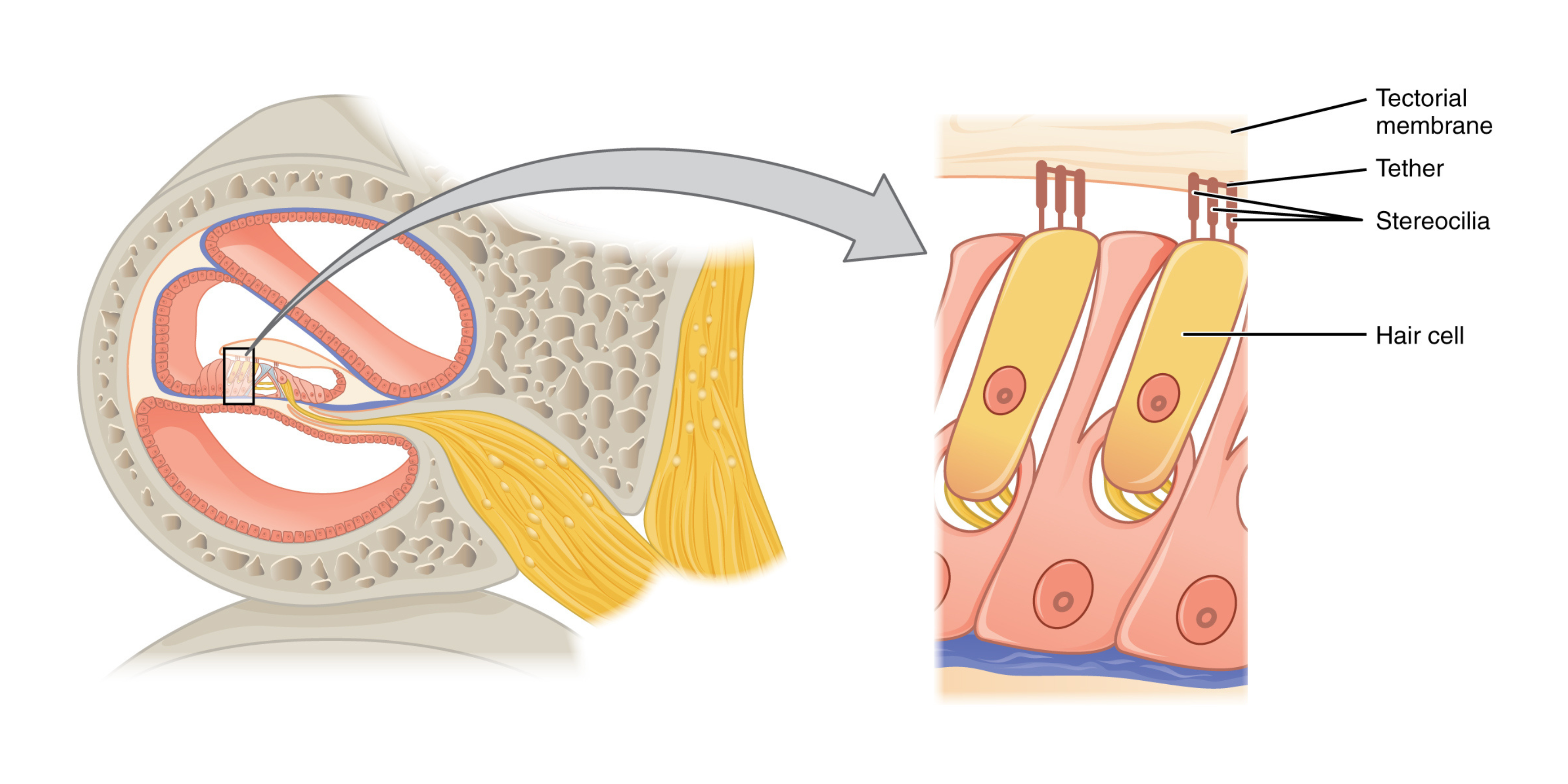
Organ of Corti
- In a cross-section of the cochlea, the cochlear duct lies between the scala vestibuli and scala tympani. The basilar membrane, which supports the organ of Corti, moves in response to the fluid waves.
- Each organ of Corti contains hair cells. Their stereocilia (microvilli-like extensions) project into the tectorial membrane. When fluid waves move the basilar membrane, the tectorial membrane slides across the stereocilia, bending them:
- Bending toward the tallest stereocilium opens ion channels, causing depolarization and nerve impulses.
- Bending away from the tallest stereocilium reduces tension, closing channels and hyperpolarizing the cell.
Frequency tuning
- Different parts of the basilar membrane resonate best at different frequencies, so hair cells in a given region respond most strongly to particular sound waves.
- Humans typically detect frequencies from about 20 Hz (low pitch) to 20 kHz (high pitch). Lower pitches produce peak vibrations closer to the cochlear apex, while higher pitches peak nearer the round and oval windows.