



Troubleshooting and evaluation
Troubleshooting artifact
Artifact is signal distortion or unwanted, erratic movement of the stylus caused by outside interference. The medical assistant needs to identify the type of artifact and then take actions to prevent the artifact. The leads can be viewed on the screen or after printing the ECG tracing. Carefully look for artifact. If an abnormal pattern persists after you’ve corrected for artifact, it may be a genuine, potentially life-threatening rhythm rather than artifact - notify the provider immediately rather than continuing to troubleshoot. The following sections discuss the appearance and causes of the common types of artifact and ways to prevent them.
Wandering baseline
Wandering baseline artifact is an upward and downward movement of the waveform. The isoelectric lines shift locations. This artifact can be the result of the following:
- Poor skin preparation: The patient has oily skin or used lotion. The electrodes are falling off.
- Old electrodes: The electrodes are dirty or expired, or the gel on the electrodes is dried.
- Placement of electrodes: The electrodes are placed on bony areas.
- Movement: The movement of breathing can also be the cause of this artifact.
- The medical assistant should take these steps:
- Clean the skin with alcohol and allow it to dry. Slightly abrade the skin with a gauze pad or fine sandpaper tape to help the electrodes stick to the skin.
- Replace the electrode with a new one. Ensure the electrodes are not expired and the gel is not dried.
- Make sure all electrodes and lead wires are firmly attached.
- Make sure the baseline filter is turned on (if it is a feature of the machine).
Somatic tremor
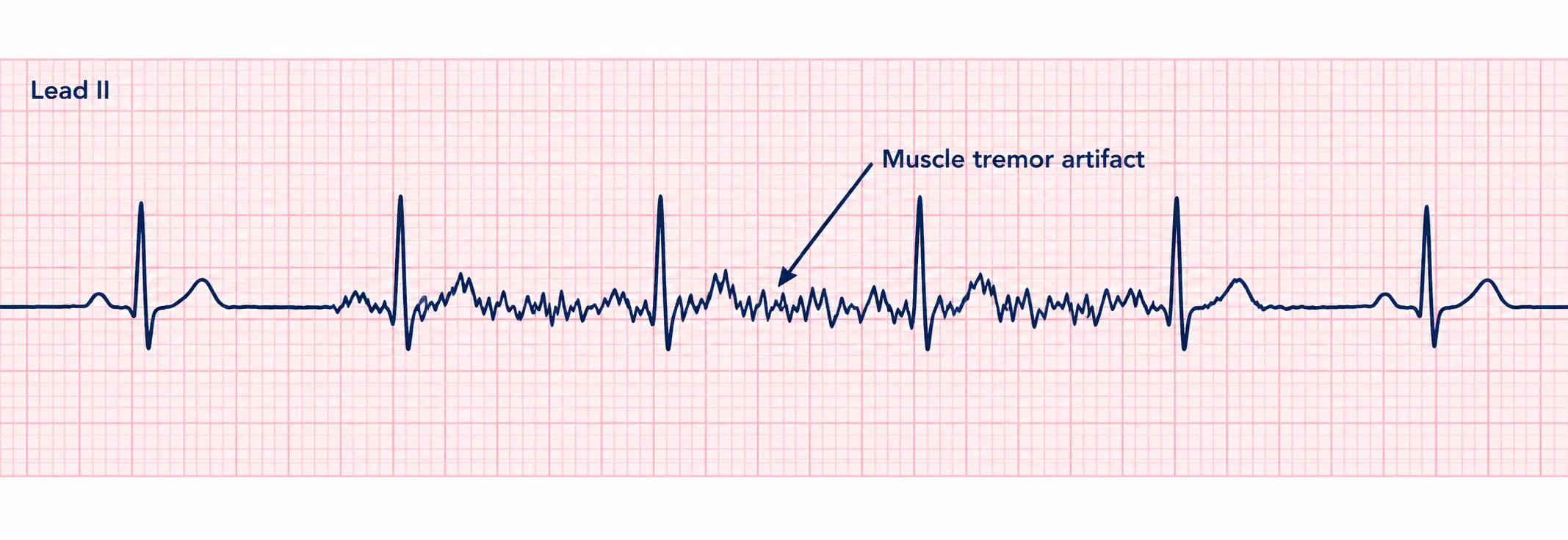
Somatic tremor artifact, also called muscle artifact, appears as jagged peaks with irregular heights and spacing. There are several causes:
- Involuntary movement: Tremors from disease conditions (e.g., Parkinson disease), shivering due to coldness
- Voluntary movement: Talking, chewing gum, supporting arms or legs because the table is too small for the person
The medical assistant should take the following steps:
- Help the patient relax. Cover the patient with a blanket if cold.
- Remind the patient not to move or talk during the tracing.
- Watch for a pattern with involuntary movements. Take the tracing when the movements lessen.
- Use a larger exam table for the procedure.
- If the machine has a muscle tremor filter, make sure it is turned on.
AC interference
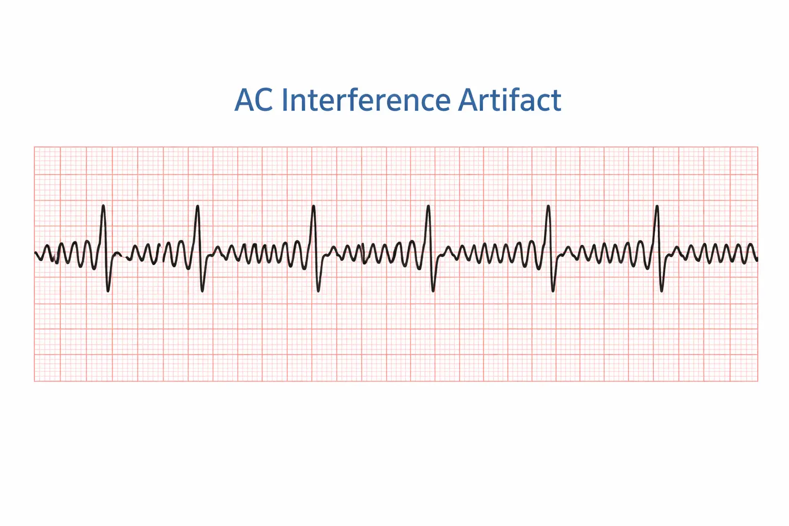
Alternating current (AC) interference artifact, also called 60-cycle interference artifact, appears as a series of small spikes that creates a thick-looking tracing. It’s caused by electrical interference from a number of sources:
- There are too many electrical devices in the area.
- The patient cable and lead wires are wrapped closely together.
- An electrical cord is under the exam table, and an electrical outlet is improperly grounded.
- A cell phone in a patient’s pocket or a smartwatch may also cause interference.
The medical assistant should do the following:
- Unplug the ECG machine.
- Unplug or remove nearby electrical devices (e.g., laptops, pagers, cell phones).
- Separate the lead wires so they do not overlap.
- Move the table away from the wall or move to another room for the procedure.
- Ensure that no cell phones are near the procedure area.
Interrupted baseline
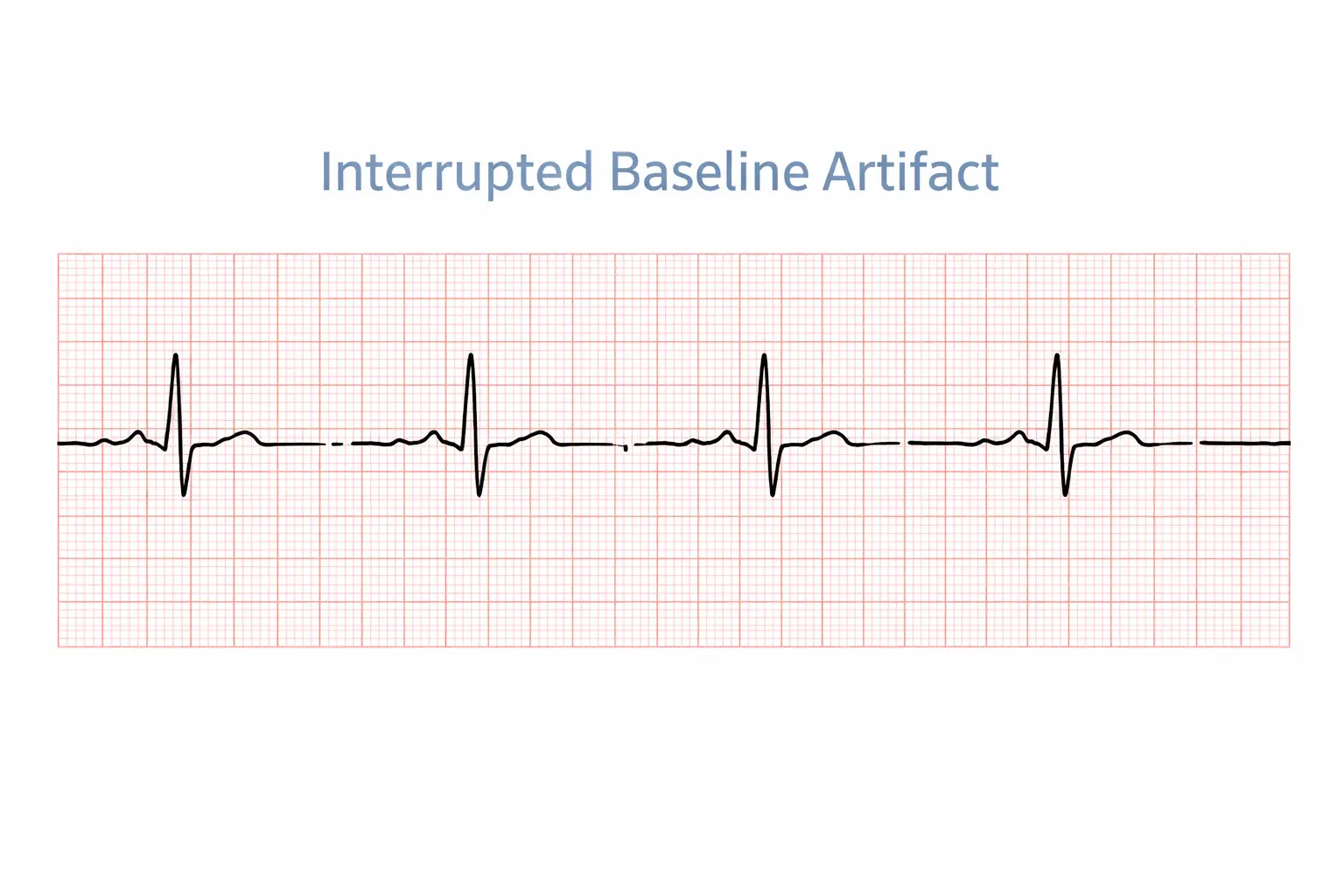
Interrupted baseline artifact (also called intermittent signal artifact) appears when the tracing looks normal at first, then disappears or becomes erratic because the electrical connection is broken - a loose or broken cable, clip, or lead wire. If the lead wire or electrode falls off completely, an isoelectric line (a flat line) will appear on the tracing.
The medical assistant should do the following:
- Check that all electrodes and lead wires are attached.
- If a broken wire is suspected, be prepared to supply a new cable.
Evaluating an ECG tracing
The provider reads the ECG, but the medical assistant must be able to determine abnormal rates and rhythms that might be life threatening. This section discusses how to determine the heart rate, evaluate the ECG tracing, and identify abnormal rhythms.
Rate
Many machines indicate the heart rate. The medical assistant can also calculate the rate from the ECG tracing. Numerous methods can be used to calculate a regular heart rate when the paper speed (chart speed) is 25 mm/second:
- Six-second method: Count the number of QRS complexes (R waves) in a 6-second strip (30 large boxes) and multiply by 10 (least accurate method). For example, complexes in seconds gives bpm.
- 1500 method: Count the number of small boxes between two consecutive R waves. Divide the number into 1500. For example, small boxes gives bpm.
- Sequence method: Count the number of large boxes between two consecutive R waves, then divide by that count to get the rate: box bpm, , , , , bpm.