



Achievable logoAchievable logo
CCMA
Sign in
Sign up
Purchase
Textbook
Practice exams
Support
How it works
Exam catalog
Mountain with a flag at the peak
Textbook
1. Medical assistant
2. Electronic records
3. Medical terminology and anatomy
4. The fundamentals of infection control
5. Introduction to vital signs
6. The patient interview and history
7. The physical examination
8. Appointment scheduling
9. Insurance billing
10. Diagnostic coding and the ICD-10-CM System
11. Procedural coding
12. Medical billing and reimbursement essentials
13. Assisting with medical specialties
14. Assisting with the musculoskeletal system
15. Assisting with the cardiovascular system
16. Assisting with the respiratory system
17. Assisting with the nervous system
18. Anatomy and physiology of the urinary system
19. Assisting in obstetrics and gynecology
20. Assisting in endocrinology
21. Assisting in ophthalmology & otolaryngology
22. Assisting in gastroenterology
23. Assisting in the immune & lymphatic systems
24. Assisting in pediatrics: the developmental stages and care
25. The medical assistant’s role in caring for the older patient
26. The role of the medical assistant in physical therapy examination and assessment
27. Preparing for minor surgery: room, solutions, and supplies
28. Introduction to the clinical laboratory
29. Urinalysis
30. Blood collection
31. Analysis of blood
32. Electrocardiography and heart structure
32.1 The principles of electrocardiography
32.2 ECG terminology, waves, and intervals
32.3 Rhythm analysis and arrhythmias
32.4 The electrocardiograph and ECG preparation
32.5 Troubleshooting and evaluation
32.6 Stress testing
33. The principles of pharmacology
34. Essential calculations and measurement systems
35. Solid, liquid, & solutions medication doses
36. Administering medications
37. Metabolism and core nutrient roles
38. Medical emergencies in the healthcare setting
Achievable logoAchievable logo
32.3 Rhythm analysis and arrhythmias
Achievable CCMA
32. Electrocardiography and heart structure
Our CCMA course is currently in development and is a work-in-progress.

Rhythm analysis and arrhythmias

7 min read
Font
Discuss
Share
Feedback

Analyzing an ECG tracing

For most medical assistants, their job description states that they need to be able to perform ECGs. Some medical assistants may work as ECG technicians. In this position, they may need to analyze an ECG tracing.

The ECG rhythm strip (lead II view) is evaluated from left to right. The following should be assessed:

  • Rate: What is the rate?
  • Rhythm: Is it regular or irregular? An irregular rhythm on an ECG tracing will have time differences between cardiac cycles. With a regular rhythm, each cardiac cycle occurs the same length of time apart.
  • Appearance of the segments, waves, and intervals.

Identifying an abnormal rhythm

When performing an ECG, the medical assistant must identify if the patient has an arrhythmia that requires immediate care by the provider. If the medical assistant identifies a life-threatening arrhythmia, it is important to keep the patient hooked up to the ECG machine and to get the provider immediately.

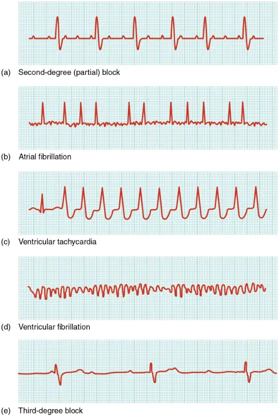
EKG tracings showing common ECG abnormalities
ECG Abnormalities
OpenStax
/
CC BY 4.0

Sinus arrhythmias

A sinus rhythm is considered normal. The electrical activity begins in the SA node and goes through the rest of the conduction system. Atrial and ventricular depolarizations occur. With sinus arrhythmias, the electrical pathway is normal. The rate or rhythm of the heartbeat is altered. The alteration may result from the SA node firing too slowly or too quickly.

  • Sinus bradycardia: The adult heart rate is below 60 beats per minute. This is a normal finding in well-conditioned athletes. It is abnormal in other individuals.
  • Sinus tachycardia: The adult heart rate is above 100 beats per minute. This is normal in a person doing aerobic exercise. It is abnormal in a resting individual.
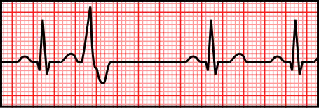
EKG tracing showing premature atrial contractions (PACs)
PAC's
Wikimedia Commons
/
Public Domain

Atrial arrhythmias

Atrial arrhythmias occur when there is a problem with the SA node starting the impulse. They can also occur due to a conduction problem in the atria.

  • Premature atrial contractions (PACs): Occur when the atria contract sooner than they should. The P wave can be abnormally shaped, or an extra P wave can be seen. PACs can be seen in people who smoke or consume large amounts of caffeine. An occasional PAC is not abnormal. More than six PACs in a minute is considered abnormal.
  • Atrial flutter: Occurs when the atria contract faster than the ventricles (up to 300 beats per minute). They become out of sync with the ventricles. Extra P waves are seen with regular QRS complexes. Atrial flutter can be caused by alcohol and stimulants (cocaine, caffeine, diet pills, and cold medications). It can also be caused by coronary heart disease, hypertension, cardiomyopathy, heart valve diseases, hyperthyroidism, obstructive pulmonary disease, and pulmonary embolism diseases. Atrial flutter is reversed with medication to slow the heart or with cardioversion (electrical shock).
EKG tracing showing atrial flutter rhythm
Atrial Flutter
Wikimedia Commons
/
CC BY-SA 4.0

Heart block

A heart block occurs when there is a disruption or slowing of the electrical impulse through the heart. Heart block can be congenital or acquired. Heart disease, surgery, or medications can cause acquired heart block. There are three types of heart block, with third-degree heart block being the most severe:

  • First-degree heart block: The impulse slows as it moves from the atria to the ventricles. This creates a longer PR segment. First-degree heart block may not cause symptoms. It may not require treatment.
  • Second-degree heart block: The impulse slows or is blocked as it moves into the ventricles. When blocked, there is no QRS complex after the P wave, and the ventricles do not contract. When the impulse slows, the PR segment is longer. This arrhythmia requires a pacemaker to help maintain the heart rate. Pacemakers are discussed in a later section of this chapter.
  • Third-degree heart block: The impulse does not reach the ventricles. As a backup system, special ventricular cells create an impulse that causes the ventricles to contract. On the ECG tracing, the P wave is faster than normal, and the QRS complex is not coordinated with the P wave. This is a life-threatening arrhythmia and requires emergency treatment and a pacemaker.
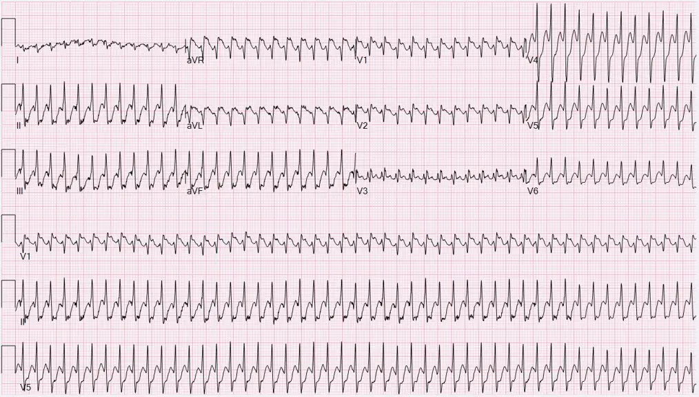
  • Ventricular tachycardia (V-tach): Occurs when the ventricles beat at a rapid rate (up to 250 beats per minute). It may be seen with multiple PVCs in a row. It may be a short run of fast beats or may last longer than 30 seconds. V-tach is a life-threatening condition. If it is not reversed with drugs or cardioversion, it can become ventricular fibrillation.
  • Ventricular fibrillation (V-fib): Occurs when the ventricles quiver uncontrollably. They are essentially ineffective at pumping any blood. The patient has no pulse, is not breathing, and is unresponsive. This is the most critical, life-threatening arrhythmia. Cardioversion with a defibrillator is necessary to restore normal function of the electrical conduction system.
  • Asystole: Results in the absence of a heartbeat. A flat line appears on the tracing
EKG tracing showing premature ventricular contractions (PVCs)
Premature ventricular contractions
Wikimedia Commons
/
Public Domain

Analyzing an ECG tracing

  • Assess rate, rhythm (regular/irregular), and appearance of segments, waves, intervals
  • Rhythm strip (lead II) evaluated left to right
  • Identify irregularities in timing between cardiac cycles

Identifying an abnormal rhythm

  • Recognize arrhythmias needing immediate provider attention
  • Keep patient connected to ECG if life-threatening arrhythmia detected

Sinus arrhythmias

  • Sinus rhythm: normal electrical pathway, altered rate or rhythm
  • Sinus bradycardia: HR < 60 bpm (normal in athletes, abnormal otherwise)
  • Sinus tachycardia: HR > 100 bpm (normal during exercise, abnormal at rest)

Atrial arrhythmias

  • Problem with SA node or atrial conduction
  • Premature atrial contractions (PACs): early atrial contraction, abnormal/extra P wave, >6/min abnormal
  • Atrial flutter: atria contract faster than ventricles (up to 300 bpm), extra P waves, causes include stimulants and heart disease, treated with medication or cardioversion

Heart block

  • Disruption/slowing of electrical impulse from atria to ventricles
  • First-degree: prolonged PR segment, usually asymptomatic
  • Second-degree: some impulses blocked (no QRS after P), requires pacemaker
  • Third-degree: no impulses reach ventricles, P and QRS uncoordinated, life-threatening, needs emergency treatment and pacemaker

Ventricular arrhythmias

  • Ventricular tachycardia (V-tach): rapid ventricular rate (up to 250 bpm), life-threatening, can progress to V-fib
  • Ventricular fibrillation (V-fib): ventricles quiver, no effective pumping, no pulse, requires immediate defibrillation
  • Asystole: absence of heartbeat, flat line on ECG

Sign up for free to take 15 quiz questions on this topic

Previous
Next  | 32.4 The electrocardiograph and ECG preparation
All rights reserved ©2016 - 2026 Achievable, Inc.

Rhythm analysis and arrhythmias

Analyzing an ECG tracing

For most medical assistants, their job description states that they need to be able to perform ECGs. Some medical assistants may work as ECG technicians. In this position, they may need to analyze an ECG tracing.

The ECG rhythm strip (lead II view) is evaluated from left to right. The following should be assessed:

  • Rate: What is the rate?
  • Rhythm: Is it regular or irregular? An irregular rhythm on an ECG tracing will have time differences between cardiac cycles. With a regular rhythm, each cardiac cycle occurs the same length of time apart.
  • Appearance of the segments, waves, and intervals.

Identifying an abnormal rhythm

When performing an ECG, the medical assistant must identify if the patient has an arrhythmia that requires immediate care by the provider. If the medical assistant identifies a life-threatening arrhythmia, it is important to keep the patient hooked up to the ECG machine and to get the provider immediately.

Sinus arrhythmias

A sinus rhythm is considered normal. The electrical activity begins in the SA node and goes through the rest of the conduction system. Atrial and ventricular depolarizations occur. With sinus arrhythmias, the electrical pathway is normal. The rate or rhythm of the heartbeat is altered. The alteration may result from the SA node firing too slowly or too quickly.

  • Sinus bradycardia: The adult heart rate is below 60 beats per minute. This is a normal finding in well-conditioned athletes. It is abnormal in other individuals.
  • Sinus tachycardia: The adult heart rate is above 100 beats per minute. This is normal in a person doing aerobic exercise. It is abnormal in a resting individual.

Atrial arrhythmias

Atrial arrhythmias occur when there is a problem with the SA node starting the impulse. They can also occur due to a conduction problem in the atria.

  • Premature atrial contractions (PACs): Occur when the atria contract sooner than they should. The P wave can be abnormally shaped, or an extra P wave can be seen. PACs can be seen in people who smoke or consume large amounts of caffeine. An occasional PAC is not abnormal. More than six PACs in a minute is considered abnormal.
  • Atrial flutter: Occurs when the atria contract faster than the ventricles (up to 300 beats per minute). They become out of sync with the ventricles. Extra P waves are seen with regular QRS complexes. Atrial flutter can be caused by alcohol and stimulants (cocaine, caffeine, diet pills, and cold medications). It can also be caused by coronary heart disease, hypertension, cardiomyopathy, heart valve diseases, hyperthyroidism, obstructive pulmonary disease, and pulmonary embolism diseases. Atrial flutter is reversed with medication to slow the heart or with cardioversion (electrical shock).

Heart block

A heart block occurs when there is a disruption or slowing of the electrical impulse through the heart. Heart block can be congenital or acquired. Heart disease, surgery, or medications can cause acquired heart block. There are three types of heart block, with third-degree heart block being the most severe:

  • First-degree heart block: The impulse slows as it moves from the atria to the ventricles. This creates a longer PR segment. First-degree heart block may not cause symptoms. It may not require treatment.
  • Second-degree heart block: The impulse slows or is blocked as it moves into the ventricles. When blocked, there is no QRS complex after the P wave, and the ventricles do not contract. When the impulse slows, the PR segment is longer. This arrhythmia requires a pacemaker to help maintain the heart rate. Pacemakers are discussed in a later section of this chapter.
  • Third-degree heart block: The impulse does not reach the ventricles. As a backup system, special ventricular cells create an impulse that causes the ventricles to contract. On the ECG tracing, the P wave is faster than normal, and the QRS complex is not coordinated with the P wave. This is a life-threatening arrhythmia and requires emergency treatment and a pacemaker.
  • Ventricular tachycardia (V-tach): Occurs when the ventricles beat at a rapid rate (up to 250 beats per minute). It may be seen with multiple PVCs in a row. It may be a short run of fast beats or may last longer than 30 seconds. V-tach is a life-threatening condition. If it is not reversed with drugs or cardioversion, it can become ventricular fibrillation.
  • Ventricular fibrillation (V-fib): Occurs when the ventricles quiver uncontrollably. They are essentially ineffective at pumping any blood. The patient has no pulse, is not breathing, and is unresponsive. This is the most critical, life-threatening arrhythmia. Cardioversion with a defibrillator is necessary to restore normal function of the electrical conduction system.
  • Asystole: Results in the absence of a heartbeat. A flat line appears on the tracing
Key points

Analyzing an ECG tracing

  • Assess rate, rhythm (regular/irregular), and appearance of segments, waves, intervals
  • Rhythm strip (lead II) evaluated left to right
  • Identify irregularities in timing between cardiac cycles

Identifying an abnormal rhythm

  • Recognize arrhythmias needing immediate provider attention
  • Keep patient connected to ECG if life-threatening arrhythmia detected

Sinus arrhythmias

  • Sinus rhythm: normal electrical pathway, altered rate or rhythm
  • Sinus bradycardia: HR < 60 bpm (normal in athletes, abnormal otherwise)
  • Sinus tachycardia: HR > 100 bpm (normal during exercise, abnormal at rest)

Atrial arrhythmias

  • Problem with SA node or atrial conduction
  • Premature atrial contractions (PACs): early atrial contraction, abnormal/extra P wave, >6/min abnormal
  • Atrial flutter: atria contract faster than ventricles (up to 300 bpm), extra P waves, causes include stimulants and heart disease, treated with medication or cardioversion

Heart block

  • Disruption/slowing of electrical impulse from atria to ventricles
  • First-degree: prolonged PR segment, usually asymptomatic
  • Second-degree: some impulses blocked (no QRS after P), requires pacemaker
  • Third-degree: no impulses reach ventricles, P and QRS uncoordinated, life-threatening, needs emergency treatment and pacemaker

Ventricular arrhythmias

  • Ventricular tachycardia (V-tach): rapid ventricular rate (up to 250 bpm), life-threatening, can progress to V-fib
  • Ventricular fibrillation (V-fib): ventricles quiver, no effective pumping, no pulse, requires immediate defibrillation
  • Asystole: absence of heartbeat, flat line on ECG

More from Electrocardiography and heart structure

  • The principles of electrocardiography
  • ECG terminology, waves, and intervals
  • The electrocardiograph and ECG preparation
  • Troubleshooting and evaluation
  • Stress testing