



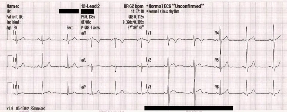
12-lead ECG and supplies
12-lead ECG
One of the most common ECG procedures in the ambulatory care setting is a resting 12-lead ECG. Performing the ECG is a task delegated by the supervising provider - what an MA is permitted to do is governed by state law and medical board rules, not just the employer’s job description. The medical assistant is responsible for the following:
- Assembling the supplies and equipment needed
- Preparing the patient for the test
- Performing the test
Before the medical assistant finishes the procedure, the ECG tracing must be examined for artifacts - any unwanted signal or distortion on the tracing that doesn’t come from the heart’s electrical activity, such as a wandering baseline or electrical interference. If any artifact is present on the tracing, the medical assistant must problem-solve the situation. Ultimately, the goal is to give the provider a clear ECG.
For a resting 12-lead ECG, the patient rests on the exam table. Ten electrodes are placed on the body. Lead wires attach to the electrodes and connect to the ECG machine (electrocardiograph). The 10 electrodes and lead wires pick up the electrical impulse from the surface of the body and carry it to the ECG machine. The machine creates the ECG tracing.
A more in-depth discussion of electrode placement and artifacts are presented later in the chapter. For now, it is important to have an idea of where the electrodes are placed on the body. An electrode is placed on each of the arms and legs. The right leg electrode is considered the ground lead electrode and is required for a clear ECG tracing. Six electrodes are placed on the chest.
Using the 10 electrodes and lead wires, the ECG machine creates 12 images, or pictures, which are called leads. Each lead picture looks different. Think of a photographer moving around a person, taking 12 pictures. Each picture looks different because of the change in the photographer’s angle. For each lead, the picture is created differently. It is important for the medical assistant to know what electrodes and lead wires create the leads when troubleshooting unclear tracings. The three types of leads - bipolar, augmented, and precordial - are discussed in the following sections.
Bipolar (or standard) leads
The bipolar or standard leads are named leads I, II, and III. They use the arm electrodes and the left leg electrode to create pictures of the vertical (or frontal) plane of the heart. (Remember, the right electrode is used as a ground and is important for all leads.)
The bipolar leads are created from a measurement of current traveling from a negative pole to the positive pole:
- Lead I: Right arm (RA) to left arm (LA)
- Lead II: Right arm (RA) to left leg (LL)
- Lead III: Left arm (LA) to left leg (LL)
If you join the endpoints (positive poles) of these three leads, you get a triangle. This triangle is known as the Einthoven triangle.
It is important to know which two electrodes create each lead when troubleshooting problems with the ECG tracing. If a lead has an artifact, the medical assistant must check the two electrodes and the lead wires that create the lead - the same principle applies to every lead type, just with a different set of electrodes.
Augmented leads
Augmented vector (aV) leads, commonly referred to as augmented leads, provide information on the vertical (frontal) plane of the heart. These unipolar leads are augmented, or increased in size, on the tracing. Augmented leads use the right arm (RA), left arm (LA), and left leg (LL) electrodes.
Each augmented lead uses all three extremity electrodes (RA, LA, and LL) to create the picture. Midpoint between two of the electrodes is the negative pole. The current is measured as it moves from the negative pole to the positive pole.
- Lead aVR (augmented Vector Right) records current traveling between the right arm and the central point between the left arm and left leg.
- Lead aVL (augmented Vector Left) records current traveling between the left arm and the central point between the right arm and left leg.
- Lead aVF (augmented Vector Foot) records current traveling between the left leg electrode and the central point between the left and right arms.
Since each augmented lead uses all three extremity electrodes, an artifact on any augmented lead means checking all three: RA, LA, and LL.
Chest (or precordial) leads
The chest (precordial) leads are also unipolar leads. Each is a positive pole. The precordial leads provide information on the horizontal (front to back) plane of the heart. The six precordial leads are labeled V1, V2, V3, V4, V5, and V6.
The six leads are numbered the same as the precordial lead wires (V1 through V6), so troubleshooting one of them means checking that same numbered electrode and lead wire - for example, an artifact on V4 means checking the V4 electrode and lead wire.
ECG supplies
The medical assistant needs to be familiar with the supplies used for ECGs. It is important to monitor the expiration dates of ECG supplies. Using expired supplies can cause unclear ECG tracings.
Thermal ECG paper
ECG machines with printers use special ECG paper. The paper can be either Z-fold paper or 8.5 × 11-inch paper. A special heat-sensitive coating on the paper allows the tracing to be “burned” onto the paper. Here are some tips for handling the ECG paper:
- Store in a dry, cool, dark location.
- Do not expose to heat, bright light, or an ultraviolet (UV) light source.
- Do not expose to alcohol, adhesives, cleaners, or solvents.
- Do not store with vinyl, shrink wrap, or plastics.
The ECG paper is universally created with small and large boxes. The small box is 1 × 1 mm, and the large box is 5 × 5 mm. The large box is made up of 25 small boxes (five rows and five columns). It has a thicker border than the small boxes. When the provider analyzes the tracing, the height and the width of the waveforms are measured with a caliper. The small and large boxes can help in the interpreting process.
The vertical lines measure the amplitude, or voltage, of the waveforms. Each small box is 0.1 mV (millivolt), and each large box is 0.5 mV. To determine the amplitude, count the small boxes vertically, starting at the baseline, to the highest/lowest point of the wave.
The horizontal lines measure the time. When the paper speed (chart speed) is set at 25 mm/second, each small box is 0.04 second, and each large box equals 0.2 second. To determine the width and time of the waveform, count the small boxes horizontally from the start to the finish of the wave. Multiply the number of small boxes by 0.04 to find the time.
Example: measuring a QRS complex
A QRS complex spans 4 small boxes on the ECG paper, recorded at the standard paper speed of 25 mm/second.
- Each small box equals second at this speed.
- Multiply the number of small boxes by the time per box: second.
Answer: The QRS complex lasts second.
Electrodes
Electrodes are single-use disposable adhesive tabs that are placed on the skin. The skin is a poor conductor of electricity. The electrodes contain an electrolyte gel that helps pick up the electrical impulses. As the impulses make their way to the surface of the body, the electrolyte gel helps to conduct the impulses into the lead wires. For a resting 12-lead ECG, the most common type of electrode is the tab variety. The snap electrode is used more often for exercise stress tests and in hospital settings.
It is important to check the expiration date of the electrodes. If they are expired, the gel may be dried out, and the conduction will be poor. Many operators’ manuals for ECG machines recommend not mixing different manufacturers’ electrodes.