


Achievable logoAchievable logo
CCMA
Sign in
Sign up
Purchase
Textbook
Practice exams
Support
How it works
Exam catalog
Mountain with a flag at the peak
Textbook
1. Medical assistant
2. Electronic records
3. Medical terminology and anatomy
4. The fundamentals of infection control
5. Introduction to vital signs
6. The patient interview and history
7. The physical examination
8. Appointment scheduling
9. Insurance billing
10. Diagnostic coding and the ICD-10-CM System
11. Procedural coding
12. Medical billing and reimbursement essentials
13. Assisting with medical specialties
14. Assisting with the musculoskeletal system
15. Assisting with the cardiovascular system
16. Assisting with the respiratory system
17. Assisting with the nervous system
18. Anatomy and physiology of the urinary system
19. Assisting in obstetrics and gynecology
20. Assisting in endocrinology
21. Assisting in ophthalmology & otolaryngology
22. Assisting in gastroenterology
23. Assisting in the immune & lymphatic systems
24. Assisting in pediatrics: the developmental stages and care
25. The medical assistant’s role in caring for the older patient
26. The role of the medical assistant in physical therapy examination and assessment
27. Preparing for minor surgery: room, solutions, and supplies
28. Introduction to the clinical laboratory
29. Urinalysis
30. Blood collection
31. Analysis of blood
32. Electrocardiography and heart structure
32.1 The principles of electrocardiography
32.2 ECG terminology, waves, and intervals
32.3 Rhythm analysis and arrhythmias
32.4 The electrocardiograph and ECG preparation
32.5 Troubleshooting and evaluation
32.6 Stress testing
33. The principles of pharmacology
34. Essential calculations and measurement systems
35. Solid, liquid, & solutions medication doses
36. Administering medications
37. Metabolism and core nutrient roles
38. Medical emergencies in the healthcare setting
Achievable logoAchievable logo
32.6 Stress testing
Achievable CCMA
32. Electrocardiography and heart structure
Our CCMA course is currently in development and is a work-in-progress.

Stress testing

10 min read
Font
Discuss
Share
Feedback

Besides the 12-lead resting ECG, an ECG can be obtained during exercise and nuclear stress tests. The following sections examine exercise stress tests and nuclear stress tests.

Exercise stress test

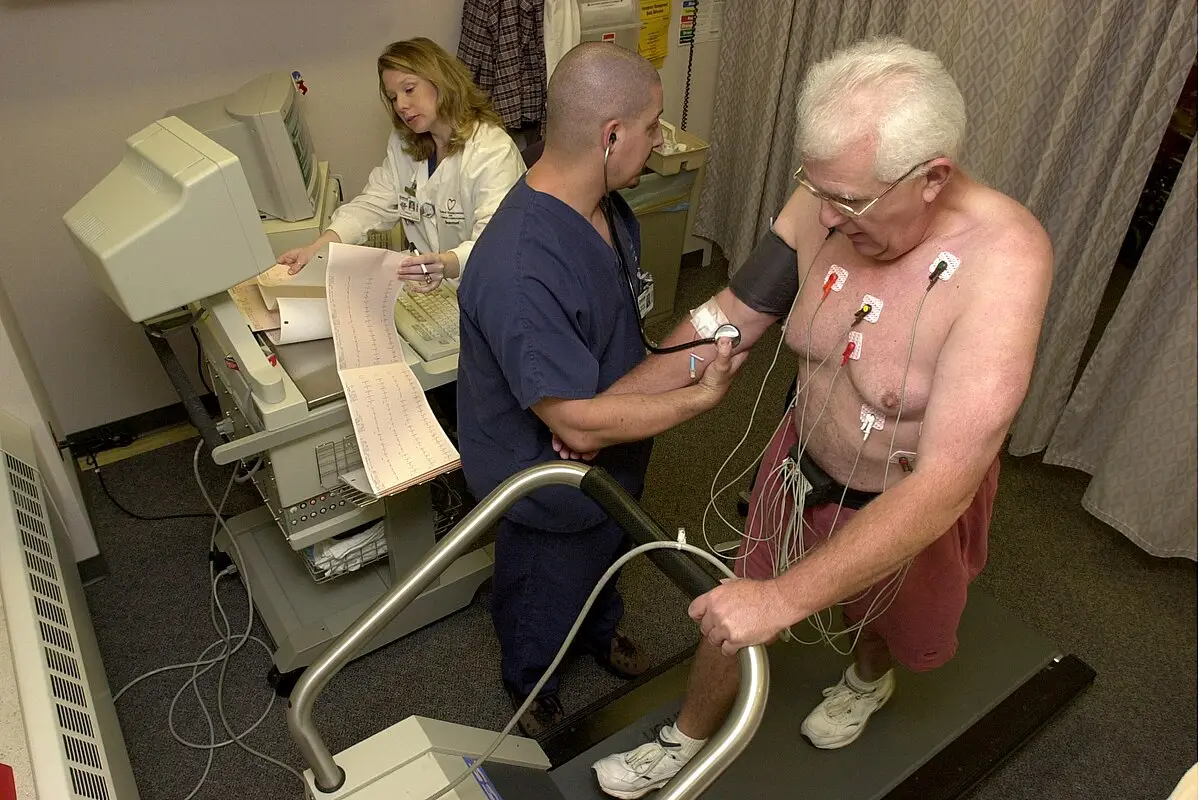
An exercise stress test records the ECG while the patient is exercising. The patient either walks on a treadmill with an incline or uses an exercise bike. During the test, a continual ECG is recorded, and the blood pressure is monitored.

The test may be ordered for these reasons:

  • The patient is starting an exercise program.
  • The patient has angina that is getting worse.
  • To evaluate the patient’s heart after an angioplasty or bypass surgery.
  • To evaluate heart rhythm changes with the stress of exercise.

Preparation for the test includes the following:

  • The patient should wear clothes and shoes for exercising.
  • The provider will indicate what daily medications should be taken prior to the test.
  • The patient should not take a dose of Viagra, Cialis, or Levitra for erectile dysfunction 48 hours before a stress test.
  • The patient should not smoke or consume caffeine or alcohol 3 hours before the test.
  • The patient signs a consent form prior to the test.

Here is what occurs during the test:

  • Electrodes and lead wires will be placed on the patient. A blood pressure cuff will be applied to the patient’s arm.
  • The patient will begin to exercise slowly, and gradually the exercise difficulty will increase.
  • The provider should always be present during the test. If the patient has chest discomfort, dizziness, palpitations, or shortness of breath, the test will stop. Emergency supplies or a crash cart should be kept nearby in case of an emergency.
Patient undergoing an exercise stress test
Exercise stress test
Wikimedia Commons
/
Public Domain

Remote ECG monitoring

With the advances in technology, several types of remote ECG monitoring can be used for patient care. The following sections discuss types of remote ECG monitoring that can be used.

Holter monitor

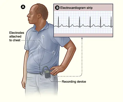
A Holter monitor is used to monitor the heart over a 24- to 48-hour period while patients go about their normal activities. Sometimes patients experience a heart issue that cannot be picked up on a resting 12-lead ECG. Having a longer monitoring period can capture the abnormalities.

Holter monitor used for extended cardiac monitoring
Holter monitor
Wikimedia Commons
/
U.S. Government Work (Public Domain)

When placing electrodes for a Holter monitor, consult the users’ guide. There are many configurations for electrode placement. Different monitors use varying numbers of electrodes. Holter monitors typically use three to seven chest electrodes. As with the resting 12-lead ECG, lead wires attach the electrodes to the small Holter monitor. The monitor runs on batteries and is small enough to fit into a pocket.

The following are patient education points for a Holter monitor test:

  • Wear the monitor continuously. The monitor will be removed during your appointment in 24 to 48 hours.
  • While wearing the Holter monitor, avoid metal detectors, large magnets, high-voltage areas, x-rays, and electric blankets. Do not get the monitor wet (e.g., no bathing, showering, or swimming).
  • Keep a detailed journal of the symptoms experienced. These symptoms could include chest pain, shortness of breath, palpitations or changes in the heartbeat, dizziness, or fainting. Document the date, time, symptoms experienced, and the activity being done at the time. (Many facilities provide patients with a journal for the documentation.)
  • During the return visit, the provider will look at the tracing for abnormalities. The journal of timed activities will help the provider identify what is occurring.
  • The patient is instructed on what to do if the electrodes or lead wires fall off.
  • The patient is instructed on who to contact with questions or if problems occur.

Cardiac event recorder

The cardiac event recorder is a portable, battery-powered ECG device. The recorder is activated by the patient when symptoms occur, and it records the ECG. Patients may wear a cardiac event recorder for 30 days. When symptoms occur only occasionally, it is difficult to capture the abnormality on a resting 12-lead ECG or a Holter monitor. The patient may also be asked to keep a journal, as with the Holter monitor. With the cardiac event recorder, patients go about their daily activities. The monitor can be removed during bathing and showering or swimming.

When patients experience symptoms, they push the recorder’s activation button. An ECG is recorded and stored. The ECG can be sent to the provider via a phone or other technology. A provider will review the tracing. If there is an emergency, the patient will be called and told to go to the nearest emergency department.

There are two types of cardiac event recorders:

  • Looping memory monitor (also called a cardiac loop recorder): A pager-sized monitor that connects to two chest electrodes by wires. The monitor continuously records the ECG. When the memory is full, it overwrites from the beginning (i.e., continues the loop of recording). When a patient has symptoms, they activate the monitor. The ECG from prior to, during, and for a short while after the event is stored. It does not overwrite the stored ECGs.
  • Symptom event monitor (also called a post-event monitor): Can be a handheld device or worn on the wrist. When symptoms are felt, the patient activates the monitor and places it on the chest. Small metal disks on the back of the monitor act as electrodes. The current ECG is recorded and stored. Unlike the looping memory monitor, the symptom event monitor cannot record and store the ECG prior to the symptoms.

Implantable loop recorder

The implantable loop recorder (also called a loop recorder implant) is a small recorder (under 2 inches) that looks like a flash drive. It is surgically implanted just under the skin in the upper left chest. It can be implanted in an ambulatory care facility.

Often the device is used for patients who have episodes of fainting, seizures, and palpitations, and other ECG tests have not been able to detect the abnormality. It also catches infrequent arrhythmias that can be missed with traditional ECG monitoring.

The device continuously records the ECG for 2 to 3 years. It records the ECG if the heart rate falls below or rises above the preset limits (set by the provider). The patient can also record the ECG if symptoms are experienced. The provider can download the saved ECG data at the next appointment.

Transtelephonic monitoring

A transtelephonic monitor (TTM) is a small device that records a patient’s ECG when the patient pushes the activation button. The TTM has four electrodes on the back of the device that can record a single lead. The ECG data is sent via phone to the base station at the provider’s office. The signal is converted to a readable tracing that can be displayed on a monitor or printed out. There are two types of TTMs:

  • TTM with internal memory: The ECG is recorded and stored when the patient pushes a button. The data can be transmitted by phone to the provider. This type of TTM is used for patients with infrequent arrhythmias.
  • TTM with no internal memory: The TTM is placed over the heart, and the phone receiver is placed over the TTM. Once the activation button is pushed, the TTM records and immediately sends the live ECG to the provider via the phone line. The data is not saved on the device. This type of TTM is used for patients with pacemakers and internal cardioverter-defibrillators. It allows the provider to monitor the patient remotely.

Portable handheld ECG monitors

There are a variety of portable handheld ECG monitors on the market. Many produce a single lead. The ECG data can then be transferred to other devices using software, Bluetooth, smartphones, or USB ports. These devices are marketed to be used at home. A person can use the device and save ECG tracings. The tracings can then be reviewed by the person’s provider at the next visit. The machines work differently, some having several electrode options. Finger contact, chest contact (bare skin just below the nipple), and regular chest electrodes can be used to create an ECG tracing.

Patient coaching

For any cardiac procedures that patients need to undergo, it is important for the medical assistant to coach the patient on the required preparation. The medical assistant should instruct the patient about what to expect during the test and then explain any follow-up required. Explaining procedures to patients can reduce the stress and confusion they experience.

Legal and ethical issues

When a medical assistant is performing an ECG, it is important that the electrodes and lead wires are placed on the patient correctly. If the arm lead wires are reversed, the P, QRS, and T waves are negative deflections in lead I. If the leg lead wires are reversed, Lead III is inverted, and the P wave is larger in lead I compared to lead II. Normally, the P wave is larger in lead II. Taking an ECG when the lead wires are reversed can lead to more cardiac testing and additional provider visits. The medical assistant has a professional responsibility to perform ECGs accurately so that the patient receives the best possible care.

Patient-centered care

When a patient is undergoing cardiac testing to rule out or rule in a heart condition, it can be a stressful experience. It is important for the medical assistant to be sensitive to the patient’s concerns and feelings. Questions from the patient and family members should be addressed, and any questions outside the medical assistant’s scope of practice should be directed to the provider.

The medical assistant should provide patients with clear directions on the cardiac procedure being done. After the procedure, the medical assistant should let patients know who will contact them about the test results. If possible, give patients a timeline so that they will know when to anticipate the notification.

Professional behaviors

Many times, patients may think that the medical assistant can read ECGs. They may ask the medical assistant if everything looks all right or if the ECG is normal. It is outside of the medical assistant’s scope of practice to read ECGs. This is the role of the provider. The medical assistant should explain to the patient that the provider will read the ECG, and the patient will be informed of the results. The medical assistant should take care not to add false hope comments, such as “I am sure the ECG will be normal.” These types of comments may give the patient a false impression of the results, and the medical assistant’s behavior is unprofessional.

Stress testing

  • Exercise stress test: ECG during exercise, monitors heart under stress
  • Indications: starting exercise, worsening angina, post-angioplasty/bypass, rhythm evaluation
  • Preparation: exercise attire, medication guidance, avoid ED meds/caffeine/alcohol, consent required

Exercise stress test procedure

  • Electrodes, lead wires, BP cuff applied; gradual exercise increase
  • Provider present; stop test for symptoms (chest pain, dizziness, etc.)
  • Emergency equipment must be available

Remote ECG monitoring

  • Used for ongoing or intermittent cardiac monitoring outside clinic

Holter monitor

  • 24–48 hour continuous ECG during normal activities
  • Avoid water, magnets, metal detectors, high voltage; keep symptom/activity journal
  • Provider reviews tracing and journal for abnormalities

Cardiac event recorder

  • Patient-activated ECG device, worn up to 30 days
  • Two types:
    • Looping memory monitor: continuous recording, stores pre- and post-event ECG
    • Symptom event monitor: records only when activated, no pre-event data
  • Data sent to provider; journal may be kept

Implantable loop recorder

  • Small device implanted under skin, upper left chest
  • Continuous ECG for 2–3 years; auto- or patient-activated recording
  • Used for infrequent, undetected arrhythmias

Transtelephonic monitoring (TTM)

  • Patient-activated, single-lead ECG sent via phone
  • Two types:
    • With internal memory: stores and transmits ECG
    • No internal memory: live ECG sent immediately, no storage
  • Used for arrhythmia or device monitoring

Portable handheld ECG monitors

  • Single-lead, home-use devices; data transferable via Bluetooth/USB
  • Multiple electrode/contact options (finger, chest, standard electrodes)
  • Tracings reviewed by provider at follow-up

Patient coaching

  • Medical assistant instructs on preparation, procedure, and follow-up
  • Clear explanations reduce patient stress and confusion

Legal and ethical issues

  • Accurate electrode/lead wire placement essential
  • Reversed leads cause abnormal ECG readings, may lead to unnecessary testing
  • Professional responsibility to ensure correct ECG performance

Patient-centered care

  • Address patient/family questions, provide clear instructions
  • Inform about result notification and expected timeline
  • Refer out-of-scope questions to provider

Professional behaviors

  • Medical assistants do not interpret ECGs; provider reads results
  • Avoid giving false reassurance or interpreting findings
  • Maintain professional boundaries and communication

Sign up for free to take 14 quiz questions on this topic

Previous
Next  | 33.1 The principles of pharmacology
All rights reserved ©2016 - 2026 Achievable, Inc.

Stress testing

Besides the 12-lead resting ECG, an ECG can be obtained during exercise and nuclear stress tests. The following sections examine exercise stress tests and nuclear stress tests.

Exercise stress test

An exercise stress test records the ECG while the patient is exercising. The patient either walks on a treadmill with an incline or uses an exercise bike. During the test, a continual ECG is recorded, and the blood pressure is monitored.

The test may be ordered for these reasons:

  • The patient is starting an exercise program.
  • The patient has angina that is getting worse.
  • To evaluate the patient’s heart after an angioplasty or bypass surgery.
  • To evaluate heart rhythm changes with the stress of exercise.

Preparation for the test includes the following:

  • The patient should wear clothes and shoes for exercising.
  • The provider will indicate what daily medications should be taken prior to the test.
  • The patient should not take a dose of Viagra, Cialis, or Levitra for erectile dysfunction 48 hours before a stress test.
  • The patient should not smoke or consume caffeine or alcohol 3 hours before the test.
  • The patient signs a consent form prior to the test.

Here is what occurs during the test:

  • Electrodes and lead wires will be placed on the patient. A blood pressure cuff will be applied to the patient’s arm.
  • The patient will begin to exercise slowly, and gradually the exercise difficulty will increase.
  • The provider should always be present during the test. If the patient has chest discomfort, dizziness, palpitations, or shortness of breath, the test will stop. Emergency supplies or a crash cart should be kept nearby in case of an emergency.

Remote ECG monitoring

With the advances in technology, several types of remote ECG monitoring can be used for patient care. The following sections discuss types of remote ECG monitoring that can be used.

Holter monitor

A Holter monitor is used to monitor the heart over a 24- to 48-hour period while patients go about their normal activities. Sometimes patients experience a heart issue that cannot be picked up on a resting 12-lead ECG. Having a longer monitoring period can capture the abnormalities.

When placing electrodes for a Holter monitor, consult the users’ guide. There are many configurations for electrode placement. Different monitors use varying numbers of electrodes. Holter monitors typically use three to seven chest electrodes. As with the resting 12-lead ECG, lead wires attach the electrodes to the small Holter monitor. The monitor runs on batteries and is small enough to fit into a pocket.

The following are patient education points for a Holter monitor test:

  • Wear the monitor continuously. The monitor will be removed during your appointment in 24 to 48 hours.
  • While wearing the Holter monitor, avoid metal detectors, large magnets, high-voltage areas, x-rays, and electric blankets. Do not get the monitor wet (e.g., no bathing, showering, or swimming).
  • Keep a detailed journal of the symptoms experienced. These symptoms could include chest pain, shortness of breath, palpitations or changes in the heartbeat, dizziness, or fainting. Document the date, time, symptoms experienced, and the activity being done at the time. (Many facilities provide patients with a journal for the documentation.)
  • During the return visit, the provider will look at the tracing for abnormalities. The journal of timed activities will help the provider identify what is occurring.
  • The patient is instructed on what to do if the electrodes or lead wires fall off.
  • The patient is instructed on who to contact with questions or if problems occur.

Cardiac event recorder

The cardiac event recorder is a portable, battery-powered ECG device. The recorder is activated by the patient when symptoms occur, and it records the ECG. Patients may wear a cardiac event recorder for 30 days. When symptoms occur only occasionally, it is difficult to capture the abnormality on a resting 12-lead ECG or a Holter monitor. The patient may also be asked to keep a journal, as with the Holter monitor. With the cardiac event recorder, patients go about their daily activities. The monitor can be removed during bathing and showering or swimming.

When patients experience symptoms, they push the recorder’s activation button. An ECG is recorded and stored. The ECG can be sent to the provider via a phone or other technology. A provider will review the tracing. If there is an emergency, the patient will be called and told to go to the nearest emergency department.

There are two types of cardiac event recorders:

  • Looping memory monitor (also called a cardiac loop recorder): A pager-sized monitor that connects to two chest electrodes by wires. The monitor continuously records the ECG. When the memory is full, it overwrites from the beginning (i.e., continues the loop of recording). When a patient has symptoms, they activate the monitor. The ECG from prior to, during, and for a short while after the event is stored. It does not overwrite the stored ECGs.
  • Symptom event monitor (also called a post-event monitor): Can be a handheld device or worn on the wrist. When symptoms are felt, the patient activates the monitor and places it on the chest. Small metal disks on the back of the monitor act as electrodes. The current ECG is recorded and stored. Unlike the looping memory monitor, the symptom event monitor cannot record and store the ECG prior to the symptoms.

Implantable loop recorder

The implantable loop recorder (also called a loop recorder implant) is a small recorder (under 2 inches) that looks like a flash drive. It is surgically implanted just under the skin in the upper left chest. It can be implanted in an ambulatory care facility.

Often the device is used for patients who have episodes of fainting, seizures, and palpitations, and other ECG tests have not been able to detect the abnormality. It also catches infrequent arrhythmias that can be missed with traditional ECG monitoring.

The device continuously records the ECG for 2 to 3 years. It records the ECG if the heart rate falls below or rises above the preset limits (set by the provider). The patient can also record the ECG if symptoms are experienced. The provider can download the saved ECG data at the next appointment.

Transtelephonic monitoring

A transtelephonic monitor (TTM) is a small device that records a patient’s ECG when the patient pushes the activation button. The TTM has four electrodes on the back of the device that can record a single lead. The ECG data is sent via phone to the base station at the provider’s office. The signal is converted to a readable tracing that can be displayed on a monitor or printed out. There are two types of TTMs:

  • TTM with internal memory: The ECG is recorded and stored when the patient pushes a button. The data can be transmitted by phone to the provider. This type of TTM is used for patients with infrequent arrhythmias.
  • TTM with no internal memory: The TTM is placed over the heart, and the phone receiver is placed over the TTM. Once the activation button is pushed, the TTM records and immediately sends the live ECG to the provider via the phone line. The data is not saved on the device. This type of TTM is used for patients with pacemakers and internal cardioverter-defibrillators. It allows the provider to monitor the patient remotely.

Portable handheld ECG monitors

There are a variety of portable handheld ECG monitors on the market. Many produce a single lead. The ECG data can then be transferred to other devices using software, Bluetooth, smartphones, or USB ports. These devices are marketed to be used at home. A person can use the device and save ECG tracings. The tracings can then be reviewed by the person’s provider at the next visit. The machines work differently, some having several electrode options. Finger contact, chest contact (bare skin just below the nipple), and regular chest electrodes can be used to create an ECG tracing.

Patient coaching

For any cardiac procedures that patients need to undergo, it is important for the medical assistant to coach the patient on the required preparation. The medical assistant should instruct the patient about what to expect during the test and then explain any follow-up required. Explaining procedures to patients can reduce the stress and confusion they experience.

Legal and ethical issues

When a medical assistant is performing an ECG, it is important that the electrodes and lead wires are placed on the patient correctly. If the arm lead wires are reversed, the P, QRS, and T waves are negative deflections in lead I. If the leg lead wires are reversed, Lead III is inverted, and the P wave is larger in lead I compared to lead II. Normally, the P wave is larger in lead II. Taking an ECG when the lead wires are reversed can lead to more cardiac testing and additional provider visits. The medical assistant has a professional responsibility to perform ECGs accurately so that the patient receives the best possible care.

Patient-centered care

When a patient is undergoing cardiac testing to rule out or rule in a heart condition, it can be a stressful experience. It is important for the medical assistant to be sensitive to the patient’s concerns and feelings. Questions from the patient and family members should be addressed, and any questions outside the medical assistant’s scope of practice should be directed to the provider.

The medical assistant should provide patients with clear directions on the cardiac procedure being done. After the procedure, the medical assistant should let patients know who will contact them about the test results. If possible, give patients a timeline so that they will know when to anticipate the notification.

Professional behaviors

Many times, patients may think that the medical assistant can read ECGs. They may ask the medical assistant if everything looks all right or if the ECG is normal. It is outside of the medical assistant’s scope of practice to read ECGs. This is the role of the provider. The medical assistant should explain to the patient that the provider will read the ECG, and the patient will be informed of the results. The medical assistant should take care not to add false hope comments, such as “I am sure the ECG will be normal.” These types of comments may give the patient a false impression of the results, and the medical assistant’s behavior is unprofessional.

Key points

Stress testing

  • Exercise stress test: ECG during exercise, monitors heart under stress
  • Indications: starting exercise, worsening angina, post-angioplasty/bypass, rhythm evaluation
  • Preparation: exercise attire, medication guidance, avoid ED meds/caffeine/alcohol, consent required

Exercise stress test procedure

  • Electrodes, lead wires, BP cuff applied; gradual exercise increase
  • Provider present; stop test for symptoms (chest pain, dizziness, etc.)
  • Emergency equipment must be available

Remote ECG monitoring

  • Used for ongoing or intermittent cardiac monitoring outside clinic

Holter monitor

  • 24–48 hour continuous ECG during normal activities
  • Avoid water, magnets, metal detectors, high voltage; keep symptom/activity journal
  • Provider reviews tracing and journal for abnormalities

Cardiac event recorder

  • Patient-activated ECG device, worn up to 30 days
  • Two types:
    • Looping memory monitor: continuous recording, stores pre- and post-event ECG
    • Symptom event monitor: records only when activated, no pre-event data
  • Data sent to provider; journal may be kept

Implantable loop recorder

  • Small device implanted under skin, upper left chest
  • Continuous ECG for 2–3 years; auto- or patient-activated recording
  • Used for infrequent, undetected arrhythmias

Transtelephonic monitoring (TTM)

  • Patient-activated, single-lead ECG sent via phone
  • Two types:
    • With internal memory: stores and transmits ECG
    • No internal memory: live ECG sent immediately, no storage
  • Used for arrhythmia or device monitoring

Portable handheld ECG monitors

  • Single-lead, home-use devices; data transferable via Bluetooth/USB
  • Multiple electrode/contact options (finger, chest, standard electrodes)
  • Tracings reviewed by provider at follow-up

Patient coaching

  • Medical assistant instructs on preparation, procedure, and follow-up
  • Clear explanations reduce patient stress and confusion

Legal and ethical issues

  • Accurate electrode/lead wire placement essential
  • Reversed leads cause abnormal ECG readings, may lead to unnecessary testing
  • Professional responsibility to ensure correct ECG performance

Patient-centered care

  • Address patient/family questions, provide clear instructions
  • Inform about result notification and expected timeline
  • Refer out-of-scope questions to provider

Professional behaviors

  • Medical assistants do not interpret ECGs; provider reads results
  • Avoid giving false reassurance or interpreting findings
  • Maintain professional boundaries and communication

More from Electrocardiography and heart structure

  • The principles of electrocardiography
  • ECG terminology, waves, and intervals
  • Rhythm analysis and arrhythmias
  • The electrocardiograph and ECG preparation
  • Troubleshooting and evaluation