


Achievable logoAchievable logo
CCMA
Sign in
Sign up
Purchase
Textbook
Practice exams
Support
How it works
Exam catalog
Mountain with a flag at the peak
Textbook
1. Medical assistant
2. Electronic records
3. Medical terminology and anatomy
4. The fundamentals of infection control
5. Introduction to vital signs
6. The patient interview and history
7. The physical examination
8. Appointment scheduling
9. Insurance billing
10. Diagnostic coding and the ICD-10-CM System
11. Procedural coding
12. Medical billing and reimbursement essentials
13. Assisting with medical specialties
14. Assisting with the musculoskeletal system
15. Assisting with the cardiovascular system
16. Assisting with the respiratory system
17. Assisting with the nervous system
18. Anatomy and physiology of the urinary system
19. Assisting in obstetrics and gynecology
20. Assisting in endocrinology
21. Assisting in ophthalmology & otolaryngology
22. Assisting in gastroenterology
23. Assisting in the immune & lymphatic systems
24. Assisting in pediatrics: the developmental stages and care
25. The medical assistant’s role in caring for the older patient
26. The role of the medical assistant in physical therapy examination and assessment
27. Preparing for minor surgery: room, solutions, and supplies
28. Introduction to the clinical laboratory
29. Urinalysis
30. Blood collection
31. Analysis of blood
32. Electrocardiography and heart structure
32.1 The principles of electrocardiography
32.2 ECG terminology, waves, and intervals
32.3 Rhythm analysis and arrhythmias
32.4 The electrocardiograph and ECG preparation
32.5 Troubleshooting and evaluation
32.6 Stress testing
33. The principles of pharmacology
34. Essential calculations and measurement systems
35. Solid, liquid, & solutions medication doses
36. Administering medications
37. Metabolism and core nutrient roles
38. Medical emergencies in the healthcare setting
Achievable logoAchievable logo
32.1 The principles of electrocardiography
Achievable CCMA
32. Electrocardiography and heart structure
Our CCMA course is currently in development and is a work-in-progress.

The principles of electrocardiography

8 min read
Font
Discuss
Share
Feedback

Heart disease is the leading cause of death in the United States. Medical assistants in primary and specialty areas often care for patients with heart disorders and must do the following:

  • Understand the cardiovascular system
  • Recognize early symptoms of potential disorders
  • Coach patients on cardiac tests and treatments ordered by providers
  • Accurately perform cardiac tests
  • Identify and troubleshoot problems when performing tests

The heart is a complex organ. Electrical impulses move through the heart, and the cells react by contracting. The contracting cells result in contraction of the chambers. The contractions cause the blood to move through the heart and out to the arteries. Both the electrical and mechanical activities of the heart can be assessed to monitor the heart’s function. When a provider needs to assess the electrical activity of the heart, an electrocardiogram is commonly ordered.

Electrocardiography is a painless test. Electrodes are placed on the body, and wires connect the electrodes to the ECG machine (electrocardiograph). Electrical impulses from the heart make their way to the surface of the skin. Think of what occurs when you throw a rock into a lake. Waves are created, and they eventually make their way to the shore. The electricity from the heart eventually makes its way to the surface of the skin. The electrodes pick up the electricity, and the electricity moves into the machine. The electrocardiograph creates a record of the impulses, which is called an electrocardiogram (ECG, EKG).

A medical assistant performs the electrocardiography. The provider reads and interprets the ECG to identify any abnormalities in the electrical conduction in the heart. It is important for the medical assistant to do the following:

  • Know the normal function of the heart
  • Perform the procedure accurately
  • Identify problems during the ECG procedure and take appropriate actions

This chapter discusses ECGs. The chapter begins with a review of the cardiovascular system. The components of the ECG and the process of obtaining an ECG follow the review.

The heart structure

The heart is divided into four chambers. Two atrial chambers receive blood from the body. Two ventricular chambers pump blood out to the body. The septum divides the right and left sides of the heart.

The tricuspid valve is found between the right atrium and the right ventricle. The pulmonary valve is between the right ventricle and the pulmonary artery. The bicuspid, or mitral, valve is found between the left atrium and left ventricle. The aortic valve is between the left ventricle and the aorta. When the valves open, blood moves to the next chamber or out of the heart through the arteries. The mechanical action of the heart and valves can be assessed by echocardiography (ECHO).

Heart wall structure

The heart wall has three layers: the epicardium, the myocardium, and the endocardium. Cardiac muscle fibers in the myocardium are electrically linked together, forming one unit. A myocardial cell forms a strong connection to the next cells through special junctions called intercalated discs. These discs allow electricity to flow freely from one cell to the next. The intercalated discs are responsible for the cell-to-cell communication that is required for coordinated muscle contraction. The intercalated discs help the muscle fibers form one unit that contracts all at once instead of a little at a time. The one-unit approach is important because the atria and ventricles need to contract in an appropriately timed and coordinated effort.

Blood flow

The right atrium receives deoxygenated blood from these structures:

  • Superior vena cava (blood comes from the head, neck, chest, and upper extremities)
  • Inferior vena cava (blood comes from the abdomen, pelvis, and lower extremities)
  • Coronary sinus (blood from the coronary veins in the heart muscle)

When the right atrial chamber contracts, the tricuspid valve (atrioventricular [AV] valve) opens. Blood empties from the right atrium into the right ventricle. When the ventricles contract, the deoxygenated blood in the right ventricle passes through the opened pulmonary valve (semilunar [SL] valve) and moves into the pulmonary artery. The pulmonary artery brings the blood to the lungs. In the lungs, the blood picks up oxygen (O2) and gives up carbon dioxide (CO2). The pulmonary vein brings the oxygenated blood back to the left atrium. When the left atrial chamber contracts, the blood is pushed past the opened mitral (or bicuspid) valve (AV valve). The blood empties into the left ventricle. When the ventricles contract, the blood in the left ventricle moves through the opened aortic valve (SL valve) and into the aorta. The aorta transports oxygenated blood to the body. The first arteries to split off the aorta are the left and right coronary arteries. These arteries bring the oxygenated blood to the heart muscles.

A complete heartbeat, or cardiac cycle, can be divided into diastole and systole phases. During the diastole phase, the heart is at rest, and the atria fill with blood. The systole phase occurs when the heart is contracting.

Heart’s conduction system

The electrical cells make up the conduction system of the heart. These cells are found throughout the myocardium. They can respond to and transmit electrical impulses to neighboring cells. The following sections review the conduction system and the cardiac cell cycles. Understanding the anatomy and physiology of the heart helps when learning electrocardiography.

Conduction pathway

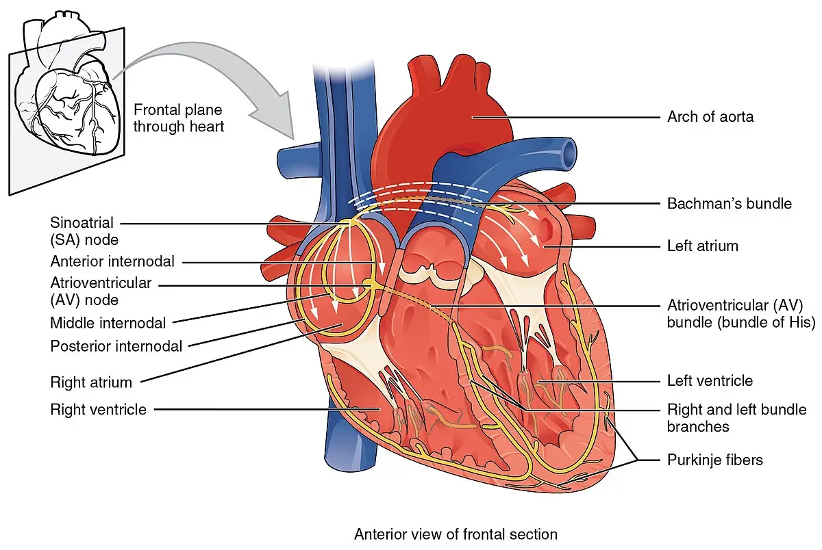
The conduction system is composed of five structures.

  • Sinoatrial (SA) node: The SA node is called the “pacemaker of the heart.” It is located in the posterior superior wall of the right atrium. The cardiac cells in the SA node generate the impulse. An impulse from the SA node starts each heartbeat. When the SA node discharges the impulse, it travels in many directions through the heart muscle. Nearby atrial cardiac cells slowly pick up the impulse. The impulse also moves quickly across special bands of tissue called intermodal tracts. The Bachmann bundle, a specialized intermodal tract, takes the impulse to the left atrium. Other intermodal tracts take the impulse quickly to the AV node.
  • Atrioventricular (AV) node: The AV node is located at the base of the interatrial septum. When the impulse reaches the AV node, it moves very slowly through the node. This slowdown allows the atrial chambers to finish contracting, moving the blood into the ventricular chambers.
  • Bundle of His (or AV bundle): The bundle of His is located in the upper interventricular septum. When the impulse leaves the AV node, it moves to the bundle of His.
  • Right and left bundle branches: The bundle branches are located in the lower interventricular septum. After the impulse passes through the bundle of His, it enters the right and left bundle branches. The right bundle branch brings the impulse to the right ventricle. The left bundle branch brings the impulse to the left ventricle.
  • Purkinje fibers: The bundle branches split into many Purkinje fibers. The Purkinje fibers transmit the impulse quickly and efficiently to the ventricular cardiac cells. This helps the ventricular chambers to contract.
Electrical conduction system of the heart shown
Cardiac conduction system
Wikimedia Commons
/
CC BY 3.0

States of cardiac cells

The cardiac cells cycle through three states, or steps, in the same sequence for each impulse.

  • Polarized state: Before the impulse hits the cardiac cell, the cell is in the resting state, or resting potential. The inside of the cardiac cell is negatively charged. Outside of the cell is positively charged. There is no electrical activity seen on the ECG tracing during the polarized state.
  • Depolarized state: When the impulse hits the cardiac cell, large numbers of positively charged sodium ions move into the cell. A small number of potassium ions move outside the cell. The movement of sodium and potassium changes the cell’s charge to positive. The change, also called action potential, allows the impulse to move through the cell. Depolarization (when the impulse hits the cell) causes electrical activity on the ECG tracing.
  • Repolarized state: After the impulse passes over the cell, the sodium and potassium ions move back to their original locations. This causes the cell’s charge to change back to a negative charge. This recovery phase is called the repolarized state. Electrical activity (less than the depolarized state) can be recorded during the repolarized state.

Remember that the impulse occurs first and very soon after the contraction starts. These are two distinct activities, yet they appear to be close together. The electrical activity from the depolarized state and repolarized state is recorded on the ECG tracing. The contractions are mechanical actions and are not recorded on the ECG tracing. An echocardiogram (ECHO) can be used to gather information on the mechanical action of the heart, such as the valves opening and closing.

Principles of Electrocardiography

  • Assesses electrical activity of the heart using an electrocardiogram (ECG/EKG)
  • Medical assistants: understand heart function, perform ECGs, identify and troubleshoot issues
  • Electrodes on skin detect electrical impulses; provider interprets ECG results

Heart Structure

  • Four chambers: right/left atria (receive blood), right/left ventricles (pump blood)
  • Valves: tricuspid (right AV), pulmonary (right SL), mitral/bicuspid (left AV), aortic (left SL)
  • Septum divides right and left sides
  • Mechanical action assessed by echocardiography (ECHO)

Heart Wall Structure

  • Three layers: epicardium (outer), myocardium (muscle), endocardium (inner)
  • Myocardial cells connected by intercalated discs for coordinated contraction
  • Intercalated discs enable rapid electrical communication

Blood Flow

  • Right atrium receives deoxygenated blood from:
    • Superior vena cava
    • Inferior vena cava
    • Coronary sinus
  • Blood path: right atrium → tricuspid valve → right ventricle → pulmonary valve → pulmonary artery → lungs → pulmonary vein → left atrium → mitral valve → left ventricle → aortic valve → aorta → body
  • Cardiac cycle: diastole (relaxation/filling), systole (contraction/pumping)

Heart’s Conduction System

  • Specialized electrical cells throughout myocardium
  • Enables initiation and transmission of electrical impulses for heartbeats

Conduction Pathway

  • Sinoatrial (SA) node: pacemaker, initiates impulse
  • Atrioventricular (AV) node: slows impulse, allows atrial contraction
  • Bundle of His: transmits impulse from AV node
  • Right and left bundle branches: carry impulse to ventricles
  • Purkinje fibers: distribute impulse to ventricular muscle cells for contraction

States of Cardiac Cells

  • Polarized state: resting, inside negative, no ECG activity
  • Depolarized state: sodium in/potassium out, inside positive, ECG activity seen
  • Repolarized state: ions return, inside negative, recovery phase, some ECG activity

Key Distinctions

  • ECG records electrical activity (depolarization/repolarization), not mechanical contractions
  • Echocardiogram (ECHO) assesses mechanical heart actions (e.g., valve movement)

Sign up for free to take 13 quiz questions on this topic

Previous
Next  | 32.2 ECG terminology, waves, and intervals
All rights reserved ©2016 - 2026 Achievable, Inc.

The principles of electrocardiography

Heart disease is the leading cause of death in the United States. Medical assistants in primary and specialty areas often care for patients with heart disorders and must do the following:

  • Understand the cardiovascular system
  • Recognize early symptoms of potential disorders
  • Coach patients on cardiac tests and treatments ordered by providers
  • Accurately perform cardiac tests
  • Identify and troubleshoot problems when performing tests

The heart is a complex organ. Electrical impulses move through the heart, and the cells react by contracting. The contracting cells result in contraction of the chambers. The contractions cause the blood to move through the heart and out to the arteries. Both the electrical and mechanical activities of the heart can be assessed to monitor the heart’s function. When a provider needs to assess the electrical activity of the heart, an electrocardiogram is commonly ordered.

Electrocardiography is a painless test. Electrodes are placed on the body, and wires connect the electrodes to the ECG machine (electrocardiograph). Electrical impulses from the heart make their way to the surface of the skin. Think of what occurs when you throw a rock into a lake. Waves are created, and they eventually make their way to the shore. The electricity from the heart eventually makes its way to the surface of the skin. The electrodes pick up the electricity, and the electricity moves into the machine. The electrocardiograph creates a record of the impulses, which is called an electrocardiogram (ECG, EKG).

A medical assistant performs the electrocardiography. The provider reads and interprets the ECG to identify any abnormalities in the electrical conduction in the heart. It is important for the medical assistant to do the following:

  • Know the normal function of the heart
  • Perform the procedure accurately
  • Identify problems during the ECG procedure and take appropriate actions

This chapter discusses ECGs. The chapter begins with a review of the cardiovascular system. The components of the ECG and the process of obtaining an ECG follow the review.

The heart structure

The heart is divided into four chambers. Two atrial chambers receive blood from the body. Two ventricular chambers pump blood out to the body. The septum divides the right and left sides of the heart.

The tricuspid valve is found between the right atrium and the right ventricle. The pulmonary valve is between the right ventricle and the pulmonary artery. The bicuspid, or mitral, valve is found between the left atrium and left ventricle. The aortic valve is between the left ventricle and the aorta. When the valves open, blood moves to the next chamber or out of the heart through the arteries. The mechanical action of the heart and valves can be assessed by echocardiography (ECHO).

Heart wall structure

The heart wall has three layers: the epicardium, the myocardium, and the endocardium. Cardiac muscle fibers in the myocardium are electrically linked together, forming one unit. A myocardial cell forms a strong connection to the next cells through special junctions called intercalated discs. These discs allow electricity to flow freely from one cell to the next. The intercalated discs are responsible for the cell-to-cell communication that is required for coordinated muscle contraction. The intercalated discs help the muscle fibers form one unit that contracts all at once instead of a little at a time. The one-unit approach is important because the atria and ventricles need to contract in an appropriately timed and coordinated effort.

Blood flow

The right atrium receives deoxygenated blood from these structures:

  • Superior vena cava (blood comes from the head, neck, chest, and upper extremities)
  • Inferior vena cava (blood comes from the abdomen, pelvis, and lower extremities)
  • Coronary sinus (blood from the coronary veins in the heart muscle)

When the right atrial chamber contracts, the tricuspid valve (atrioventricular [AV] valve) opens. Blood empties from the right atrium into the right ventricle. When the ventricles contract, the deoxygenated blood in the right ventricle passes through the opened pulmonary valve (semilunar [SL] valve) and moves into the pulmonary artery. The pulmonary artery brings the blood to the lungs. In the lungs, the blood picks up oxygen (O2) and gives up carbon dioxide (CO2). The pulmonary vein brings the oxygenated blood back to the left atrium. When the left atrial chamber contracts, the blood is pushed past the opened mitral (or bicuspid) valve (AV valve). The blood empties into the left ventricle. When the ventricles contract, the blood in the left ventricle moves through the opened aortic valve (SL valve) and into the aorta. The aorta transports oxygenated blood to the body. The first arteries to split off the aorta are the left and right coronary arteries. These arteries bring the oxygenated blood to the heart muscles.

A complete heartbeat, or cardiac cycle, can be divided into diastole and systole phases. During the diastole phase, the heart is at rest, and the atria fill with blood. The systole phase occurs when the heart is contracting.

Heart’s conduction system

The electrical cells make up the conduction system of the heart. These cells are found throughout the myocardium. They can respond to and transmit electrical impulses to neighboring cells. The following sections review the conduction system and the cardiac cell cycles. Understanding the anatomy and physiology of the heart helps when learning electrocardiography.

Conduction pathway

The conduction system is composed of five structures.

  • Sinoatrial (SA) node: The SA node is called the “pacemaker of the heart.” It is located in the posterior superior wall of the right atrium. The cardiac cells in the SA node generate the impulse. An impulse from the SA node starts each heartbeat. When the SA node discharges the impulse, it travels in many directions through the heart muscle. Nearby atrial cardiac cells slowly pick up the impulse. The impulse also moves quickly across special bands of tissue called intermodal tracts. The Bachmann bundle, a specialized intermodal tract, takes the impulse to the left atrium. Other intermodal tracts take the impulse quickly to the AV node.
  • Atrioventricular (AV) node: The AV node is located at the base of the interatrial septum. When the impulse reaches the AV node, it moves very slowly through the node. This slowdown allows the atrial chambers to finish contracting, moving the blood into the ventricular chambers.
  • Bundle of His (or AV bundle): The bundle of His is located in the upper interventricular septum. When the impulse leaves the AV node, it moves to the bundle of His.
  • Right and left bundle branches: The bundle branches are located in the lower interventricular septum. After the impulse passes through the bundle of His, it enters the right and left bundle branches. The right bundle branch brings the impulse to the right ventricle. The left bundle branch brings the impulse to the left ventricle.
  • Purkinje fibers: The bundle branches split into many Purkinje fibers. The Purkinje fibers transmit the impulse quickly and efficiently to the ventricular cardiac cells. This helps the ventricular chambers to contract.

States of cardiac cells

The cardiac cells cycle through three states, or steps, in the same sequence for each impulse.

  • Polarized state: Before the impulse hits the cardiac cell, the cell is in the resting state, or resting potential. The inside of the cardiac cell is negatively charged. Outside of the cell is positively charged. There is no electrical activity seen on the ECG tracing during the polarized state.
  • Depolarized state: When the impulse hits the cardiac cell, large numbers of positively charged sodium ions move into the cell. A small number of potassium ions move outside the cell. The movement of sodium and potassium changes the cell’s charge to positive. The change, also called action potential, allows the impulse to move through the cell. Depolarization (when the impulse hits the cell) causes electrical activity on the ECG tracing.
  • Repolarized state: After the impulse passes over the cell, the sodium and potassium ions move back to their original locations. This causes the cell’s charge to change back to a negative charge. This recovery phase is called the repolarized state. Electrical activity (less than the depolarized state) can be recorded during the repolarized state.

Remember that the impulse occurs first and very soon after the contraction starts. These are two distinct activities, yet they appear to be close together. The electrical activity from the depolarized state and repolarized state is recorded on the ECG tracing. The contractions are mechanical actions and are not recorded on the ECG tracing. An echocardiogram (ECHO) can be used to gather information on the mechanical action of the heart, such as the valves opening and closing.

Key points

Principles of Electrocardiography

  • Assesses electrical activity of the heart using an electrocardiogram (ECG/EKG)
  • Medical assistants: understand heart function, perform ECGs, identify and troubleshoot issues
  • Electrodes on skin detect electrical impulses; provider interprets ECG results

Heart Structure

  • Four chambers: right/left atria (receive blood), right/left ventricles (pump blood)
  • Valves: tricuspid (right AV), pulmonary (right SL), mitral/bicuspid (left AV), aortic (left SL)
  • Septum divides right and left sides
  • Mechanical action assessed by echocardiography (ECHO)

Heart Wall Structure

  • Three layers: epicardium (outer), myocardium (muscle), endocardium (inner)
  • Myocardial cells connected by intercalated discs for coordinated contraction
  • Intercalated discs enable rapid electrical communication

Blood Flow

  • Right atrium receives deoxygenated blood from:
    • Superior vena cava
    • Inferior vena cava
    • Coronary sinus
  • Blood path: right atrium → tricuspid valve → right ventricle → pulmonary valve → pulmonary artery → lungs → pulmonary vein → left atrium → mitral valve → left ventricle → aortic valve → aorta → body
  • Cardiac cycle: diastole (relaxation/filling), systole (contraction/pumping)

Heart’s Conduction System

  • Specialized electrical cells throughout myocardium
  • Enables initiation and transmission of electrical impulses for heartbeats

Conduction Pathway

  • Sinoatrial (SA) node: pacemaker, initiates impulse
  • Atrioventricular (AV) node: slows impulse, allows atrial contraction
  • Bundle of His: transmits impulse from AV node
  • Right and left bundle branches: carry impulse to ventricles
  • Purkinje fibers: distribute impulse to ventricular muscle cells for contraction

States of Cardiac Cells

  • Polarized state: resting, inside negative, no ECG activity
  • Depolarized state: sodium in/potassium out, inside positive, ECG activity seen
  • Repolarized state: ions return, inside negative, recovery phase, some ECG activity

Key Distinctions

  • ECG records electrical activity (depolarization/repolarization), not mechanical contractions
  • Echocardiogram (ECHO) assesses mechanical heart actions (e.g., valve movement)

More from Electrocardiography and heart structure

  • ECG terminology, waves, and intervals
  • Rhythm analysis and arrhythmias
  • The electrocardiograph and ECG preparation
  • Troubleshooting and evaluation
  • Stress testing