T and B lymphocytes
Both T and B lymphocytes originate from a common lymphoid progenitor in the bone marrow. After that, they develop in different places:
- T lymphocytes continue their development in the thymus.
- B lymphocytes develop in the bone marrow (and in the liver during fetal life).
Development of T cells: T cells can be divided into two lineages based on their surface receptors:
- alpha, beta T cells
- gamma, delta T cells
Alpha, beta T cells are further divided into CD4 and CD8 T cells based on which class of MHC they recognize:
- CD4 T cells bind to MHC class II.
- CD8 T cells bind to MHC class I.
Early in development, T cells do not express either CD4 or CD8. These are called “double negative” T cells. Later, thymocytes may express both CD4 and CD8; these are called “double positive” T cells, and they are precursors to CD4 and CD8 T cells.
Location in the thymus changes as T cells mature:
- Double negative and double positive cells are located in the cortex of the thymus.
- Single positive cells are located in the medulla, from where they enter the bloodstream.
Surface markers also change with maturation:
- CD44, c-Kit, and CD 25 (receptor for IL 2) are seen in early T cells.
- CD3 and CD2 are expressed in more mature T cell lineages.
The hormones thymosin and thymopoietin help T cell differentiation in the thymus.
During thymic development, T cells are tested for how they respond to self MHC and self antigens:
- Only thymocytes whose receptors can interact with self MHC are selected for survival. This is called “positive selection”.
- Thymocytes that react against self-antigens undergo “negative selection” and are eliminated.
A transcriptional regulator called AIRE (Autoimmune Regulator) enhances the synthesis of an array of self-proteins to be displayed on thymic epithelial cells, which helps establish T cell tolerance. Mutations in AIRE cause Autoimmune polyendocrinopathy.
Different thymic cells support these selection steps:
- Thymic cortical epithelial cells have branching processes that contact developing T cells and expose them to MHC molecules, playing an important role in positive selection.
- Dendritic cells regulate negative selection of self-reactive T cells.
Mature, single positive CD4 or CD8 T cells then leave the thymus:
- CD4+ T cells are also called helper T cells.
- CD8+ T cells are also called cytotoxic T cells.
The CD4 molecule is composed of four domains: D1,2,3 and 4. Domains D1 and D2 are involved in binding to the MHC class II molecule.
A CD8 molecule is a heterodimer of one alpha and one beta chain or two alpha chains that are linked to each other. CD8 binds to the alpha 3 domain on MHC I.
CD4 and CD8 act as co-receptors and bind to MHC II or I, respectively. This binding is essential for an effective immune response.
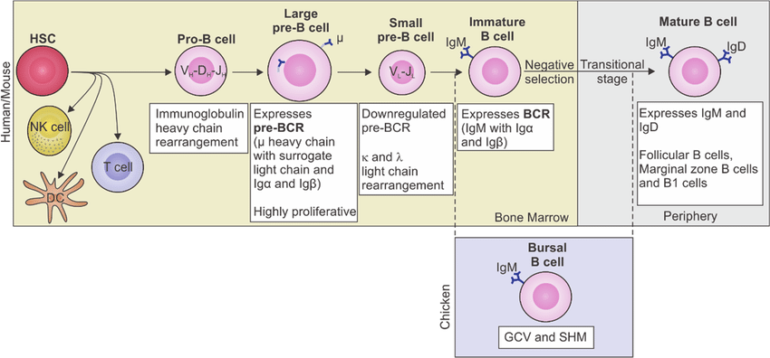
Development of B cells: Bone marrow stromal cells are essential in the early stages of B cell development. Stromal cells secrete several growth factors like stem cell factor, cytokines and adhesion molecules to help B cell differentiation.
As B cells mature, their surface immunoglobulins change:
- Immature B cells have IgM on their surface.
- Mature B cells have IgM and IgD on their surface.
B cell surface proteins CD45R and CD19 are expressed in all stages of B cell development. Self-reactive B cells undergo clonal deletion.