Physiology of cardiac system
Anatomy
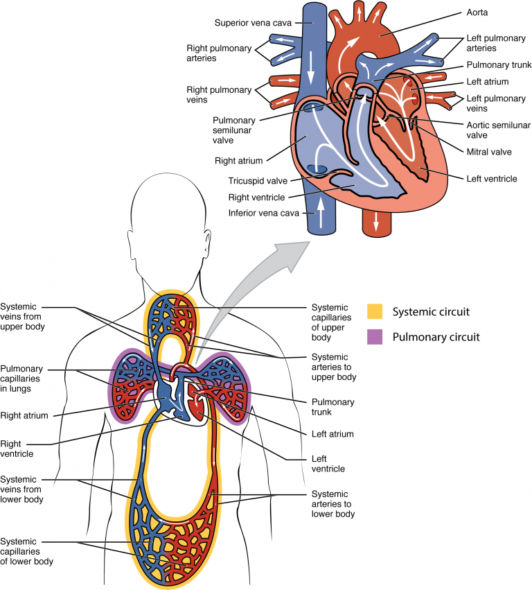
The heart is a muscle that pumps blood throughout the body’s systems. The left side of the heart pumps oxygenated blood from the lungs to all body systems via the arterial supply. Blood then returns to the right side of the heart via the venous system, where waste products are excreted by various organs. The right side then pumps deoxygenated blood into the lungs, where carbon dioxide (CO2) is released, and oxygen is absorbed. This cycle repeats with each heartbeat.
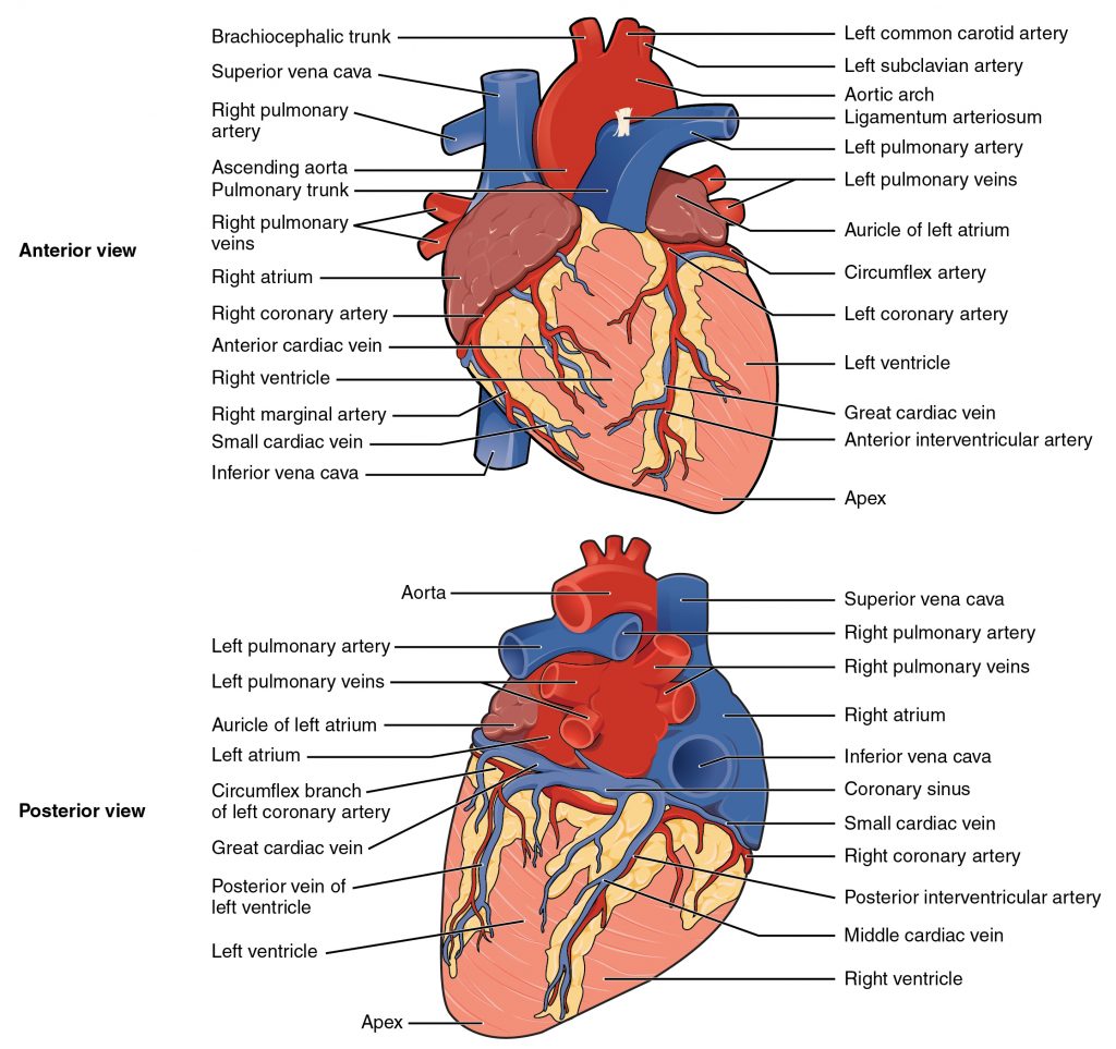
The heart consists of:
- External walls
- Blood-filled chambers
- Valves that allow blood to flow through the chambers
- Blood vessels that carry blood to the other structures
- Electrical signaling conduction system
The three walls of the heart are:
- Endocardium; innermost layer
- Myocardium: muscular middle layer
- Epicardium: outermost layer
Chambers of the heart:
- Right atrium
- Right ventricle
- Left atrium
- Left ventricle
Valves of the heart:
- Atrioventricular valves:
- Tricuspid valve: lies between the right atrium and the right ventricle
- Mitral valve: lies between the left atrium and left ventricle
- Semilunar valves:
- Aortic valve: oxygenated blood flows from the left ventricle to the aorta
- Pulmonary valve: deoxygenated blood flows from the right ventricle to the pulmonary artery
Blood vessels:
- Arteries: carry oxygenated blood
- Veins: carry deoxygenated blood
- Capillaries: small blood vessels for the exchange of deoxygenated and oxygenated blood
Chambers of the heart
The heart is separated into four chambers that work to ensure blood flow occurs in the right direction. The left and right sides of the heart work separately to ensure blood reaches either the lungs or the body system. The right side of the heart pumps deoxygenated blood to the lungs, where gas exchange occurs. The left side pumps oxygenated blood to the rest of the body.
Blood flow through the heart: step-by-step
- Deoxygenated blood returns to the heart:
- Blood low in oxygen returns from the body to the heart through two major veins:
- Superior vena cava (SVC): returns blood from the upper body
- Inferior vena cava (IVC): returns blood from the lower body
- Both veins empty into the right atrium.
- Blood low in oxygen returns from the body to the heart through two major veins:
- Right atrium
- Deoxygenated blood enters the right atrium, the upper right chamber of the heart.
- Blood then flows through the tricuspid valve into the right ventricle.ventricle.
- Right ventricle
- The right ventricle pumps deoxygenated blood through the pulmonary semilunar valve into the pulmonary arteries.
- Pulmonary arteries to the lungs
- The pulmonary arteries carry deoxygenated blood from the heart to the lungs.
- This is an important exception because most arteries carry oxygenated blood.
- The pulmonary arteries carry deoxygenated blood from the heart to the lungs.
- Lungs oxygenate the blood
- In the lungs, carbon dioxide (CO₂) is removed and oxygen (O₂) is added to the blood through gas exchange at the alveoli.
- The blood is now oxygenated.
- Left atrium
- Oxygen-rich blood returns to the heart through the pulmonary veins and enters the left atrium, the upper left chamber of the heart.
- This is another important exception because pulmonary veins carry oxygenated blood.
- Blood then passes through the mitral (bicuspid) valve into the left ventricle.
- Oxygen-rich blood returns to the heart through the pulmonary veins and enters the left atrium, the upper left chamber of the heart.
- Left ventricle
- The left ventricle pumps oxygenated blood through the aortic semilunar valve into the aorta.
- The left ventricle has the thickest myocardium because it must generate enough force to pump blood throughout the entire body.
- Aorta to the body
- The aorta distributes oxygen-rich blood to the systemic circulation, supplying tissues and organs throughout the body.
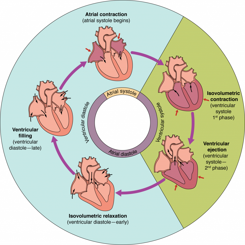
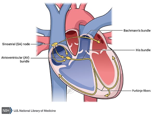
Electrical signals in the heart
Electrical signals within the heart act as a pacemaker to the heart by assisting with controlling heart rate, coordinating the heart chambers (atria and ventricles), adapting to the changing needs of the body, and ensuring appropriate circulation.
Crucial factors of cardiac function
The amount of blood pumped with each heartbeat is crucial to maintaining the vitality of the lungs and the peripheral system. The principles that are important in this process are preload and afterload. If there is dysfunction in either of these components, then the individual may experience stroke, myocardial infarction, or death.