




Electrocardiograms
How to read electrocardiograms
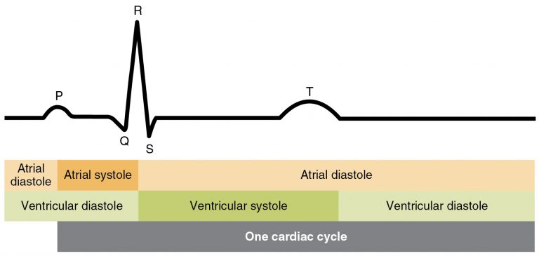
Important considerations for reading electrocardiograms
- Is the rhythm regular and consistent?
- Is the rate fast, slow, or normal?
- Are P waves present and look the same?
- Is the PR interval normal and consistent?
- Are the QRS complexes normal and immediately follow the P wave?
- Is the T wave present and normal in appearance?
- When viewing a multiple lead EKG strip view lead II or the line where the pattern can be defined clearly.
- When a lead that is normally upright shows its P waves, QRS complexes, and T waves all inverted (lead I when the arm electrodes are swapped, for example), the cause is usually improper lead placement, not a heart problem: have the electrodes checked and focus on the pattern listed above 1-6. Some leads, such as aVR, are normally inverted, while a newly inverted T wave on its own can be a sign of ischemia and should be reported to the supervising PT.
Calculation of heart rate with EKG
Calculation of heart rate using an EKG should only be done when the rhythm is consistent and predictable, allowing for easy calculations.
Calculations as follows for a 6-second strip method:
Count the number of QRS complexes in 6 seconds and multiply by 10
Example:
7 QRS peaks noted in 6 seconds x 10 = 70 bpm (heart rate). This is in the normal range of 60-100.
Common EKGs
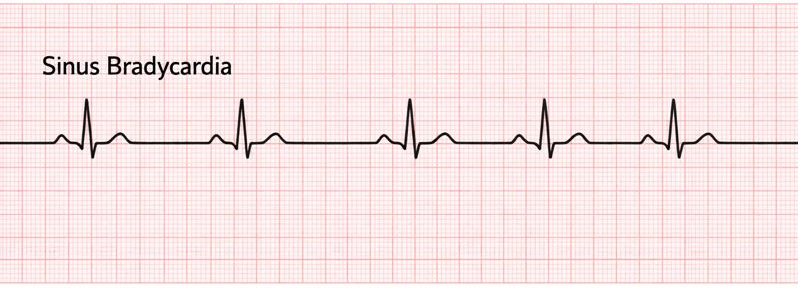
Sinus bradycardia (slow heart rate, below 60 beats per minute at rest)
PT implications
- Common to see this rhythm when working with those taking beta-blockers and in the elderly population
- If present, ok to proceed with initiation of activity and continuance of activity,
- Will need to use the perceived exertion scale (RPE) to assess tolerance to activity, since beta-blockers blunt the normal heart rate response to exertion
- Monitor for symptoms of dizziness, lightheadedness, and pallor
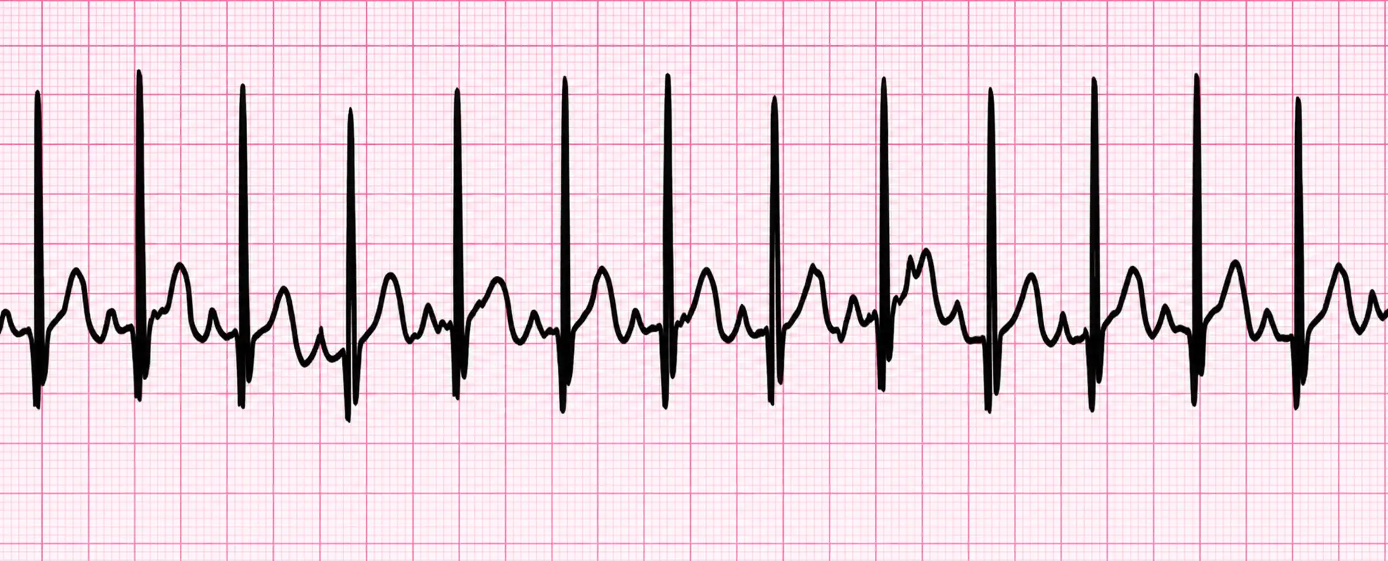
Sinus tachycardia (fast heart rate, > 100 beats per minute at rest)
PT implications
- Common to observe this rhythm during exercise
- If present before exercise notify supervising PT
- Pay attention if the individual develops a headache, dizziness, or lightheadedness with tachycardia
- If these symptoms present, the therapist will stop the activity and reassess all vitals
A-fibrillation (A-fib)
An irregular, tachycardic heart rhythm in which the atrium contracts erratically. On the EKG, discrete P waves are absent and are replaced by chaotic fibrillatory (f) waves.
PT implications
If not previously recognized, stop activity and notify
If identified previously:
- Vital sign monitoring
- Continuous or frequent monitoring of heart rate, blood pressure, and oxygen saturation is essential.
- Pulse may be irregular, so use auscultation or ECG for accuracy rather than the radial pulse alone.
- Be aware that heart rate may be elevated or vary unpredictably.
- Exercise prescription
- Begin with low to moderate intensity activities; avoid high-intensity or sudden exertion early in rehab.
- Use Rate of Perceived Exertion (RPE) rather than heart rate alone due to variability.
- Monitor for signs of exercise intolerance: dizziness, fatigue, palpitations, chest pain, or dyspnea.
A-flutter
A regular, tachycardic contraction of the atrium; constant atrial contraction produces a sawtooth pattern of flutter (F) waves in place of P waves.
PT implications
If not previously recognized, stop activity and notify
If identified previously, follow the same vital sign monitoring and exercise progression guidance as atrial fibrillation (above), with these flutter-specific notes:
- A-flutter typically presents with a regular but rapid atrial rate (240-400 bpm); ventricular response may be regular or irregular depending on AV block.
- Patients may suddenly shift into more dangerous rhythms (e.g., atrial fibrillation or rapid ventricular response), so use ECG or telemetry when possible, especially in acute care settings.
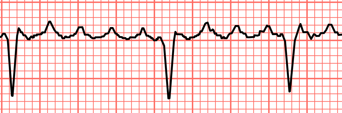
Premature ventricular contraction (PVC)
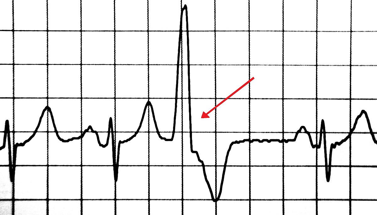
An early, wide, bizarre QRS complex from an ectopic ventricular focus, occurring before the next expected sinus beat and typically followed by a compensatory pause.
PT implications
- Singular PVC at rest and exercise (no restriction): initiate or continue with activity, as this can be an anomaly
- Can be caused by stress, caffeine, or idiopathic causes
- Couplet (2) (precaution): stop and rest for 5 minutes; resume exercise only if resolved
- Three or more PVCs in a row at rest or with exercise (contraindication): do not initiate activity and discontinue exercise, as this could lead to fatal heart rhythms
- Bigeminy - a PVC every other heartbeat (contraindication): do not initiate activity and discontinue exercise, as this could lead to fatal heart rhythms
- Monitor for symptoms: heart fluttering, heart pounding, missed heartbeats
Ventricular tachycardia
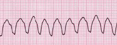
A rapid heart rhythm originating from the ventricles. The ventricles contract at a fast rate and may not fill effectively, which can significantly reduce cardiac output and impair blood flow to the body.
PT implications
- Medical emergency and CPR initiated
Ventricular fibrillation
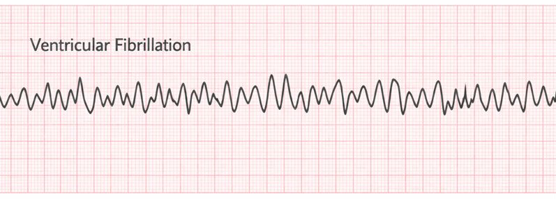
Ventricular fibrillation is a life-threatening dysrhythmia characterized by rapid, chaotic electrical activity causing ineffective quivering of the ventricles. Because there is no coordinated ventricular contraction, the heart is unable to produce effective cardiac output or blood flow to the body.
PT implications
- Medical emergency and CPR initiated