



Scoliosis, autism, and birth injuries
These pediatric conditions affect the spine, development, and the infant at birth; as with the hip and knee conditions, the PTA carries out the plan of care established by the supervising PT.
Scoliosis
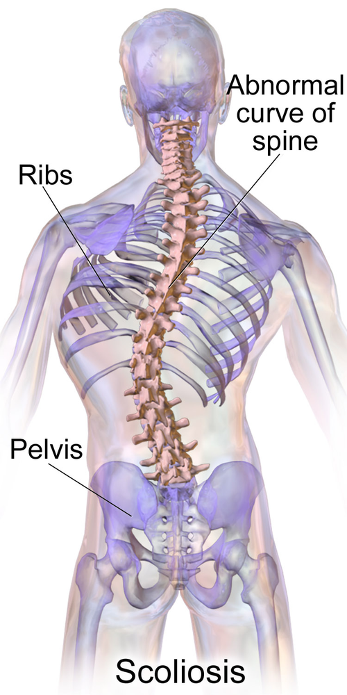
Most idiopathic cases are diagnosed during adolescence via physical examination and X-ray imaging. The severity of scoliosis is defined by the degree of curve - this also guides which interventions may be performed.
Cobb angle (severity of scoliosis)
- Mild scoliosis: Cobb angle of 10 to 20 degrees (a curve under 10 degrees is not considered scoliosis)
- No intervention or conservative management
- Moderate scoliosis: Cobb angle between 20 and 40 degrees
- Bracing and physical therapy may be indicated
- Severe scoliosis: Cobb angle greater than 40 degrees
- Surgical intervention is generally recommended once the curve exceeds about 45-50 degrees; curves of 40-45 degrees in a still-growing child may still be braced; large curves, generally greater than 50 degrees, can begin to compromise the respiratory system
A younger age or a lower Risser sign (the most common radiographic marker of skeletal maturity) means more spinal growth remains and a greater chance of curve progression, so a growing child with a curve of about 25 to 40 degrees is a candidate for bracing.
Symptoms
- Uneven shoulders
- One shoulder blade that appears more prominent than the other
- Uneven waist
- One hip is higher than the other
- One side of the rib cage jutting forward
Physical therapy interventions for scoliosis
- Address muscle imbalances that may be present due to curvature, per the plan of care
- For functional scoliosis caused by leg-length discrepancy, apply and reinforce use of prescribed shoe inserts
- Reinforce the prescribed bracing wear schedule (e.g., TLSO/Boston brace) and scoliosis-specific exercises per the plan of care
- Reinforce use of prescribed durable medical equipment, monitor breathing in large curves, and report changes in fit, tolerance, or respiratory status to the supervising PT
- Inspect the skin under the brace for redness or breakdown, and report it to the supervising PT
Autism spectrum disorder
Etiology is unknown, but there is a strong genetic predisposition, along with some prenatal or perinatal environmental factors that may contribute. Diagnosis is made through observation and standardized diagnostic criteria, and can reliably be made by age 2.
Symptoms
- Sensory integration dysfunction
- Hyposensitivity (sensory seeking) or hypersensitivity (sensory avoidant)
- Difficulty with verbal and non-verbal communication
- Impaired coordination
- Balance deficits
- Occasional strength and range of motion deficits
- Developmental delay
Physical therapy interventions for autism spectrum disorder
- Address developmental delay
- Improve strength
- Improve balance and coordination
- Sensory integration activities
- Be aware that new people and situations can cause either withdrawal behavior or increased aggression
Brachial plexus injuries
Common causes of brachial plexus injuries include shoulder dystocia, breech birth, assisted birth techniques such as forceps or vacuum, increased birth weight, multiple birth (twins or more), or gestational diabetes. Diagnosis is based on physical examination of upper extremity movement and primitive reflexes, with x-ray or MRI as needed. A clavicle fracture, the most common newborn fracture, can also leave the arm moving less and the Moro reflex absent on that side, but palpation finds tenderness, crepitus and irregularity along the clavicle, and radiographs confirm the fracture.
Severity of brachial plexus injuries
- Traction
- Stretching of the nerve with spontaneous recovery
- Rupture
- Nerve is torn but remains attached to the spinal cord; may require surgical intervention
- Avulsion
- Nerve is completely torn from the spinal cord; permanent disability may result after surgical intervention
Common brachial plexus injuries
| Condition | Nerve roots | Posture/presentation | Preserved function | Typical complication |
|---|---|---|---|---|
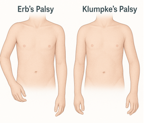
| Erb’s palsy | C5-C6 | “Waiter’s tip”: shoulder adducted and internally rotated, elbow extended, forearm pronated, wrist flexed | Hand and grasp function preserved | Shoulder girdle immobility leading to subluxation |
| Klumpke’s palsy | C8-T1 | Claw hand (intrinsic hand muscles and finger flexors/extensors affected); Horner syndrome possible with T1 involvement | Functional use of the shoulder and elbow | Contractures of the hand |
| Global palsy | C5-T1 | Total arm paralysis | None | Permanent disability common after surgical intervention |
Physical therapy interventions for brachial plexus injuries
- Partial immobilization for 1-2 weeks of the injured extremity
- Constraint-induced movement therapy: restrain the non-injured arm to encourage use of the injured arm
- Range of motion to avoid contractures
- Age-appropriate movements to decrease the likelihood of developmental delay
- Parent education on positioning and handling of the infant
Torticollis
Torticollis can be caused by difficulties at birth, large infant weight, breech birth, or improper positioning after birth. Diagnosis is confirmed via physical examination.
Symptoms
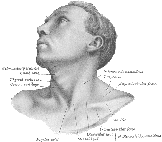
- Persistent head tilt to one side
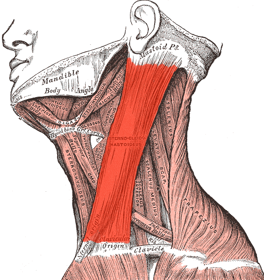
- Difficulty turning the head toward the side of the tilt (the involved side), because the tight SCM holds the chin rotated away
- A lump or knot may be felt in the tight neck muscle
- Facial asymmetry may develop over time
- Flat head on the side opposite the muscle tightness may present (plagiocephaly)
Physical therapy interventions for torticollis
- Stretching of the contracted sternocleidomastoid muscle
- Stretching involves lateral flexion away from the involved side and rotation toward the involved side (e.g., for right torticollis: left lateral flexion and right rotation), which reverses the muscle’s actions
- Stop stretching if the infant shows distress or a color change, and report it to the supervising PT
- Positioning to allow for prolonged stretching
- Addressing any developmental delay issues
- Collect and report objective measures (e.g., cervical range of motion, degree of head tilt) to the supervising PT rather than progressing the plan independently
- Stretching and positioning are the standard treatment for congenital muscular torticollis; surgery on the sternocleidomastoid is suggested only if nonsurgical options do not correct it, and botulinum toxin may be injected into the stiff muscle when torticollis is difficult to treat