Nervous system
Major functions
- Major functions of the nervous system include controlling and integrating body processes, responding to external stimuli, and coordinating both sensory (afferent) input and motor (efferent) output. This organization also supports higher-level integrative and cognitive activities needed for complex behaviors.
Nervous system organization
The vertebrate nervous system has 2 divisions:
- CNS- the brain and spinal cord
- PNS- all other neural elements
- Within the PNS, the somatic nervous system controls voluntary movement of skeletal muscles, while the autonomic nervous system regulates involuntary functions of visceral organs.
Autonomic nervous system
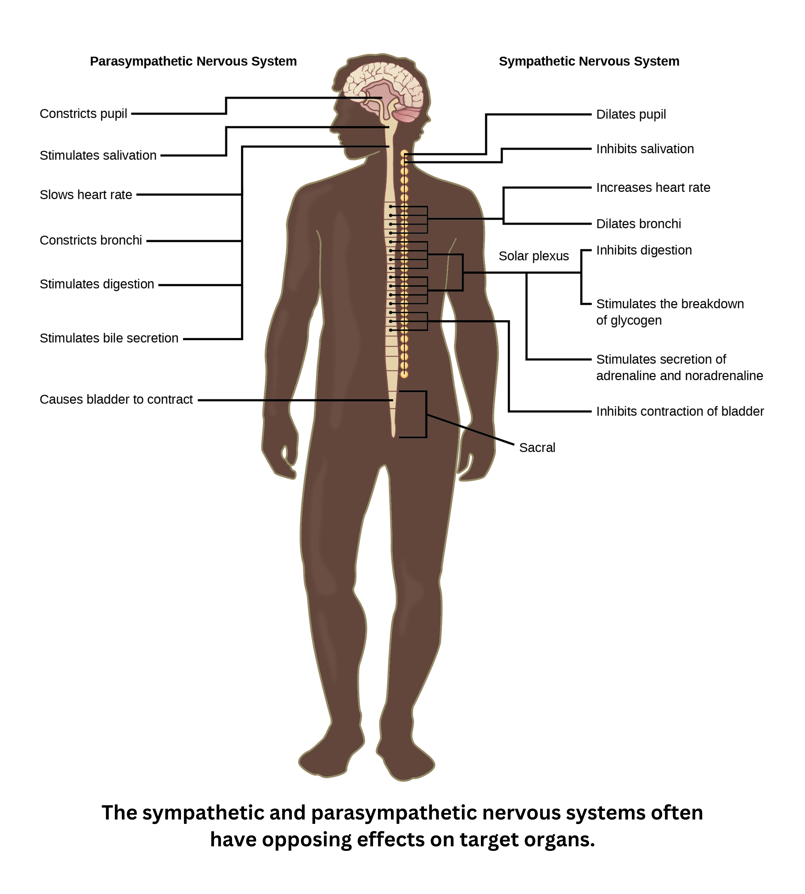
The autonomic nervous system (ANS) has two divisions:
- Sympathetic (fight or flight): increases heart rate and blood pressure, redirects blood to muscles, dilates pupils, and breaks down glycogen to release glucose.
- Parasympathetic (rest): reduces heart rate and blood pressure, directs blood toward digestion, constricts pupils, and converts glucose to glycogen for storage.
These divisions are often described as antagonistic because they tend to produce opposite effects.
Within the ANS:
- Sensor neurons or affectors detect changes and relay information to the CNS.
- Effector neurons carry commands from the CNS to target tissues.
Reflex arcs and feedback loops
Positive feedback loops amplify an initial event. Examples include uterine contractions triggering oxytocin release (which intensifies contractions) and activated platelets attracting more platelets during clot formation.
Negative feedback loops counteract an event. One example is blood pressure regulation: a drop in blood pressure triggers ADH release to raise it, while an increase in blood pressure reduces ADH secretion.
A reflex arc is typically a rapid form of negative feedback. A typical reflex arc includes:
- A receptor detecting a stimulus
- A sensor neuron transmitting signals to an integration center (often in the spinal cord)
- A motor neuron directing the response
- An effector (such as a muscle)
Common examples include the knee-jerk reflex and the withdrawal reflex. These are negative feedback mechanisms that help protect the body.
The Golgi tendon reflex prevents excessive muscle tension by reducing contraction when forces become too high.
Most spinal reflexes can occur without direct input from the brain. However, efferent control means the brain can sometimes override a reflex when a conscious decision is made (for example, holding still or not yelling when getting your ears pierced).
Nerve cell
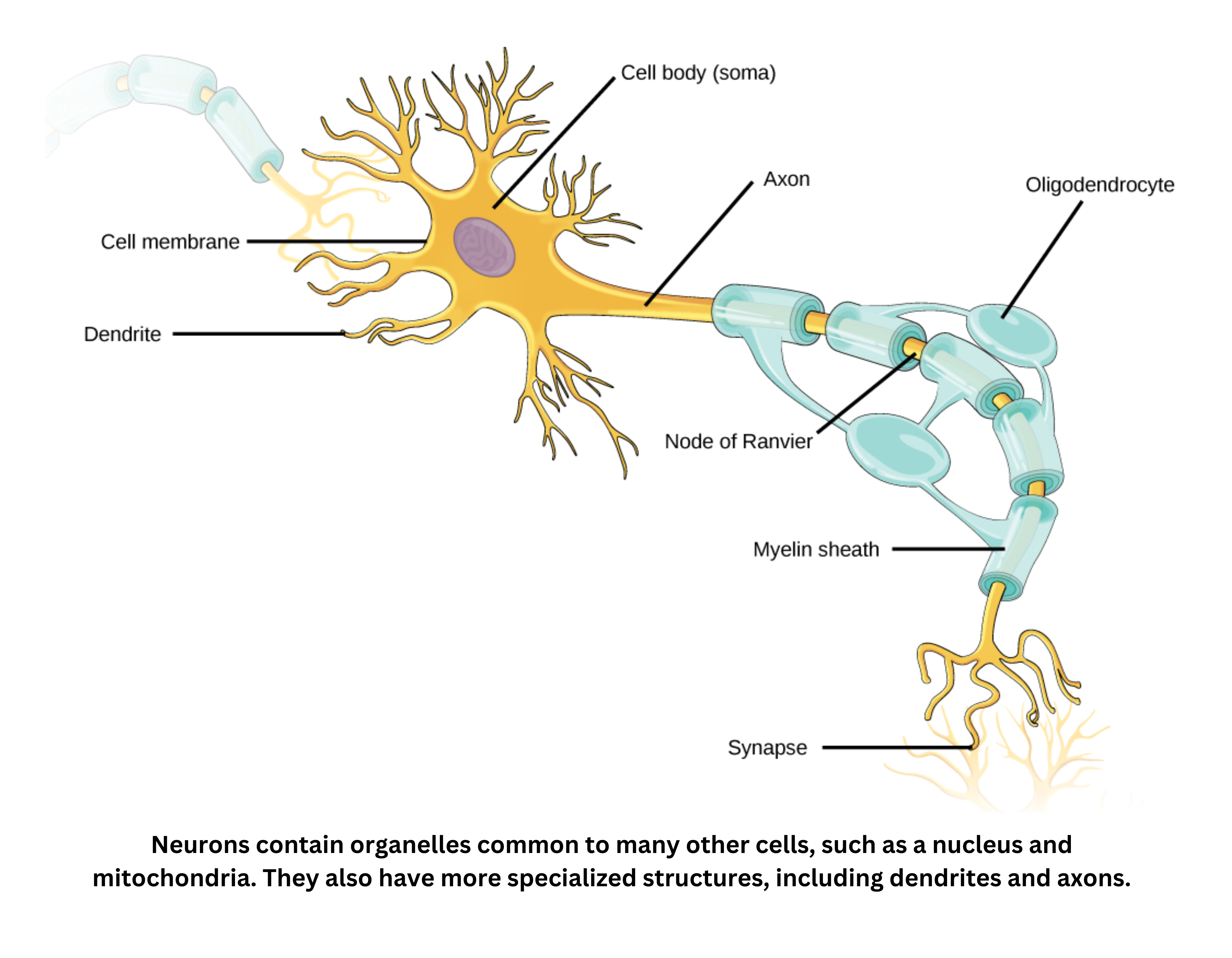
Cell body: site of nucleus, organelles
A nerve cell (neuron) includes a cell body, which contains the nucleus and other organelles. The cell body synthesizes many proteins, supported by abundant rough endoplasmic reticulum and Golgi complexes.
Attached to the cell body are dendrites, branching structures that form the neuron’s receptive region. Their branching increases surface area for incoming signals.
Extending from the cell body is a single axon, which carries electrical impulses toward the axon terminals - also called synaptic knobs or boutons - where neurotransmitters are released.
The axon may be wrapped in a myelin sheath, produced by Schwann cells in the peripheral nervous system and by oligodendrocytes in the central nervous system. Myelin is a fatty insulating layer that covers the axon in segments, leaving gaps called nodes of Ranvier. Because these nodes lack myelin, the action potential can jump from node to node, which greatly speeds conduction.
A synapse is a specialized junction that allows an impulse to pass from one neuron to another.
Signals may pass from a presynaptic axon terminal to:
- A postsynaptic dendrite (axodendritic)
- A postsynaptic cell body (axosomatic)
- In rare cases, another axon (axoaxonic)
When an action potential reaches the presynaptic terminal, it triggers neurotransmitter release into the synaptic cleft by exocytosis. This happens when calcium enters the presynaptic terminal, causing vesicles in the synaptic knob to fuse with the presynaptic membrane.
Neurotransmitters then diffuse across the cleft and bind receptors on the postsynaptic membrane. This binding opens ligand-gated ion channels, producing a local change in membrane potential called a graded potential. If the graded potential is strong enough to reach threshold, it triggers a new action potential in the postsynaptic neuron.
Neurotransmitters (for example, acetylcholine, norepinephrine, dopamine, and serotonin) are removed or broken down to prevent continual stimulation.
With continuous synaptic activity, neurotransmitter stores can become temporarily depleted, leading to short-term synaptic “fatigue.” Even with these synaptic steps, the postsynaptic action potential is still all-or-nothing. Once triggered, it has a consistent size, helping preserve signal fidelity as it travels through the nervous system.
Threshold and all-or-none behavior: If a stimulus raises the membrane potential from its resting value () past a threshold (typically ), an action potential occurs. Because the response is all-or-nothing, once threshold is crossed, the spike has a consistent magnitude whether the stimulus barely reaches threshold or exceeds it by a large amount.