Nerve cells, electrochemistry and biosignalling
Excitatory and inhibitory nerve fibers
- Excitatory inputs move the membrane potential closer to threshold, making depolarization more likely.
- Inhibitory inputs make the membrane potential more negative, making depolarization less likely.
Several subthreshold excitatory inputs can summate so that, together, they reach threshold. If inhibitory signals occur at the same time, they can reduce or cancel the effect of excitatory inputs.
High frequency firing can also promote reaching threshold. When action potentials occur close together, some depolarization from the previous event can persist, so each new input adds to the remaining depolarization and can gradually bring the membrane potential to threshold.
Glial cells, neuroglia
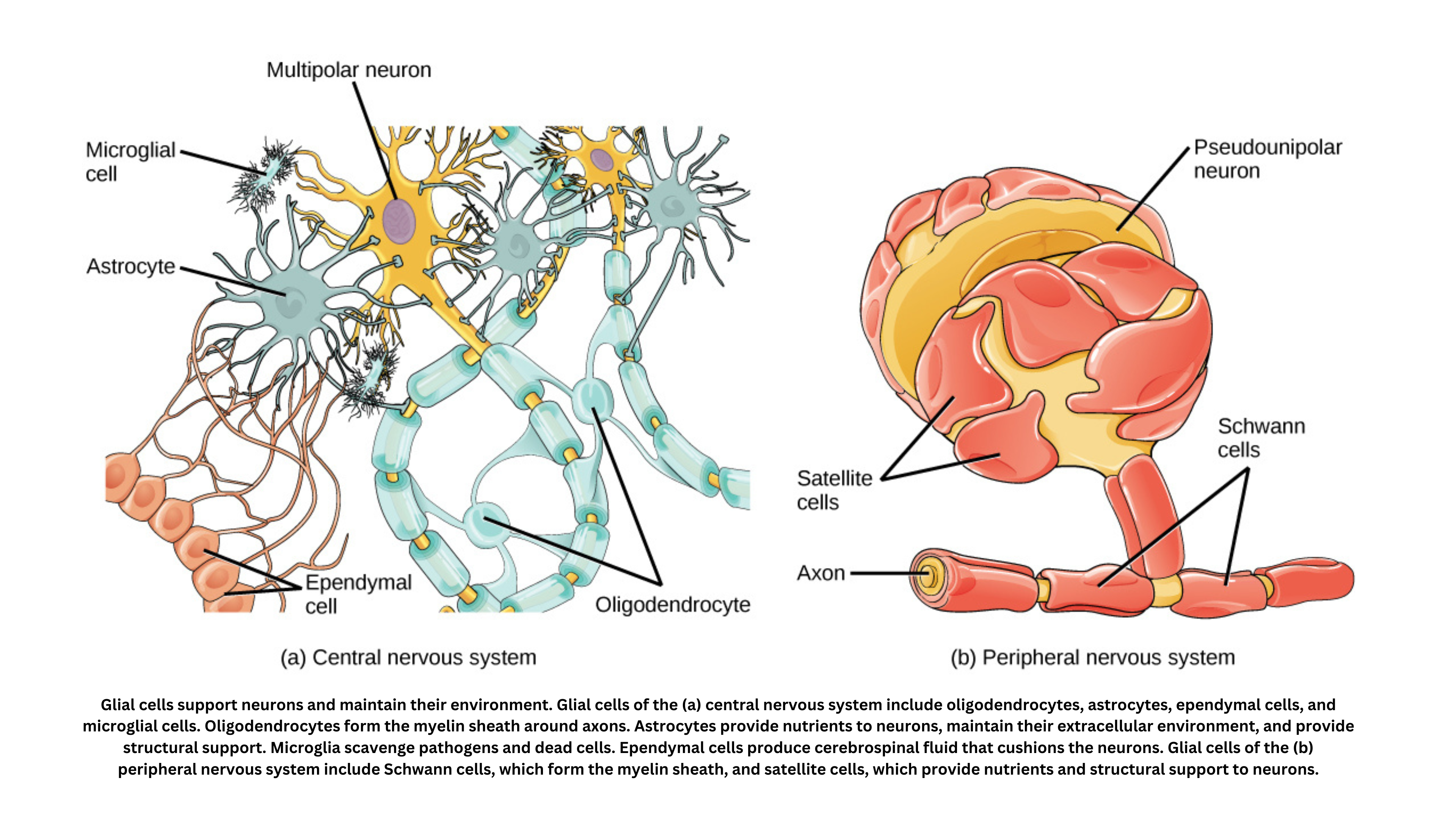
Glial cells - also called neuroglia - are non-neuronal cells with essential support roles in both the central and peripheral nervous systems. They were once described as the “glue” that holds neurons together (which is where the term “neuroglia” comes from), but they’re now understood to be active, indispensable contributors to nervous system health and function.
Key glial types and their functions
Astrocytes: Star-shaped and abundant, they balance ions, maintain the blood-brain barrier, support neurons, modulate synapses, and aid in repair. Oligodendrocytes & Schwann Cells: In the CNS, oligodendrocytes myelinate axons for fast impulse transmission; Schwann cells perform this role in the PNS. Microglia: Acting as the nervous system’s immune cells, they patrol for pathogens and damage, coordinating inflammatory responses. Ependymal Cells: These line the brain’s ventricles and spinal cord canal, producing and regulating cerebrospinal fluid to cushion and clear waste.
Glial cells aren’t just structural support for the nervous system - they also:
- Maintain homeostasis: By regulating the chemical composition around neurons, glia help preserve the environment needed for reliable neuronal signaling.
- Modulate synaptic function: Astrocytes, in particular, can influence how neurons communicate by controlling neurotransmitter levels at synapses.
- Aid in repair and regeneration: After injury, certain glial cells help clear debris and create conditions that support neuronal recovery, although this response can also contribute to scarring or other complications.
- Contribute to neurodevelopment: During development, glial cells guide the formation of neural circuits and support the migration of neurons to their proper locations.
Electrochemistry
Concentration cells and electron flow
- In a concentration cell, a difference in ion concentration drives the redox reaction. The electrodes are identical, so their standard potentials cancel out. However, the half-cell with the higher ion concentration has a greater chemical potential. Electrons move from the electrode in the less concentrated solution (lower chemical potential) to the electrode in the more concentrated solution (higher chemical potential). This electron flow pushes the system toward equilibrium by reducing the concentration difference between the two half-cells.
The Nernst equation
- The Nernst equation relates the cell’s electrode potential to ion concentrations and temperature:
= - ln Q
Where:
- is the electrode potential,
- is the standard electrode potential,
- is the universal gas constant,
- is the absolute temperature in Kelvin,
- is the number of electrons transferred,
- is Faraday’s constant, and
- is the reaction quotient representing the ratio of ion concentrations.
This form shows that even a small difference in ion concentration (Q) can generate a measurable voltage, because the potential depends logarithmically on the concentration ratio.
Biosignalling (BC)
Biosignalling describes how cells detect, process, and respond to external signals. Many signalling pathways begin with specialized proteins embedded in the cell membrane, including the following.
Gated ion channels: These channels control the flow of ions into and out of cells, which can change electrical activity and trigger cellular responses. Common types include:
- Voltage-gated channels: Open or close in response to changes in the electrical potential across the cell membrane. This mechanism is essential in neurons and muscle cells, where rapid voltage changes trigger signal transmission.
- Ligand-gated channels: Respond to the binding of specific molecules (ligands), such as neurotransmitters. Ligand binding causes a conformational change that opens the channel, allowing ions to pass and altering the cell’s electrical state.
Receptor enzymes:
- These receptors combine signal detection with enzymatic activity. When a ligand binds, it activates an intrinsic catalytic function that can modify other proteins (often through phosphorylation). This can trigger a cascade of intracellular signals, producing a coordinated response to the original stimulus.
G Protein-Coupled Receptors (GPCRs):
- One of the largest families of cell-surface receptors, GPCRs mediate many physiological processes. When a ligand binds, the receptor changes shape and activates an associated G protein. The activated G protein then interacts with intracellular effectors, starting a chain of events that can alter cellular metabolism, gene expression, or other functions.