


Achievable logoAchievable logo
CCMA
Sign in
Sign up
Purchase
Textbook
Practice exams
Support
How it works
Exam catalog
Mountain with a flag at the peak
Textbook
1. Medical assistant
2. Electronic records
3. Medical terminology and anatomy
4. The fundamentals of infection control
5. Introduction to vital signs
6. The patient interview and history
7. The physical examination
8. Appointment scheduling
9. Insurance billing
10. Diagnostic coding and the ICD-10-CM System
11. Procedural coding
12. Medical billing and reimbursement essentials
13. Assisting with medical specialties
14. Assisting with the musculoskeletal system
15. Assisting with the cardiovascular system
16. Assisting with the respiratory system
17. Assisting with the nervous system
18. Anatomy and physiology of the urinary system
19. Assisting in obstetrics and gynecology
20. Assisting in endocrinology
21. Assisting in ophthalmology & otolaryngology
21.1 Anatomy and physiology of the eye
21.2 Major ocular disorders: cataracts, glaucoma, and retinal disease
21.3 Refractive errors and common eye infections
21.4 Diseases and disorders of the ear
21.5 The medical assistant's role in eye examinations
21.6 The medical assistant's role in ear examinations and treatments
22. Assisting in gastroenterology
23. Assisting in the immune & lymphatic systems
24. Assisting in pediatrics: the developmental stages and care
25. The medical assistant’s role in caring for the older patient
26. The role of the medical assistant in physical therapy examination and assessment
27. Preparing for minor surgery: room, solutions, and supplies
28. Introduction to the clinical laboratory
29. Urinalysis
30. Blood collection
31. Analysis of blood
32. Electrocardiography and heart structure
33. The principles of pharmacology
34. Essential calculations and measurement systems
35. Solid, liquid, & solutions medication doses
36. Administering medications
37. Metabolism and core nutrient roles
38. Medical emergencies in the healthcare setting
Achievable logoAchievable logo
21.1 Anatomy and physiology of the eye
Achievable CCMA
21. Assisting in ophthalmology & otolaryngology
Our CCMA course is currently in development and is a work-in-progress.

Anatomy and physiology of the eye

8 min read
Font
Discuss
Share
Feedback

A medical assistant is responsible for performing a wide variety of procedures in an ophthalmologic or otorhinolaryngologic practice. First, the medical assistant must be familiar with the normal anatomy and physiology of the eyes, ears, nose, and throat. With an understanding of how these specialty sensory organs function, the medical assistant can master the skills needed to become a valuable asset to providers who specialize in the treatment of eye and ear disorders.

Ophthalmology is the science of the eye and its disorders and diseases. A physician who specializes in the diagnosis and treatment of disorders and diseases of the eye is an ophthalmologist. An ophthalmologist is a licensed medical physician who can diagnose eye disorders, prescribe medication, conduct eye screenings, prescribe glasses or contact lenses, and perform optic surgery. An optometrist is not a medical doctor, but this individual is licensed and has earned a degree as a doctor of optometry (OD). An optometrist can perform eye examinations, diagnose vision problems and eye diseases, prescribe ophthalmic medications, and treat visual defects through corrective lenses and eye exercises. Opticians are trained to fill prescriptions written by ophthalmologists and optometrists for corrective lenses by grinding the lenses and dispensing eyewear.

Otorhinolaryngology is the medical specialty that deals with the ear, nose, and throat. It frequently is referred to as otolaryngology or even as a single specialty of otology or laryngology. Usually, the specialty otorhinolaryngology is referred to simply as ear, nose, and throat (ENT). An otolaryngologist is a licensed medical physician who is trained in the medical and surgical management and treatment of patients with diseases and disorders of the ear, nose, throat, and related structures of the head and neck.

Anatomy and physiology of the eye

The eye can be divided into the ocular adnexa, the structures that surround and support the function of the eyeball, and the structures of the eye itself. The anatomy of the ocular adnexa and the eyeball will be discussed in the following sections.

Ocular adnexa

Each of our eyes is encased in a protective, bony socket called the orbit. Our binocular vision sends two slightly different images to the brain that produce depth of vision. Within the orbit, a cushion of fatty tissue protects the eyeball. Only about one-sixth of the eye is outside the orbit. The eyelid helps protect the eye from trauma. The eyebrows help keep irritants out of the eyes. The eyelashes line the margins of the eyelids and help trap foreign particles (Figure 31.1).

The conjunctiva is a thin mucous membrane that lines the eyelid. It covers the outside of the eyeball except for the most central portion. The cornea covers the center of the eye. The mucus secreted from the conjunctiva helps keep the eye moist. The eye blinks about every 2 to 3 seconds. The meibomian glands are located along the edge of the eyelids near the lashes. These glands secrete an oil that is part of the eye’s tears. This oil helps prevent tears from drying up too quickly, creating dry eyes. Blinking causes the lacrimal gland, located in the superior outer portion of the upper eyelid to secrete tears. Tears move across the eyes, cleansing and moistening the surface of the eye. Tears drain into the lacrimal canals in the medial corner of the eye. The tears then drain into the nasal cavity through the nasolacrimal duct. This is why, when a person cries, the excess tears ultimately empty into the nose, producing a watery nasal discharge.

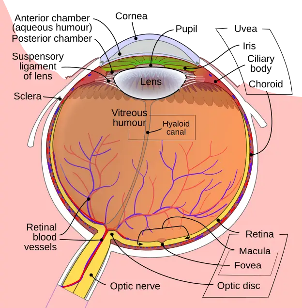
Anatomical structures of the eye labeled
The anatomy of the eye
Wikimedia Commons
/
CC BY-SA 3.0

Eyeball

The eyeball consists of three layers. The outermost layer is made up of the white, opaque sclera (SKLAYR a) and the transparent cornea. The sclera is a tough, fibrous lining that protects the entire eyeball lying within the orbit. It is also known as the white of the eye. The transparent cornea covers the exposed one-sixth of the eyeball. The cornea acts like a window that allows light to enter the eye. The cornea also refracts, or bends the direction of light rays, after they enter the eye.

The middle layer is made up of the choroid, the iris, and the ciliary body. The iris is the colored portion of the eye. It is doughnut shaped, with an opening in the center called the pupil. The iris contains muscles that regulate the size of the pupil depending on the intensity of light. It becomes smaller in bright light and opens wider in dim light. The ciliary body contains both the ciliary muscle, which regulates the shape of the lens and the ciliary processes, which secrete aqueous humor. The choroid is the posterior portion of the middle layer of the eye. It is the eye’s vascular layer. It contains many blood vessels that supply nutrients to the outer layers of the retina. The choroid also has a brown pigment that absorbs excess light rays that could interfere with vision.

The inner layer of the eye includes the retina in the posterior portion and the lens in the anterior portion. The delicate tissue of the retina is composed of light-sensitive neurons called rods and cones. Rods are highly sensitive to light and can function in dim light. Cones function in bright light and detect color. Rods and cones convert light into nerve impulses. These impulses travel through the optic nerve to the brain, where they are converted into images.

The lens is a transparent, biconvex body that helps focus light after it passes through the cornea. The lens and the ciliary body divide the eye into two cavities. The posterior cavity, which is between the lens and the retina, contains the transparent, gel-like vitreous humor. The vitreous humor maintains the shape of the posterior eyeball. The anterior cavity, between the cornea and the lens, is filled with aqueous humor, which is continuously produced by the ciliary processes. Aqueous humor helps maintain normal pressure within the eye and provides nutrients to the lens and the cornea.

Physiology of the eye

Vision requires light and depends on the proper functioning of all parts of the eye. A visual impulse begins with the passage of light through the cornea, where the light is refracted; it then passes through the aqueous humor and the pupil into the lens. The ciliary muscle adjusts the curvature of the lens to again refract the light rays so that they pass into the retina, triggering the photoreceptor cells of the rods and cones. At this point, the light energy is converted into an electrical impulse, which is sent through the optic nerve to the visual cortex of the occipital lobe of the brain; there, the light impulse is interpreted, and a picture is created.

Functions of the major parts of the eye

Structure Function
Sclera External protection
Cornea Light refraction
Choroid Blood supply
Iris Light absorption and regulation of pupil width
Ciliary body Secretion of vitreous fluid; changes the shape of the lens
Lens Light refraction
Retinal layer Light receptor that transforms optic signals into nerve impulses
Rods Distinguish light from dark and perceive shape and movement
Cones Color vision
Central fovea Area of sharpest vision
Macula lutea Center of the retina; contains the fovea centralis, the area of most highly acute vision
External ocular muscles Move the eyeball
Optic nerve One of a pair of nerves that transmit visual stimuli to (cranial nerve II) the brain
Lacrimal glands Produce tears
Eyelid P+A1:protects eye

Ophthalmology and Otorhinolaryngology Overview

  • Ophthalmology: study/treatment of eye disorders (ophthalmologist = MD, optometrist = OD, optician = eyewear specialist)
  • Otorhinolaryngology (ENT): medical specialty for ear, nose, throat disorders (otolaryngologist = MD)

Ocular Adnexa

  • Orbit: bony socket, fatty tissue cushions eyeball
  • Eyelids, eyebrows, eyelashes: protect eye, trap irritants
  • Conjunctiva: mucous membrane, keeps eye moist
  • Meibomian glands: secrete oil for tears, prevent dry eyes
  • Lacrimal glands/canals: produce/drain tears, nasolacrimal duct connects to nasal cavity

Eyeball Anatomy

  • Three layers:
    • Outer: sclera (white, protection), cornea (transparent, refracts light)
    • Middle: choroid (vascular, nutrients), iris (colored, regulates pupil), ciliary body (muscle, lens shape, aqueous humor)
    • Inner: retina (rods/cones, light-sensitive), lens (focuses light)
  • Cavities:
    • Posterior: vitreous humor (shape)
    • Anterior: aqueous humor (pressure, nutrients)

Physiology of the Eye

  • Light path: cornea → aqueous humor → pupil → lens → retina
  • Ciliary muscle: adjusts lens for focus (accommodation)
  • Retina: rods (dim light, shape/movement), cones (color, bright light)
  • Optic nerve: transmits impulses to brain for image processing

Major Eye Structures and Functions

  • Sclera: external protection
  • Cornea/lens: light refraction
  • Choroid: blood supply
  • Iris: regulates pupil, absorbs light
  • Ciliary body: lens shape, aqueous humor secretion
  • Retina: transforms light to nerve impulses
  • Rods/cones: light/dark, color vision
  • Central fovea/macula lutea: sharpest vision
  • External ocular muscles: eye movement
  • Optic nerve: visual signal transmission
  • Lacrimal glands: tear production
  • Eyelid: protection

Ear Anatomy

  • Outer ear: auricle (pinna), external auditory canal (collects sound, cerumen/hair for protection)
  • Middle ear: tympanic membrane (eardrum), ossicles (malleus, incus, stapes), eustachian tube (pressure equalization)
  • Inner ear: cochlea (hearing, organ of Corti), semicircular canals/vestibule (equilibrium, balance)

Physiology of the Ear

  • Sound path: auricle → auditory canal → tympanic membrane → ossicles → oval window → cochlea
  • Organ of Corti: hair cells bend, initiate auditory impulse
  • Auditory impulse: cochlear nerve → cranial nerve VIII → medulla → thalamus → auditory cortex (temporal lobe)
  • Semicircular canals/vestibule: detect head position, maintain balance (dynamic/static equilibrium)

Life Span Changes: Children

  • Vision: develops post-birth, depth perception by age 2, screenings start age 3-5
  • Common eye disorder: conjunctivitis (pinkeye), highly contagious
  • Eustachian tube: shorter/more horizontal in children, increases ear infection risk
  • Hearing: present at birth, UNHS detects early hearing loss

Life Span Changes: Older Adults

  • Eye: presbyopia (farsightedness with age), ARMD and cataracts (common blindness causes), diabetic retinopathy
  • Ear: presbycusis (age-related hearing loss), hearing aids may help quality of life

Sign up for free to take 17 quiz questions on this topic

Previous
Next  | 21.2 Major ocular disorders: cataracts, glaucoma, and retinal disease
All rights reserved ©2016 - 2026 Achievable, Inc.

Anatomy and physiology of the eye

A medical assistant is responsible for performing a wide variety of procedures in an ophthalmologic or otorhinolaryngologic practice. First, the medical assistant must be familiar with the normal anatomy and physiology of the eyes, ears, nose, and throat. With an understanding of how these specialty sensory organs function, the medical assistant can master the skills needed to become a valuable asset to providers who specialize in the treatment of eye and ear disorders.

Ophthalmology is the science of the eye and its disorders and diseases. A physician who specializes in the diagnosis and treatment of disorders and diseases of the eye is an ophthalmologist. An ophthalmologist is a licensed medical physician who can diagnose eye disorders, prescribe medication, conduct eye screenings, prescribe glasses or contact lenses, and perform optic surgery. An optometrist is not a medical doctor, but this individual is licensed and has earned a degree as a doctor of optometry (OD). An optometrist can perform eye examinations, diagnose vision problems and eye diseases, prescribe ophthalmic medications, and treat visual defects through corrective lenses and eye exercises. Opticians are trained to fill prescriptions written by ophthalmologists and optometrists for corrective lenses by grinding the lenses and dispensing eyewear.

Otorhinolaryngology is the medical specialty that deals with the ear, nose, and throat. It frequently is referred to as otolaryngology or even as a single specialty of otology or laryngology. Usually, the specialty otorhinolaryngology is referred to simply as ear, nose, and throat (ENT). An otolaryngologist is a licensed medical physician who is trained in the medical and surgical management and treatment of patients with diseases and disorders of the ear, nose, throat, and related structures of the head and neck.

Anatomy and physiology of the eye

The eye can be divided into the ocular adnexa, the structures that surround and support the function of the eyeball, and the structures of the eye itself. The anatomy of the ocular adnexa and the eyeball will be discussed in the following sections.

Ocular adnexa

Each of our eyes is encased in a protective, bony socket called the orbit. Our binocular vision sends two slightly different images to the brain that produce depth of vision. Within the orbit, a cushion of fatty tissue protects the eyeball. Only about one-sixth of the eye is outside the orbit. The eyelid helps protect the eye from trauma. The eyebrows help keep irritants out of the eyes. The eyelashes line the margins of the eyelids and help trap foreign particles (Figure 31.1).

The conjunctiva is a thin mucous membrane that lines the eyelid. It covers the outside of the eyeball except for the most central portion. The cornea covers the center of the eye. The mucus secreted from the conjunctiva helps keep the eye moist. The eye blinks about every 2 to 3 seconds. The meibomian glands are located along the edge of the eyelids near the lashes. These glands secrete an oil that is part of the eye’s tears. This oil helps prevent tears from drying up too quickly, creating dry eyes. Blinking causes the lacrimal gland, located in the superior outer portion of the upper eyelid to secrete tears. Tears move across the eyes, cleansing and moistening the surface of the eye. Tears drain into the lacrimal canals in the medial corner of the eye. The tears then drain into the nasal cavity through the nasolacrimal duct. This is why, when a person cries, the excess tears ultimately empty into the nose, producing a watery nasal discharge.

Eyeball

The eyeball consists of three layers. The outermost layer is made up of the white, opaque sclera (SKLAYR a) and the transparent cornea. The sclera is a tough, fibrous lining that protects the entire eyeball lying within the orbit. It is also known as the white of the eye. The transparent cornea covers the exposed one-sixth of the eyeball. The cornea acts like a window that allows light to enter the eye. The cornea also refracts, or bends the direction of light rays, after they enter the eye.

The middle layer is made up of the choroid, the iris, and the ciliary body. The iris is the colored portion of the eye. It is doughnut shaped, with an opening in the center called the pupil. The iris contains muscles that regulate the size of the pupil depending on the intensity of light. It becomes smaller in bright light and opens wider in dim light. The ciliary body contains both the ciliary muscle, which regulates the shape of the lens and the ciliary processes, which secrete aqueous humor. The choroid is the posterior portion of the middle layer of the eye. It is the eye’s vascular layer. It contains many blood vessels that supply nutrients to the outer layers of the retina. The choroid also has a brown pigment that absorbs excess light rays that could interfere with vision.

The inner layer of the eye includes the retina in the posterior portion and the lens in the anterior portion. The delicate tissue of the retina is composed of light-sensitive neurons called rods and cones. Rods are highly sensitive to light and can function in dim light. Cones function in bright light and detect color. Rods and cones convert light into nerve impulses. These impulses travel through the optic nerve to the brain, where they are converted into images.

The lens is a transparent, biconvex body that helps focus light after it passes through the cornea. The lens and the ciliary body divide the eye into two cavities. The posterior cavity, which is between the lens and the retina, contains the transparent, gel-like vitreous humor. The vitreous humor maintains the shape of the posterior eyeball. The anterior cavity, between the cornea and the lens, is filled with aqueous humor, which is continuously produced by the ciliary processes. Aqueous humor helps maintain normal pressure within the eye and provides nutrients to the lens and the cornea.

Physiology of the eye

Vision requires light and depends on the proper functioning of all parts of the eye. A visual impulse begins with the passage of light through the cornea, where the light is refracted; it then passes through the aqueous humor and the pupil into the lens. The ciliary muscle adjusts the curvature of the lens to again refract the light rays so that they pass into the retina, triggering the photoreceptor cells of the rods and cones. At this point, the light energy is converted into an electrical impulse, which is sent through the optic nerve to the visual cortex of the occipital lobe of the brain; there, the light impulse is interpreted, and a picture is created.

Functions of the major parts of the eye

Structure Function
Sclera External protection
Cornea Light refraction
Choroid Blood supply
Iris Light absorption and regulation of pupil width
Ciliary body Secretion of vitreous fluid; changes the shape of the lens
Lens Light refraction
Retinal layer Light receptor that transforms optic signals into nerve impulses
Rods Distinguish light from dark and perceive shape and movement
Cones Color vision
Central fovea Area of sharpest vision
Macula lutea Center of the retina; contains the fovea centralis, the area of most highly acute vision
External ocular muscles Move the eyeball
Optic nerve One of a pair of nerves that transmit visual stimuli to (cranial nerve II) the brain
Lacrimal glands Produce tears
Eyelid P+A1:protects eye
Key points

Ophthalmology and Otorhinolaryngology Overview

  • Ophthalmology: study/treatment of eye disorders (ophthalmologist = MD, optometrist = OD, optician = eyewear specialist)
  • Otorhinolaryngology (ENT): medical specialty for ear, nose, throat disorders (otolaryngologist = MD)

Ocular Adnexa

  • Orbit: bony socket, fatty tissue cushions eyeball
  • Eyelids, eyebrows, eyelashes: protect eye, trap irritants
  • Conjunctiva: mucous membrane, keeps eye moist
  • Meibomian glands: secrete oil for tears, prevent dry eyes
  • Lacrimal glands/canals: produce/drain tears, nasolacrimal duct connects to nasal cavity

Eyeball Anatomy

  • Three layers:
    • Outer: sclera (white, protection), cornea (transparent, refracts light)
    • Middle: choroid (vascular, nutrients), iris (colored, regulates pupil), ciliary body (muscle, lens shape, aqueous humor)
    • Inner: retina (rods/cones, light-sensitive), lens (focuses light)
  • Cavities:
    • Posterior: vitreous humor (shape)
    • Anterior: aqueous humor (pressure, nutrients)

Physiology of the Eye

  • Light path: cornea → aqueous humor → pupil → lens → retina
  • Ciliary muscle: adjusts lens for focus (accommodation)
  • Retina: rods (dim light, shape/movement), cones (color, bright light)
  • Optic nerve: transmits impulses to brain for image processing

Major Eye Structures and Functions

  • Sclera: external protection
  • Cornea/lens: light refraction
  • Choroid: blood supply
  • Iris: regulates pupil, absorbs light
  • Ciliary body: lens shape, aqueous humor secretion
  • Retina: transforms light to nerve impulses
  • Rods/cones: light/dark, color vision
  • Central fovea/macula lutea: sharpest vision
  • External ocular muscles: eye movement
  • Optic nerve: visual signal transmission
  • Lacrimal glands: tear production
  • Eyelid: protection

Ear Anatomy

  • Outer ear: auricle (pinna), external auditory canal (collects sound, cerumen/hair for protection)
  • Middle ear: tympanic membrane (eardrum), ossicles (malleus, incus, stapes), eustachian tube (pressure equalization)
  • Inner ear: cochlea (hearing, organ of Corti), semicircular canals/vestibule (equilibrium, balance)

Physiology of the Ear

  • Sound path: auricle → auditory canal → tympanic membrane → ossicles → oval window → cochlea
  • Organ of Corti: hair cells bend, initiate auditory impulse
  • Auditory impulse: cochlear nerve → cranial nerve VIII → medulla → thalamus → auditory cortex (temporal lobe)
  • Semicircular canals/vestibule: detect head position, maintain balance (dynamic/static equilibrium)

Life Span Changes: Children

  • Vision: develops post-birth, depth perception by age 2, screenings start age 3-5
  • Common eye disorder: conjunctivitis (pinkeye), highly contagious
  • Eustachian tube: shorter/more horizontal in children, increases ear infection risk
  • Hearing: present at birth, UNHS detects early hearing loss

Life Span Changes: Older Adults

  • Eye: presbyopia (farsightedness with age), ARMD and cataracts (common blindness causes), diabetic retinopathy
  • Ear: presbycusis (age-related hearing loss), hearing aids may help quality of life

More from Assisting in ophthalmology & otolaryngology

  • Major ocular disorders: cataracts, glaucoma, and retinal disease
  • Refractive errors and common eye infections
  • Diseases and disorders of the ear
  • The medical assistant's role in eye examinations
  • The medical assistant's role in ear examinations and treatments