



Anatomy and physiology of the ear and lifespan changes
Anatomy and physiology of the ear
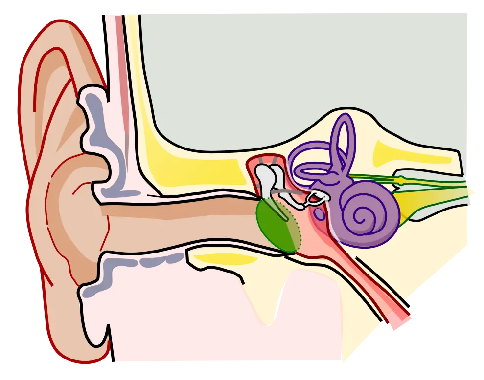
The visible portion of the ear is only a small part of the actual organ of hearing. Most of the sensory structure lies hidden in the temporal bone of the skull. The ear is divided into the outer, middle, and inner ear. The anatomy of the ear is discussed in the following sections.
Outer (external) ear
The outer ear consists of the auricle, or pinna. This is the fleshy part of the ear that can be seen on the side of the head. The next structure is the external auditory canal, the tube that extends from the auricle into the tympanic membrane (eardrum).
The auricle collects sound waves and sends them into the auditory canal. The skin that lines the auditory canal contains numerous hair follicles and many nerve endings. Earwax, or cerumen, is secreted by modified sweat glands within the external auditory canal. Both the hair and the waxy cerumen help prevent foreign objects from reaching the eardrum. The canal has a slightly curved shape and is approximately 1 inch (2.5 cm) long.
Middle ear
The middle ear is an air-filled cavity that contains three tiny bones called the ossicles: malleus, incus, and stapes. Tiny ligaments link these three tiny bones to form a bridge from the tympanic membrane to the inner ear. The ossicles transmit sound to the inner ear through the movement of the stapes. Within the middle ear is an opening for the eustachian tube. This is a connection between the ears and the throat. This connection helps equalize pressure within the middle ear. Without equalized pressure in the ear, hearing would not be possible.
The tympanic membrane is a thin, disc-shaped tissue that seals off the outer ear from the middle ear. Sound waves conducted through the external auditory canal hit the tympanic membrane and cause it to vibrate. The vibrations are picked up by the three ossicles, which mechanically transmit and amplify the movement across the middle ear to the oval window. The oval window is a membrane that connects the middle ear and the inner ear. At the oval window, this mechanical energy is transferred into the fluid of the inner ear, and the resulting fluid motion excites the receptors, generating sensorineural impulses.
Inner ear
Once sound is conducted to the oval window, it is transmitted to a structure called the labyrinth, or the inner ear. The inner ear is divided into the cochlea and the semicircular canals, which are joined by the vestibule. The semicircular canals and vestibule function to maintain equilibrium. The cochlea is responsible for the sense of hearing.
The organ of Corti, which contains the receptors for sound, is located within the cochlea. It is made up of hairlike sensory cells surrounded by sensory nerve fibers that form the cochlear branch of the eighth cranial nerve. Sound impulses cause the hairs to bend and rub against the nerve fibers, which initiate stimuli to travel through the cochlear nerve into the brain for sound interpretation.
The semicircular canals are responsible for evaluating rotational movement of the head (dynamic equilibrium). The three canals are positioned at right angles to one another on different planes. When the head turns rapidly, these fluid-filled canals must rapidly adjust and send the information to the central nervous system (CNS). The CNS then interprets the information and initiates the desired response to maintain balance. The semicircular canals detect dynamic equilibrium. Within the vestibule, two saclike structures function to establish the body’s static equilibrium by sensing the position of the head in relation to the pull of gravity. With repetitive or excessive stimulation to the equilibrium receptors, some people become nauseated and may vomit. This condition is known as motion sensitivity or motion sickness.
Physiology of the ear
Hearing starts with the sound waves reaching the tympanic membrane. Those sound waves cause the tympanic membrane to vibrate, which causes the ossicles to transmit the waves to the oval window. It is at the inner ear that the sensorineural impulses reach the cochlea.
This activates the cochlear nerve, which carries the resulting auditory impulse onto the eighth cranial nerve. The eighth cranial nerve transmits the auditory impulse to the medulla oblongata. The impulses then travel to the thalamus and on to the auditory cortex of the temporal lobe of the brain. The brain then interprets the auditory impulse into audible sound and speech patterns.
Life span changes
As we age, changes occur in the functioning of the eyes and ears, and diseases and disorders of the eyes and ears may develop. The following sections discuss some of those changes affecting the ear.
Changes across the lifespan
A child’s eustachian tube is shorter, wider, and more horizontal than an adult’s, letting pathogens from the throat reach the middle ear more easily - this is why children have a higher rate of ear infections (otitis media) than adults. Hearing sensitivity also changes with age: over time, the hair cells of the organ of Corti diminish in number, and the resulting decline in high-frequency hearing is known as presbycusis.