



Achievable logoAchievable logo
CCMA
Sign in
Sign up
Purchase
Textbook
Practice exams
Support
How it works
Resources
Exam catalog
Mountain with a flag at the peak
Textbook
Introduction
1. Medical assistant
2. Electronic records
3. Medical terminology and anatomy
4. The fundamentals of infection control
5. Introduction to vital signs
6. The patient interview and history
7. The physical examination
8. Appointment scheduling
9. Insurance billing
10. Diagnostic coding and the ICD-10-CM System
11. Procedural coding
12. Medical billing and reimbursement essentials
13. Assisting with medical specialties
14. Assisting with the musculoskeletal system
15. Assisting with the cardiovascular system
16. Assisting with the respiratory system
17. Assisting with the nervous system
18. Anatomy and physiology of the urinary system
19. Assisting in obstetrics and gynecology
20. Assisting in endocrinology
21. Assisting in ophthalmology & otolaryngology
21.1 Anatomy and physiology of the eye
21.2 Anatomy and physiology of the ear and lifespan changes
21.3 Major ocular disorders: cataracts, glaucoma, and retinal disease
21.4 Refractive errors and common eye infections
21.5 Diseases and disorders of the ear
21.6 The medical assistant's role in eye examinations
21.7 The medical assistant's role in vision screening tests
21.8 The medical assistant's role in ear examinations and treatments
21.9 Patient coaching, legal issues, and professional behaviors in eye and ear care
22. Assisting in gastroenterology
23. Assisting in the immune & lymphatic systems
24. Assisting in pediatrics: the developmental stages and care
25. The medical assistant’s role in caring for the older patient
26. The role of the medical assistant in physical therapy examination and assessment
27. Preparing for minor surgery: room, solutions, and supplies
28. Introduction to the clinical laboratory
29. Urinalysis
30. Blood collection
31. Analysis of blood
32. Electrocardiography and heart structure
33. The principles of pharmacology
34. Essential calculations and measurement systems
35. Solid, liquid, & solutions medication doses
36. Administering medications
37. Metabolism and core nutrient roles
38. Medical emergencies in the healthcare setting
Achievable logoAchievable logo
21.7 The medical assistant's role in vision screening tests
Achievable CCMA
21. Assisting in ophthalmology & otolaryngology

The medical assistant's role in vision screening tests

5 min read
Font
Discuss
Share
Feedback

Interpreting snellen results

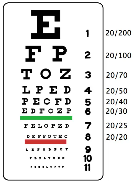
  • The patient always stands 20 feet from the chart.
  • Each result is a record of how well the patient can see compared with normal vision.
  • Example: A patient with a 20/40 reading can see that line correctly standing at 20 feet, but an individual with normal vision can see the same line correctly at 40 feet, so the patient’s vision is not as acute as someone with normal vision.
  • Example: A patient with a 20/15 reading can see that line accurately standing at 20 feet, but a person with normal vision must stand at 15 feet to have the same vision, meaning the patient’s vision is better than someone with normal vision.
Snellen eye chart used to test visual acuity
Snellen charts
Wikimedia Commons
/
Public Domain

Near visual acuity

Near visual acuity can be tested with the near vision acuity chart. This test is given to screen for presbyopia or hyperopia. If the patient wears corrective lenses, they should be worn during the test. The size of the type on the card varies from a 66-point font (newspaper headlines) to a 3-point font (medicine bottle labels). The test should be given in a well-lit room, with the patient holding the card approximately 14 to 16 inches away. As with the Snellen examination, the near visual acuity test is given for each eye, starting with the right eye. The eye not being tested should be covered with an occluder but left open. The patient should be monitored for indications of difficulty, such as squinting or tearing. The patient reads the card, starting at the top until reaching the smallest print that can be read. The medical assistant should document the number at which the patient had no more than two errors for each eye and the two eyes together, whether corrective lenses were worn, and any signs of eyestrain.

Ishihara color vision test

Defects in color vision are classified as congenital or acquired. Congenital defects are caused by an inherited color vision defect and are found most often in males. Acquired defects are caused by eye injury or disease. The Ishihara test is a simple, convenient, and accurate procedure that detects total color blindness, in addition to the red-green blindness prevalent in congenital blindness. The test assesses the perception of primary colors and shades of colors.

The test booklet contains polychromatic plates made of colored dots in numeric patterns. The numbers are one color, and the background dots are a different color. Patients with average visual acuity can read the number within the dot matrix without difficulty. Patients with color vision defects are unable to read the number, or they see a totally different number. A section of plates is included that contains colored line trails through a background of dots. These plates are designed to be used with children and adults who are unable to read numbers. In this situation, the patient uses a finger to follow the dotted trail through the picture.

The test should be administered in a quiet room that is well illuminated by sunlight, not by artificial lighting. If this cannot be done, the best situation possible is created by adjusting lights to resemble the effect of natural daylight. The test uses 14 color plates. The basic test consists of plates 1 through 11. Plates 12 through 14 are used if the patient appears to be having difficulty with red-green differentiation. The medical assistant records the number of plates read correctly. If the score is 10 or higher, the patient is within the average range. If the score is 7 or lower, the patient is suspected of having a color deficiency, and the ophthalmologist performs additional assessment tests using more precise color vision testing equipment.

Ishihara color vision test plates used to detect color blindness
Ishihara color vision test
Wikimedia Commons
/
Public Domain

Sign up for free to take 5 quiz questions on this topic

Previous
Next  | 21.8 The medical assistant's role in ear examinations and treatments
All rights reserved ©2016 - 2026 Achievable, Inc.

The medical assistant's role in vision screening tests

Interpreting snellen results

  • The patient always stands 20 feet from the chart.
  • Each result is a record of how well the patient can see compared with normal vision.
  • Example: A patient with a 20/40 reading can see that line correctly standing at 20 feet, but an individual with normal vision can see the same line correctly at 40 feet, so the patient’s vision is not as acute as someone with normal vision.
  • Example: A patient with a 20/15 reading can see that line accurately standing at 20 feet, but a person with normal vision must stand at 15 feet to have the same vision, meaning the patient’s vision is better than someone with normal vision.

Near visual acuity

Near visual acuity can be tested with the near vision acuity chart. This test is given to screen for presbyopia or hyperopia. If the patient wears corrective lenses, they should be worn during the test. The size of the type on the card varies from a 66-point font (newspaper headlines) to a 3-point font (medicine bottle labels). The test should be given in a well-lit room, with the patient holding the card approximately 14 to 16 inches away. As with the Snellen examination, the near visual acuity test is given for each eye, starting with the right eye. The eye not being tested should be covered with an occluder but left open. The patient should be monitored for indications of difficulty, such as squinting or tearing. The patient reads the card, starting at the top until reaching the smallest print that can be read. The medical assistant should document the number at which the patient had no more than two errors for each eye and the two eyes together, whether corrective lenses were worn, and any signs of eyestrain.

Ishihara color vision test

Defects in color vision are classified as congenital or acquired. Congenital defects are caused by an inherited color vision defect and are found most often in males. Acquired defects are caused by eye injury or disease. The Ishihara test is a simple, convenient, and accurate procedure that detects total color blindness, in addition to the red-green blindness prevalent in congenital blindness. The test assesses the perception of primary colors and shades of colors.

The test booklet contains polychromatic plates made of colored dots in numeric patterns. The numbers are one color, and the background dots are a different color. Patients with average visual acuity can read the number within the dot matrix without difficulty. Patients with color vision defects are unable to read the number, or they see a totally different number. A section of plates is included that contains colored line trails through a background of dots. These plates are designed to be used with children and adults who are unable to read numbers. In this situation, the patient uses a finger to follow the dotted trail through the picture.

The test should be administered in a quiet room that is well illuminated by sunlight, not by artificial lighting. If this cannot be done, the best situation possible is created by adjusting lights to resemble the effect of natural daylight. The test uses 14 color plates. The basic test consists of plates 1 through 11. Plates 12 through 14 are used if the patient appears to be having difficulty with red-green differentiation. The medical assistant records the number of plates read correctly. If the score is 10 or higher, the patient is within the average range. If the score is 7 or lower, the patient is suspected of having a color deficiency, and the ophthalmologist performs additional assessment tests using more precise color vision testing equipment.

More from Assisting in ophthalmology & otolaryngology

  • Anatomy and physiology of the eye
  • Anatomy and physiology of the ear and lifespan changes
  • Major ocular disorders: cataracts, glaucoma, and retinal disease
  • Refractive errors and common eye infections
  • Diseases and disorders of the ear