Mycoplasma pneumoniae
These bacteria don’t have a cell wall. Because of this:
- They can’t be stained with the Gram stain.
- They aren’t killed by cell wall-active antibiotics such as penicillins and cephalosporins.
They’re the only bacteria that contain cholesterol in their cell membrane. Morphologically, they’re pleomorphic and may show “gliding” motility.
It’s the most common cause of atypical pneumonia. It typically presents with fever, headache, sore throat, cough, etc. It has also been associated with the development of SJS (Stevens-Johnson syndrome), GBS, and cardiac arrhythmias.
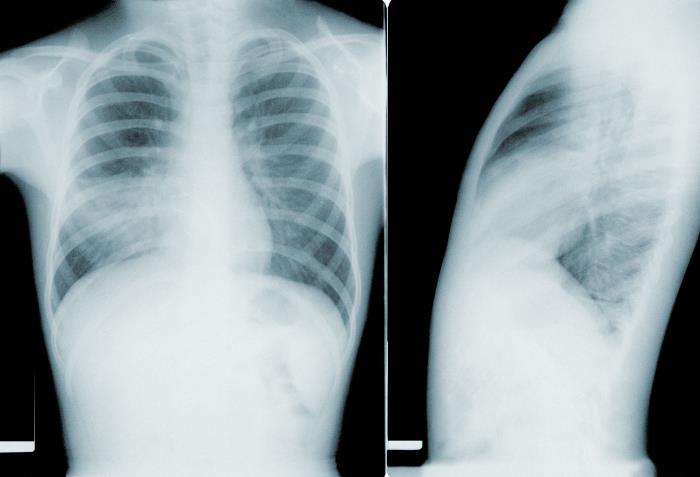
This image depicted two chest x-rays, which revealed pathologic changes in a patient’s lung fields due to a condition known as mycoplasma pneumonia, caused by a Mycoplasma pneumoniae bacterial infection. Note on the anteroposterior (AP) view on the left, the generalized infiltrate permeating both lung fields, and consolidation in the region of the right lower lobe, as well as bilateral hilar adenopathy. In the left lateral view on the right, you can see that the consolidation occupied more of the posterior aspect of the lung fields, almost obliterating a view of the spinal column.
The cold agglutinin test is positive in Mycoplasma infections. This test detects an IgM antibody that agglutinates human “O” blood group antigens at 4°C.
Complement fixation test, indirect haemagglutination test, or enzyme immunoassay are more specific for diagnosis. PCR can be used for DNA detection. Antigen can also be detected by immunofluorescence, CIEP, enzyme immunoassay, or immunoblot tests.
Mycoplasma need sterols in the culture medium to grow. They produce “fried egg” colonies on Mycoplasma agar. These colonies stain blue with Dienes stain, which is poured over the colony.