Translation
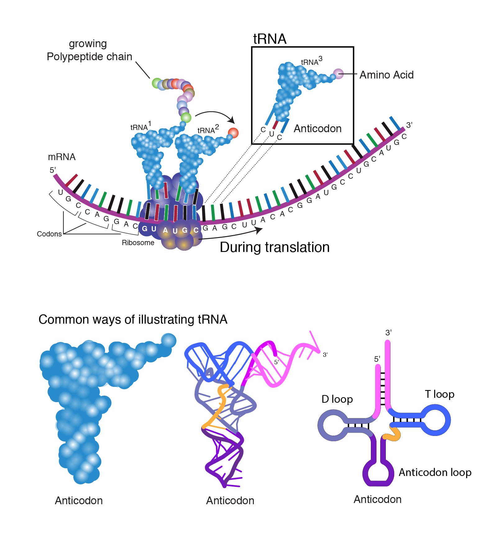
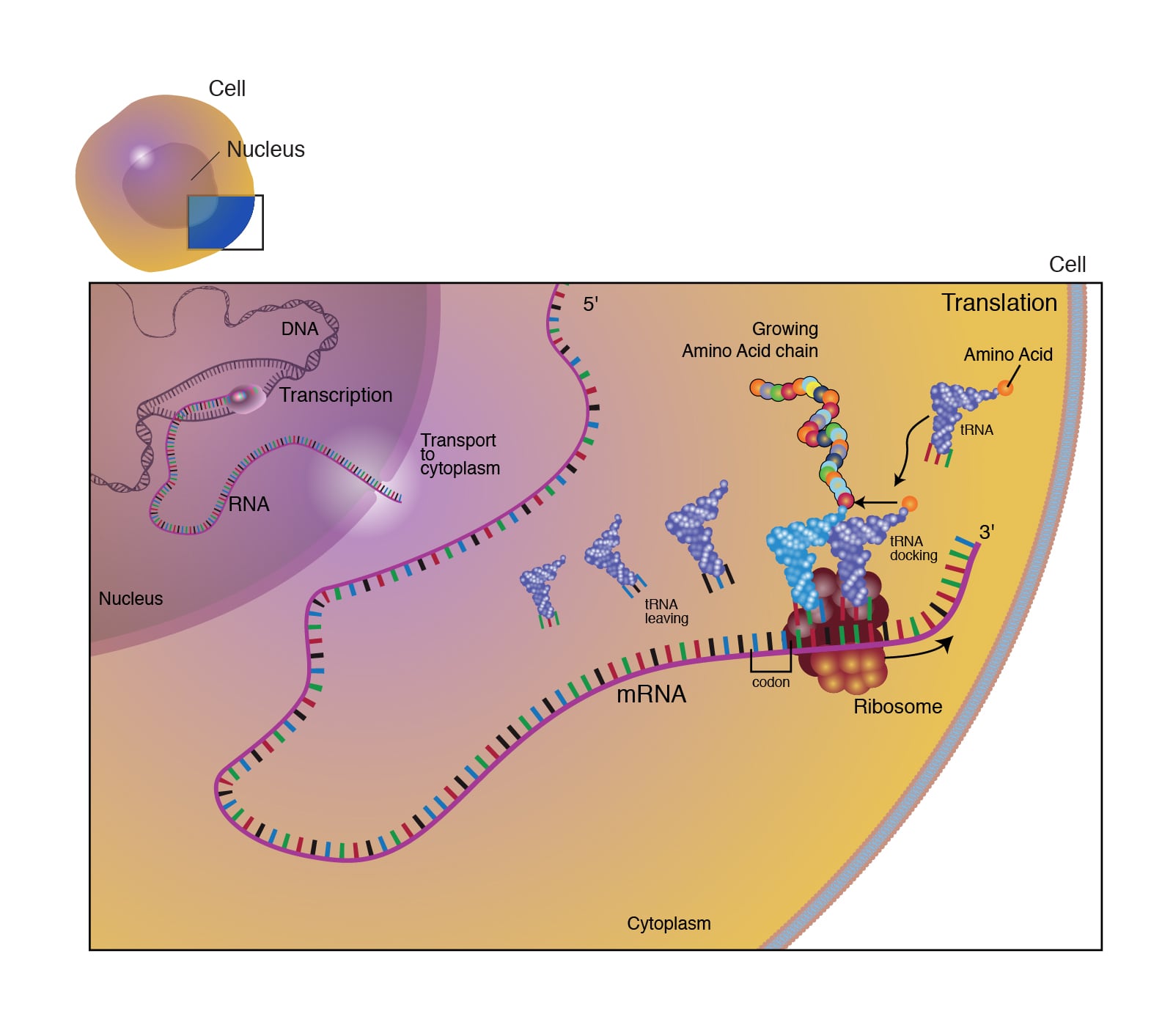
Translation is the process of converting the nucleotide sequence of an mRNA molecule into an amino acid sequence during protein synthesis. The ribosome reads the mRNA in groups of three bases (codons) to assemble a protein.
- The 40S ribosomal subunit binds mRNA and aminoacyl-tRNA and helps locate the AUG start codon on the mRNA.
- The 60S ribosomal subunit has peptidyl transferase activity.
Translation has three stages as follows:
-
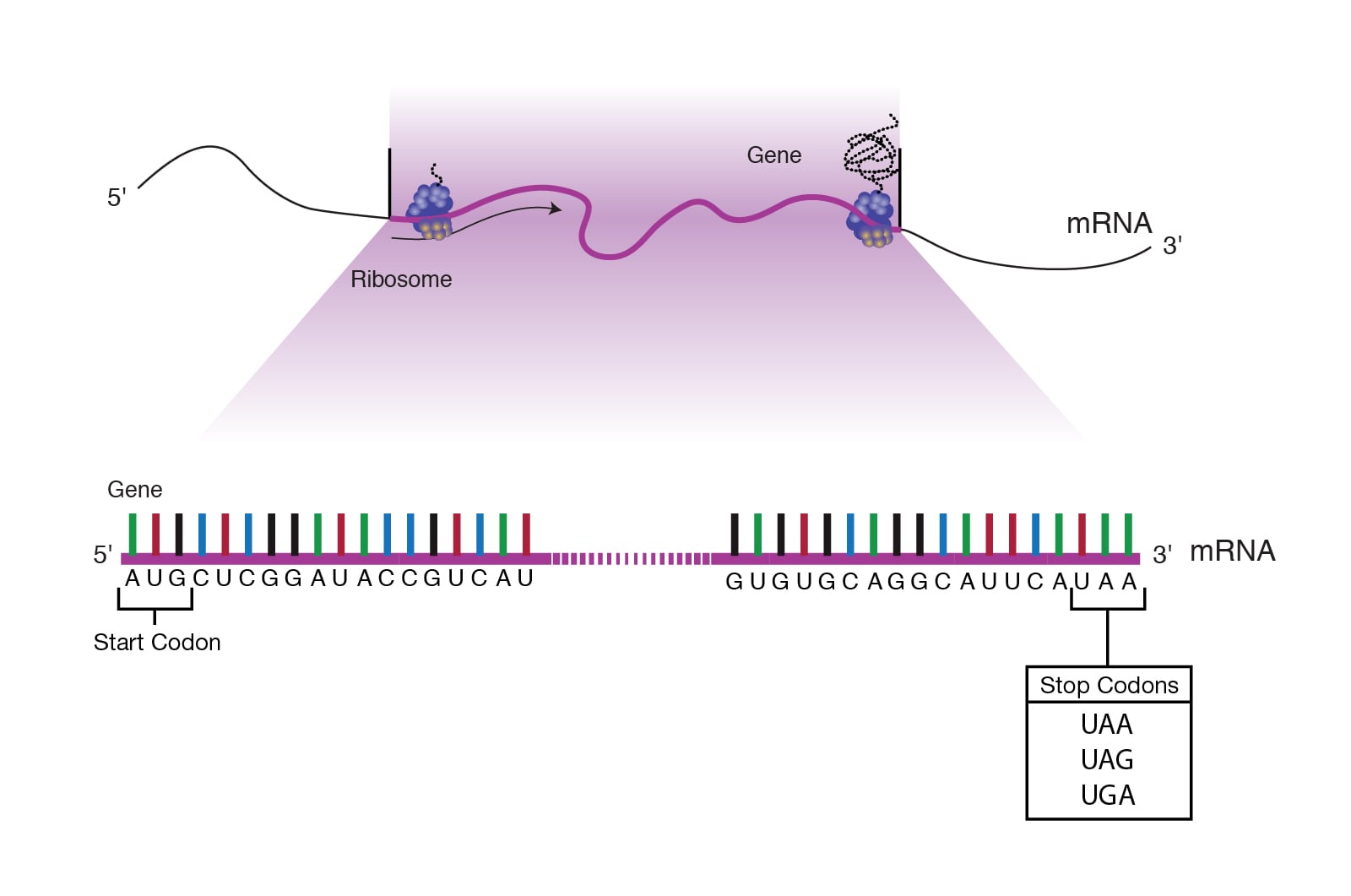
Initiation: Near the 5’ end of mRNA is a region called the untranslated region (UTR), also known as the leader sequence. This portion of mRNA lies between the first nucleotide that is transcribed and the start codon (AUG) of the coding region. The leader sequence is important because it contains a ribosome-binding site.
First, three initiation factor proteins (IF1, IF2, and IF3) bind to the small ribosomal subunit. This preinitiation complex, along with a methionine-carrying tRNA, then binds to the mRNA near the AUG start codon, forming the initiation complex on the mRNA.
The small ribosomal subunit has three binding sites:
- A site (amino acid site)
- P site (polypeptide site)
- E site (exit site)
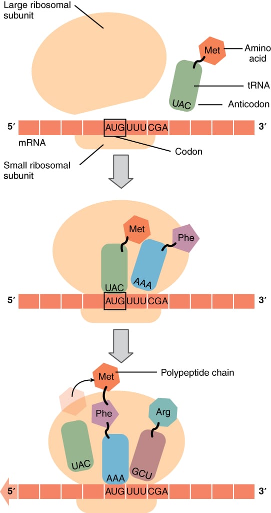
The initiator tRNA carrying methionine binds to the AUG start codon at the ribosome’s P site. This methionine becomes the first amino acid incorporated into the growing polypeptide chain. Once the initiation complex is formed, the large ribosomal subunit binds, which causes the release of the initiation factors.
The large ribosomal subunit also contributes to the three tRNA binding sites:
- The A (amino acid) site is where the aminoacyl-tRNA anticodon base pairs with the mRNA codon, ensuring the correct amino acid is added.
- The P (polypeptide) site is where the growing polypeptide chain is held and where the amino acid is transferred from its tRNA to the chain.
- The E (exit) site is where the “empty” tRNA sits before it is released back into the cytoplasm to bind another amino acid.
The initiator methionine tRNA is the only aminoacyl-tRNA that can bind directly to the P site to start translation. At this point, the A site is aligned with the second mRNA codon. The ribosome is now ready to bind the second aminoacyl-tRNA at the A site, and the first peptide bond will form between this amino acid and the initiator methionine.
- Elongation: During elongation, the ribosome moves along the mRNA in the 5’ to 3’ direction. This movement requires elongation factor G and is called translocation. After translocation:
- A new tRNA-amino acid complex binds in the A site.
- A peptide bond forms between the amino acids in the P and A sites via the peptidyl transferase activity of the 60S ribosomal subunit.
After the peptide bond forms, the ribosome translocates again. This shift moves the tRNA that no longer carries an amino acid into the E site, where it is released into the cytoplasm to pick up another amino acid. The A site becomes empty and ready to receive the next aminoacyl-tRNA. This cycle repeats until all codons in the mRNA have been read.
- Termination: Translation ends when a stop codon (UUA, UGA, or UAG) is reached. No tRNAs recognize stop codons. Instead, release factors bind and promote release of the polypeptide and mRNA, followed by dissociation of the ribosomal subunits.
Post-translational modifications: Proteins and polypeptides are modified after translation to become fully functional. Following modifications are seen :
- Prosthetic groups are added by covalent bonds
- Glycosylation involves addition of sugar groups to amino acids like serine, threonine, asparagine to form blood group antigens, membrane proteins etc.
- Proteolytic cleavage and activation of enzymes like trypsinogen to trypsin, clotting factors and hormones like proinsulin.
- Hydroxylation of lysine and proline in collagen chains
- Gamma carboxylation of glutamine residues in Vit K dependent clotting factors
- Phosphorylation of rate limiting enzymes of metabolism like glycogen synthase.