Pathophysiology of pulmonary system
External structures
- Provide the external borders for the lungs
- Borders composed of:
- Sternum
- Provides anatomical landmarks for auscultation
- Rib cage
- 12 ribs and adjacent intercostal musculature assist with movement of air
- Provide anatomical landmarks for auscultation
- Vertebral column
- Provides posterior border
- Shoulder girdle
- Provides attachment for accessory muscles to support in movement of air
- Sternum
Internal structures:
-
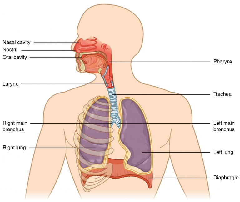
Upper airways
- Entry point for air
- Comprised of nose, mouth, pharynx, and larynx
-
Lower airways
- Continue the conduction of air where gas exchange occurs
- Comprised of trachea, bronchioles, alveolar ducts, and alveolar sacs
-
Internal supporting structures
- Each lung is divided into lobes and further subdivided into bronchopulmonary segments.
- The right lung has three lobes—upper, middle, and lower—and a total of 10 bronchopulmonary segments:
- Upper lobe: 3 segments- apical, posterior, anterior
- Middle lobe: 2 segments- lateral, medial
- Lower lobe: 5 segments- superior, medial basal, anterior basal, lateral basal, posterior basal
- The right lung has three lobes—upper, middle, and lower—and a total of 10 bronchopulmonary segments:
- The left lung has two lobes—upper and lower—and typically 8 bronchopulmonary segments:
- Upper lobe: 4 segments -apical, posterior, anterior, and lingular superior/inferior
- Lower lobe: 4 segments- superior, anteriomedial basal, lateral basal, and posterior basal
- Each lung is divided into lobes and further subdivided into bronchopulmonary segments.
-
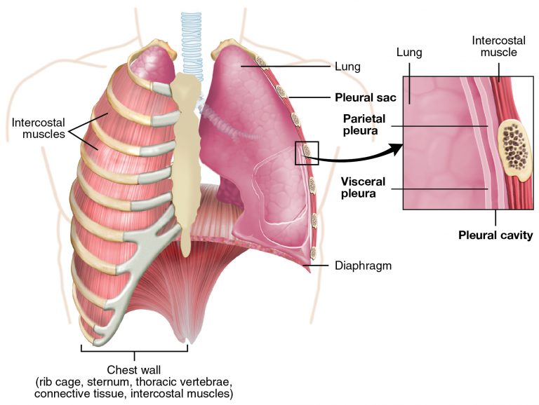
Pleura
- Parietal pleura located on the outer surface and provides barrier to thoracic cage, diaphragm, and mediastinal border
- Visceral pleura located on the inner surface and provides barrier to lung
Muscles and mechanics of ventilation
Primary muscles of inspiration
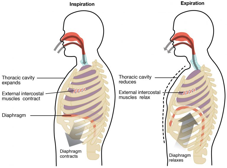
- Diaphragm
- When contracted, muscle is pulled downward- inhalation
- Leads to protruded abdominal cavity
- When relaxed, muscle is in dome shape- exhalation
- Takes the natural shape of lower rib cage
- When contracted, muscle is pulled downward- inhalation
- Intercostals
- Aid in movement of rib cage
- Accessory muscles
- Scalenes
- Sternocleidomastoid
- Trapezius
- Serratus anterior
- Pectoralis muscles
Primary muscles for exhalation
- Passive recoil of activated inspiratory muscles
- Accessory muscles that can be utilized during disease state or exercise
- Quadratus lumborum
- Abdominal muscles
- Sections of intercostals
Mechanism of breathing
- Inspiration
- Activation of muscles to provide sufficient pressure gradient to allow for air to enter the lungs
- Causes the movement of bony thorax outward
- Exhalation
- Passive recoil of the activated muscle to allow air to leave the lungs
- Causes the movement of bony thorax inward