


Achievable logoAchievable logo
CCMA
Sign in
Sign up
Purchase
Textbook
Practice exams
Support
How it works
Exam catalog
Mountain with a flag at the peak
Textbook
1. Medical assistant
2. Electronic records
3. Medical terminology and anatomy
4. The fundamentals of infection control
5. Introduction to vital signs
6. The patient interview and history
7. The physical examination
8. Appointment scheduling
9. Insurance billing
10. Diagnostic coding and the ICD-10-CM System
11. Procedural coding
12. Medical billing and reimbursement essentials
13. Assisting with medical specialties
14. Assisting with the musculoskeletal system
15. Assisting with the cardiovascular system
16. Assisting with the respiratory system
17. Assisting with the nervous system
18. Anatomy and physiology of the urinary system
19. Assisting in obstetrics and gynecology
20. Assisting in endocrinology
21. Assisting in ophthalmology & otolaryngology
22. Assisting in gastroenterology
23. Assisting in the immune & lymphatic systems
24. Assisting in pediatrics: the developmental stages and care
25. The medical assistant’s role in caring for the older patient
26. The role of the medical assistant in physical therapy examination and assessment
27. Preparing for minor surgery: room, solutions, and supplies
28. Introduction to the clinical laboratory
29. Urinalysis
30. Blood collection
30.1 Phlebotomy procedures
30.2 Evacuated collection tubes and order of draw
30.3 Needle safety and post exposure follow-up
30.4 Routine venipuncture
30.5 Potential problems associated with venipuncture
30.6 Capillary puncture: indications, equipment, and procedure
30.7 Capillary puncture: processing, legal, and professional issues
31. Analysis of blood
32. Electrocardiography and heart structure
33. The principles of pharmacology
34. Essential calculations and measurement systems
35. Solid, liquid, & solutions medication doses
36. Administering medications
37. Metabolism and core nutrient roles
38. Medical emergencies in the healthcare setting
Achievable logoAchievable logo
30.2 Evacuated collection tubes and order of draw
Achievable CCMA
30. Blood collection
Our CCMA course is currently in development and is a work-in-progress.

Evacuated collection tubes and order of draw

8 min read
Font
Discuss
Share
Feedback

Evacuated collection tubes

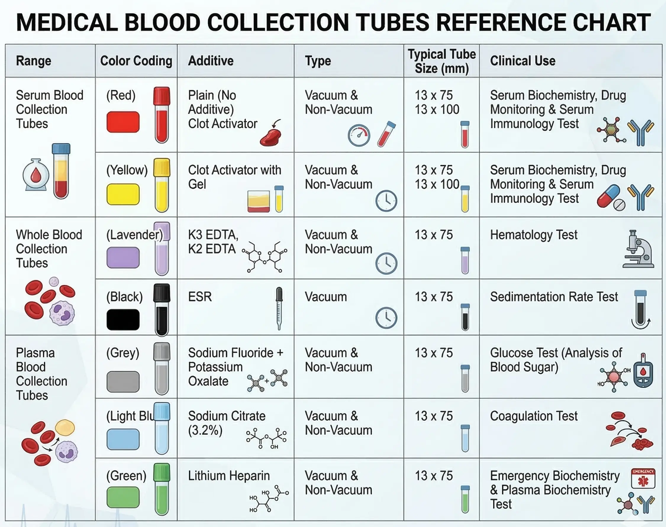
The most common collection system is the evacuated tube system (these tubes are also called vacuum tubes; a particular brand of vacuum tubes is the Vacutainer). It consists of evacuated tubes of various sizes that have color-coded stoppers. The colored stoppers indicate the tube’s contents. Venipuncture tubes must be either shatter-resistant glass or plastic. More traditional rubber tube stoppers have been replaced by safer plastic Hemogard colored tops. Hemogard tops have the advantage of not splattering blood when removed from the tube. Each of the colored stoppers and the chemical additives, anticoagulants , clot activators , or thixotropic gel that are contained in each type of tube. The different stopper colors indicate the unique contents of that color-coded tube. The vacuum in each tube draws a measured amount of blood into the tube. Tube volumes range from 2 to 15 mL.

The size of the tube to be drawn depends on several factors. Each test performed in the laboratory requires a specific amount of blood. Blood volumes can often be combined, which reduces the number of tubes that must be drawn. For example, both a complete blood count and an erythrocyte sedimentation rate test are performed on a sample from a lavender-topped tube. The combined volume for both tests can be drawn with one lavender-topped tube, so you do not need to draw two tubes. When in doubt, refer to the appropriate laboratory manual for questions regarding specimen requirements and handling. Another option would be to call the laboratory. Keep in mind that blood is approximately half cells and half liquid. If a test requires 2 mL of serum, at least 4 mL of blood must be collected.

Tube additives

Most vacuum tubes contain an additive. The plain, red-topped tube contains nothing. It is a plain tube with no additives, anticoagulants, or clot activator. A gold-topped tube contains silica to activate clotting and an inert thixotropic gel to separate serum and cells after the sample is centrifuged. Most other color-topped plastic tubes contain some form of anticoagulant, which prevents blood from clotting. The additive may be a powder, a liquid visible in the tube, or a liquid sprayed inside the tube by the manufacturer and allowed to dry. The choice of anticoagulant depends on the test to be completed.

Anticoagulant additives prevent blood from clotting, which allows the contents of the tube to be used in two ways. First, the sample can be used as whole blood; second, the sample can be centrifuged, and the liquid portion, called plasma , can be used for testing. An example of a test that uses whole blood would be a complete blood count (CBC). An example of a test that uses plasma would be a coagulation study.

Color-coded blood collection tube stoppers and additives
Color coded stoppers and their additives

Ethylenediaminetetraacetic acid (EDTA) is the anticoagulant found in the lavender-topped tube. It prevents platelet clumping and preserves the appearance of blood cells for microscopic examination.

Clot activators promote blood clotting in the tubes with either a marbled red-gray or a gold top. For example, silica particles in the gold tubes enhance clotting by providing a surface for platelet activation. Thrombin from the platelets quickly promotes clotting and is used in tubes drawn for chemistry testing or in the event a sample is needed from a patient taking a prescribed anticoagulant, such as heparin.

If blood clots and then is centrifuged, the liquid portion is referred to as serum . Without a clot activator, blood clots in 15 to 60 minutes, after which it must be centrifuged. The serum must be quickly separated from the cells because cells may continue to metabolize glucose or may release substances that interfere with testing.

Thixotropic gel can be found in some tubes, including serum separator tubes (SSTs). SST tubes are identified by the marbled red-gray rubber stopper or the gold Hemogard top. The plasma separator tube (PST) also contains thixotropic gel. The tubes have a marbled green-gray top, and the Hemogard tubes have a light green top. Thixotropic gel, a synthetic, chemically neutral gel, has a density that is between that of red cells and plasma or serum. The gel settles between the plasma/serum and cells during centrifugation. The gel forms a barrier that makes it easy to pour off the liquid portion of the sample without cells contaminating the specimen.

It is important to avoid a “short draw” (i.e., a tube that is not completely filled). Some tubes are designed to fill only partially, according to their preset vacuum. Having the proper ratio of blood to additive is crucial. Always check the tube for the expiration date. Outdated tubes may have a diminished vacuum, or the additive may have degraded and not be as effective.

Order of the draw

If more than one tube must be drawn during a venipuncture, a specified order must be followed. Carryover of additives from one tube to the next could cause sample alteration and incorrect results. CLSI has established a set of standards outlining the order to follow for a multitube draw. The same order applies to filling tubes when blood is collected in a syringe.

  1. Blood culture bottles: These bottles are filled first because they are sterile and should not be contaminated by the other tubes. In addition to blood culture bottles, a yellow-topped tube can be drawn for blood cultures for mycobacteria, fungi, and acid-fast bacilli. These tubes are not drawn frequently in an ambulatory setting. If they are ordered, they would be drawn in the same place as blood culture bottles in the order of the draw.
  2. Light blue top: These tubes, which contain sodium citrate, are next because other anticoagulants might contaminate the sample collected for coagulation studies. If no blood culture has been ordered, CLSI recommends that blood for the light blue–topped tube be drawn first if routine coagulation testing has been ordered. Examples of coagulation tests include prothrombin time with international normalized ratio (PT/INR) and activated partial thromboplastin time (APTT). See Chapter 34 for more details about coagulation testing. Some laboratories recommend that a red-topped “waste” tube with no additives be partially filled before the light blue–topped tube. This is done to remove any tissue thromboplastin that may be released during the venipuncture. Thromboplastin interferes with coagulation testing. CLSI also recommends that when a winged infusion (butterfly assembly) is used, blood should be drawn into the red-topped tube even if the order does not call for it. This is done to fill the tubing’s dead air space with blood before drawing the light blue–topped sodium citrate tube. A light blue tube must have an exact blood-to-citrate dilution. It is not necessary to completely fill the waste tube because it will be discarded.
  3. Red top or gold top: Red serum tubes without clot activator or gold serum tubes with clot activator are filled next. These tubes are drawn to test serum after the specimens have clotted and been centrifuged. SSTs with thixotropic gel are also drawn at this time. SST tubes have a marbled red-gray stopper or a gold Hemogard stopper.
  4. Green top: These tubes are drawn next because the plasma in their anticoagulated specimen is used for testing when STAT results are needed (usually in chemistry). The dark green tops contain no thixotropic gel. The tubes with marbled green-gray tops and light green Hemogard tops both contain gel to help separate the plasma from the cells when centrifuged.
  5. Lavender top: These tubes are drawn next. They contain an EDTA anticoagulant that preserves blood cell size and shape, otherwise known as morphology. EDTA tubes are drawn near the end of the draw because the additive interferes with chemistry and coagulation specimens.
  6. Gray top: This tube is drawn last, and the blood is used to test glucose or blood alcohol levels. Its additives may elevate electrolyte levels and damage cells if passed into the other tubes.

It is important to gently mix the contents of the tubes after collection by inverting them (do not shake the tubes). Most tubes should be inverted eight times, but clot activator tubes should be inverted five times, and sodium citrate three to four times.

Evacuated collection tubes

  • Most common blood collection system; uses color-coded, vacuum-sealed tubes
  • Tube stopper color indicates contents (additives, anticoagulants, clot activators, gels)
  • Tube size/volume chosen based on test requirements; combine tests when possible

Tube additives

  • Most tubes contain additives; red top = no additive, gold top = clot activator + thixotropic gel
  • Anticoagulants (e.g., EDTA in lavender top) prevent clotting for whole blood/plasma tests
  • Clot activators (e.g., silica in gold top) speed up clotting for serum tests
  • Thixotropic gel (in SST/PST tubes) forms barrier after centrifugation for easy serum/plasma separation
  • Proper blood-to-additive ratio essential; avoid short draws and expired tubes

Order of the draw

  • Specific sequence prevents additive carryover and sample contamination:
      1. Blood culture bottles/yellow top (sterile)
      1. Light blue top (sodium citrate, coagulation studies)
      1. Red/gold top (serum, SST)
      1. Green top (heparin, plasma for STAT chemistry)
      1. Lavender top (EDTA, hematology)
      1. Gray top (glucose/alcohol, last)
  • Gently invert tubes to mix; number of inversions varies by tube/additive

Needles and supplies used in phlebotomy

  • Needles: sterile, safety device, inspected before use
  • Needle parts: hub (attaches to holder/syringe), shaft (length), bevel (angled tip), lumen (gauge = diameter)
    • Higher gauge = smaller lumen; match gauge to tube size and vein type
  • Multisample needles: double-pointed, allow multiple tubes per venipuncture
  • Needle holders: secure needle, translucent, discard with needle attached after use (OSHA requirement)
  • Syringes: used for fragile veins; transfer blood to tubes immediately to prevent clotting
  • Winged infusion sets (butterfly): for small/fragile veins, pediatric/hand draws; often used with syringes for gentle vacuum

Sign up for free to take 17 quiz questions on this topic

Previous
Next  | 30.3 Needle safety and post exposure follow-up
All rights reserved ©2016 - 2026 Achievable, Inc.

Evacuated collection tubes and order of draw

Evacuated collection tubes

The most common collection system is the evacuated tube system (these tubes are also called vacuum tubes; a particular brand of vacuum tubes is the Vacutainer). It consists of evacuated tubes of various sizes that have color-coded stoppers. The colored stoppers indicate the tube’s contents. Venipuncture tubes must be either shatter-resistant glass or plastic. More traditional rubber tube stoppers have been replaced by safer plastic Hemogard colored tops. Hemogard tops have the advantage of not splattering blood when removed from the tube. Each of the colored stoppers and the chemical additives, anticoagulants , clot activators , or thixotropic gel that are contained in each type of tube. The different stopper colors indicate the unique contents of that color-coded tube. The vacuum in each tube draws a measured amount of blood into the tube. Tube volumes range from 2 to 15 mL.

The size of the tube to be drawn depends on several factors. Each test performed in the laboratory requires a specific amount of blood. Blood volumes can often be combined, which reduces the number of tubes that must be drawn. For example, both a complete blood count and an erythrocyte sedimentation rate test are performed on a sample from a lavender-topped tube. The combined volume for both tests can be drawn with one lavender-topped tube, so you do not need to draw two tubes. When in doubt, refer to the appropriate laboratory manual for questions regarding specimen requirements and handling. Another option would be to call the laboratory. Keep in mind that blood is approximately half cells and half liquid. If a test requires 2 mL of serum, at least 4 mL of blood must be collected.

Tube additives

Most vacuum tubes contain an additive. The plain, red-topped tube contains nothing. It is a plain tube with no additives, anticoagulants, or clot activator. A gold-topped tube contains silica to activate clotting and an inert thixotropic gel to separate serum and cells after the sample is centrifuged. Most other color-topped plastic tubes contain some form of anticoagulant, which prevents blood from clotting. The additive may be a powder, a liquid visible in the tube, or a liquid sprayed inside the tube by the manufacturer and allowed to dry. The choice of anticoagulant depends on the test to be completed.

Anticoagulant additives prevent blood from clotting, which allows the contents of the tube to be used in two ways. First, the sample can be used as whole blood; second, the sample can be centrifuged, and the liquid portion, called plasma , can be used for testing. An example of a test that uses whole blood would be a complete blood count (CBC). An example of a test that uses plasma would be a coagulation study.

Ethylenediaminetetraacetic acid (EDTA) is the anticoagulant found in the lavender-topped tube. It prevents platelet clumping and preserves the appearance of blood cells for microscopic examination.

Clot activators promote blood clotting in the tubes with either a marbled red-gray or a gold top. For example, silica particles in the gold tubes enhance clotting by providing a surface for platelet activation. Thrombin from the platelets quickly promotes clotting and is used in tubes drawn for chemistry testing or in the event a sample is needed from a patient taking a prescribed anticoagulant, such as heparin.

If blood clots and then is centrifuged, the liquid portion is referred to as serum . Without a clot activator, blood clots in 15 to 60 minutes, after which it must be centrifuged. The serum must be quickly separated from the cells because cells may continue to metabolize glucose or may release substances that interfere with testing.

Thixotropic gel can be found in some tubes, including serum separator tubes (SSTs). SST tubes are identified by the marbled red-gray rubber stopper or the gold Hemogard top. The plasma separator tube (PST) also contains thixotropic gel. The tubes have a marbled green-gray top, and the Hemogard tubes have a light green top. Thixotropic gel, a synthetic, chemically neutral gel, has a density that is between that of red cells and plasma or serum. The gel settles between the plasma/serum and cells during centrifugation. The gel forms a barrier that makes it easy to pour off the liquid portion of the sample without cells contaminating the specimen.

It is important to avoid a “short draw” (i.e., a tube that is not completely filled). Some tubes are designed to fill only partially, according to their preset vacuum. Having the proper ratio of blood to additive is crucial. Always check the tube for the expiration date. Outdated tubes may have a diminished vacuum, or the additive may have degraded and not be as effective.

Order of the draw

If more than one tube must be drawn during a venipuncture, a specified order must be followed. Carryover of additives from one tube to the next could cause sample alteration and incorrect results. CLSI has established a set of standards outlining the order to follow for a multitube draw. The same order applies to filling tubes when blood is collected in a syringe.

  1. Blood culture bottles: These bottles are filled first because they are sterile and should not be contaminated by the other tubes. In addition to blood culture bottles, a yellow-topped tube can be drawn for blood cultures for mycobacteria, fungi, and acid-fast bacilli. These tubes are not drawn frequently in an ambulatory setting. If they are ordered, they would be drawn in the same place as blood culture bottles in the order of the draw.
  2. Light blue top: These tubes, which contain sodium citrate, are next because other anticoagulants might contaminate the sample collected for coagulation studies. If no blood culture has been ordered, CLSI recommends that blood for the light blue–topped tube be drawn first if routine coagulation testing has been ordered. Examples of coagulation tests include prothrombin time with international normalized ratio (PT/INR) and activated partial thromboplastin time (APTT). See Chapter 34 for more details about coagulation testing. Some laboratories recommend that a red-topped “waste” tube with no additives be partially filled before the light blue–topped tube. This is done to remove any tissue thromboplastin that may be released during the venipuncture. Thromboplastin interferes with coagulation testing. CLSI also recommends that when a winged infusion (butterfly assembly) is used, blood should be drawn into the red-topped tube even if the order does not call for it. This is done to fill the tubing’s dead air space with blood before drawing the light blue–topped sodium citrate tube. A light blue tube must have an exact blood-to-citrate dilution. It is not necessary to completely fill the waste tube because it will be discarded.
  3. Red top or gold top: Red serum tubes without clot activator or gold serum tubes with clot activator are filled next. These tubes are drawn to test serum after the specimens have clotted and been centrifuged. SSTs with thixotropic gel are also drawn at this time. SST tubes have a marbled red-gray stopper or a gold Hemogard stopper.
  4. Green top: These tubes are drawn next because the plasma in their anticoagulated specimen is used for testing when STAT results are needed (usually in chemistry). The dark green tops contain no thixotropic gel. The tubes with marbled green-gray tops and light green Hemogard tops both contain gel to help separate the plasma from the cells when centrifuged.
  5. Lavender top: These tubes are drawn next. They contain an EDTA anticoagulant that preserves blood cell size and shape, otherwise known as morphology. EDTA tubes are drawn near the end of the draw because the additive interferes with chemistry and coagulation specimens.
  6. Gray top: This tube is drawn last, and the blood is used to test glucose or blood alcohol levels. Its additives may elevate electrolyte levels and damage cells if passed into the other tubes.

It is important to gently mix the contents of the tubes after collection by inverting them (do not shake the tubes). Most tubes should be inverted eight times, but clot activator tubes should be inverted five times, and sodium citrate three to four times.

Key points

Evacuated collection tubes

  • Most common blood collection system; uses color-coded, vacuum-sealed tubes
  • Tube stopper color indicates contents (additives, anticoagulants, clot activators, gels)
  • Tube size/volume chosen based on test requirements; combine tests when possible

Tube additives

  • Most tubes contain additives; red top = no additive, gold top = clot activator + thixotropic gel
  • Anticoagulants (e.g., EDTA in lavender top) prevent clotting for whole blood/plasma tests
  • Clot activators (e.g., silica in gold top) speed up clotting for serum tests
  • Thixotropic gel (in SST/PST tubes) forms barrier after centrifugation for easy serum/plasma separation
  • Proper blood-to-additive ratio essential; avoid short draws and expired tubes

Order of the draw

  • Specific sequence prevents additive carryover and sample contamination:
      1. Blood culture bottles/yellow top (sterile)
      1. Light blue top (sodium citrate, coagulation studies)
      1. Red/gold top (serum, SST)
      1. Green top (heparin, plasma for STAT chemistry)
      1. Lavender top (EDTA, hematology)
      1. Gray top (glucose/alcohol, last)
  • Gently invert tubes to mix; number of inversions varies by tube/additive

Needles and supplies used in phlebotomy

  • Needles: sterile, safety device, inspected before use
  • Needle parts: hub (attaches to holder/syringe), shaft (length), bevel (angled tip), lumen (gauge = diameter)
    • Higher gauge = smaller lumen; match gauge to tube size and vein type
  • Multisample needles: double-pointed, allow multiple tubes per venipuncture
  • Needle holders: secure needle, translucent, discard with needle attached after use (OSHA requirement)
  • Syringes: used for fragile veins; transfer blood to tubes immediately to prevent clotting
  • Winged infusion sets (butterfly): for small/fragile veins, pediatric/hand draws; often used with syringes for gentle vacuum

More from Blood collection

  • Phlebotomy procedures
  • Needle safety and post exposure follow-up
  • Routine venipuncture
  • Potential problems associated with venipuncture
  • Capillary puncture: indications, equipment, and procedure