Sensory receptors
Depending on what type of stimulus activates a receptor, sensory receptors can be classified as follows:
Electromagnetic receptors - photoreceptors, thermoreceptors. Photoreceptors are rods and cones of the retina. Thermoreceptors are slowly adapting receptors. They are of two types: warm and cold. Cold receptors mainly sense temperatures between 25-30°C. Warm receptors detect temperatures in the range of 30-45°C.
Mechanoreceptors - They are activated by pressure or changes in pressure. Examples include receptors for hearing, touch, balance, and osmoreceptors. They can be very rapidly adapting, rapidly adapting, or slowly adapting. Very rapidly and rapidly adapting mechanoreceptors detect changes in velocity, while slowly adapting receptors detect the intensity and duration of the stimulus. Following are the types of mechanoreceptors:
- Pacinian corpuscles: These are the most rapidly adapting, encapsulated corpuscles found in the deep dermis, muscle, joint capsules, and subcutaneous tissue. They respond to vibration, deep pressure, and tapping.
- Meissner’s corpuscles: These are rapidly adapting, high-precision, encapsulated corpuscles found in the papillary dermis of non-hairy skin, especially the fingertips, lips, etc. They sense two-point discrimination, light touch, tapping, and low-frequency vibrations.
- Baroreceptors: These are free nerve endings located in the carotid sinuses and aortic arch that respond to stretch of the vessel wall. Increased stretch (as in high blood pressure) increases firing, and decreased stretch reduces firing.
- Hair cells: Hair cells of the Organ of Corti (hearing) and vestibular hair cells (maintenance of balance) are mechanoreceptors.
- Free nerve endings: Free nerve endings located in the dermis, cornea, tongue, and joint capsules sense pain, temperature, and mechanical deformation.
- Merkel’s discs: These are slowly adapting receptors present in non-hairy skin (at the dermal-epidermal junction) and mucosal membranes. They detect low-frequency vibrations and vertical indentations. A similar receptor in hairy skin is called a tactile disc.
- Ruffini’s corpuscles: These are bulbous corpuscles located in the dermis and joint capsules. They detect stretch and joint rotation.
- Hair follicle plexus: These are rapidly adapting nerve fibers wrapped around the hair follicle in the dermis. They detect hair movement, including the direction of movement.
- Muscle spindle: These are spindle-like structures composed of intrafusal muscle fibers arranged parallel to extrafusal muscle fibers. They detect muscle contraction and stretch.
- Golgi tendon organ: This is a stretch receptor arranged in series with extrafusal fibers to detect tendon stretch and muscle contraction. It is located in the tendon adjacent to the myotendinous junctions.
- Osmoreceptors: Osmoreceptors respond to changes in osmotic pressure. They are activated by membrane stretch, which then leads to changes in ion permeability of the cell. They are present in the vascular organ of lamina terminalis (VOLT) in the CNS, hypothalamus, area postrema, portal circulation, macula densa, etc.
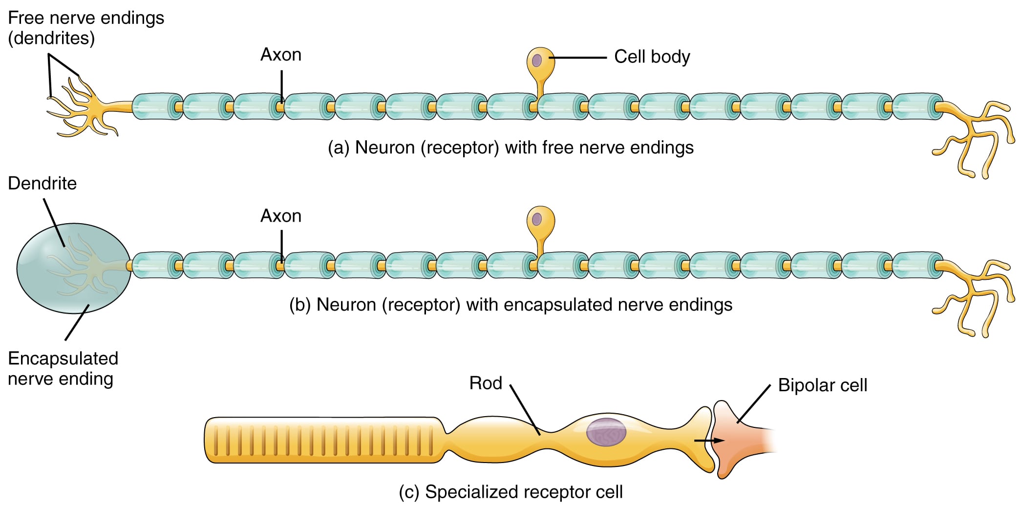
Receptor cell types can be classified on the basis of their structure. Sensory neurons can have either (a) free nerve endings or (b) encapsulated endings. Photoreceptors in the eyes, such as rod cells, are examples of © specialized receptor cells. These cells release neurotransmitters onto a bipolar cell, which then synapses with the optic nerve neurons.
Chemoreceptors - Chemoreceptors detect chemical stimuli. Odorant receptors and gustatory receptors are involved in the sensations of smell and taste, respectively. Similarly, arterial PO2 receptors in the carotid and aortic bodies and pH-sensing receptors in the medulla are also chemoreceptors.
Nociceptors: These respond to noxious stimuli that produce tissue damage, such as extremes of temperature, high pressure, and chemical damage. Nociceptors can be thermal, mechanical, or polymodal. Thermal nociceptors are innervated by A-delta myelinated nerve fibers and respond to mechanical stimuli such as sharp pain and high pressure. Polymodal receptors are innervated by unmyelinated type C nerve fibers and respond to mechanical, chemical, extreme hot, and extreme cold stimuli. Transient receptor potential (TRP) ion channels on nociceptors are activated in response to these stimuli. In addition, inflammatory mediators such as histamine (from mast cells), bradykinin, substance P, and prostaglandins can directly or indirectly activate nociceptors, causing pain and hyperalgesia. Glutamate, substance P, calcitonin, and somatostatin are used as neurotransmitters.
Most receptor molecules are tuned to a single sensory modality, but some are polymodal (activated by multiple types of sensory stimuli). Activation of receptor molecules by an adequate stimulus initiates a signal transduction process in the sensory receptor. During this process, the physical or chemical signal is amplified and converted into an electrical signal that depolarizes or hyperpolarizes the cell. This change in the receptor’s membrane potential is called the receptor potential.
Stimulus intensity is encoded by:
- activation of a larger number of receptors in response to more intense stimuli
- changes in firing rates
- activation of different types of receptors (such as nociceptors)
The properties of the stimulus (such as strength and duration) are translated into a specific temporal pattern of action potentials, which is then sensed by the brain.
When a constant stimulus is applied for a longer period of time, receptors adapt. This means the frequency of action potentials decreases even though the stimulus is still present. Phasic receptors adapt rapidly, while tonic receptors adapt slowly.
Types of nerve fibres: There are two different classification systems for nerve fibres: Lloyd-Hunt and Erlanger-Gasser. In general, myelinated nerves and larger fibers conduct faster than unmyelinated nerves and small-diameter fibers. The table below lists nerve fibres in descending order of conduction velocity and diameter.
| Lloyd-Hunt type | Erlanger-Gasser type | Myelination | Sensory receptors innervated |
| Ia | A alpha | Yes | Muscle spindles, alpha motor neurons |
| Ib | A alpha | Yes | Golgi tendon organs, alpha motor neurons |
| II | A beta | Yes | Muscle spindles, mechanoreceptors of skin for touch, pressure. |
| - | A gamma | Yes | Efferents from gamma motor neurons to muscle spindles |
| III | A delta | Yes | Free nerve endings for touch and pressure, nociceptors, cold thermoreceptors, fast pain |
| IV | C | No | Nociceptors, slow pain, warm thermoreceptors, olfaction |
Please refer to CNS anatomy for details on cerebral cortex and cerebellum structure and organization.