Pediatric disorders
Legg-Calvé-Perthes disease
Legg-Calvé-Perthes disease is an idiopathic avascular necrosis (osteonecrosis) of the femoral epiphysis. It usually affects children ages 4 to 10 and is more common in boys.
Children typically present with:
- A limp
- Pain in the hip, thigh, or knee
On exam:
- The knee exam is normal
- The ipsilateral hip has limited and painful rotation and abduction
- Trendelenburg test may be positive
It’s often associated with coagulopathies such as thrombophilia. Diagnosis is confirmed by MRI.
Treatment is typically:
- Observation in children <8 years of age
- Femoral and/or pelvic osteotomy in children >8 years of age
Slipped capital femoral epiphysis (SCFE)
SCFE is characterized by slipping of the metaphysis in relation to the epiphysis. It usually affects children ages 11 to 14, is more common in obese children and boys, and is bilateral in 20-40%.
Adolescents typically present with:
- A limp
- Hip, groin, or knee pain
On exam:
- The knee exam is normal
- The hip is often preferentially held in abduction and external rotation
- Trendelenburg may be positive
X-ray findings:
- Klein’s line does not intersect the outer part of the femoral head
Treatment is percutaneous pin fixation.
Developmental or congenital hip dysplasia
This is a developmental disorder in which the acetabulum is shallow, leading to subluxation or dislocation of the hip.
Risk factors include:
- Female sex
- Firstborn status
- Breech presentation
- Family history
- Oligohydramnios
Common findings include:
- Limping
- Waddling gait
- Uneven skin folds in the inguinal area
- Unequal lengths of the lower limbs
On physical exam, the Ortolani and Barlow maneuvers may be positive.
Ortolani maneuver:
- Hold the contralateral hip still
- Abduct the thigh of the hip being tested and gently pull anteriorly
- A palpable (and sometimes audible) “clunk” is a positive test
Barlow maneuver:
- Adduct the hip while pushing the thigh posteriorly
- If the hip goes out of the socket, it’s “dislocatable,” and the test is positive
Diagnosis is confirmed by:
- Ultrasound until age 4 months
- Radiographs after age 4 months
Treatment:
- <6 months: Pavlik harness
-
6 months: closed reduction or open surgery
Osgood-Schlatter disease
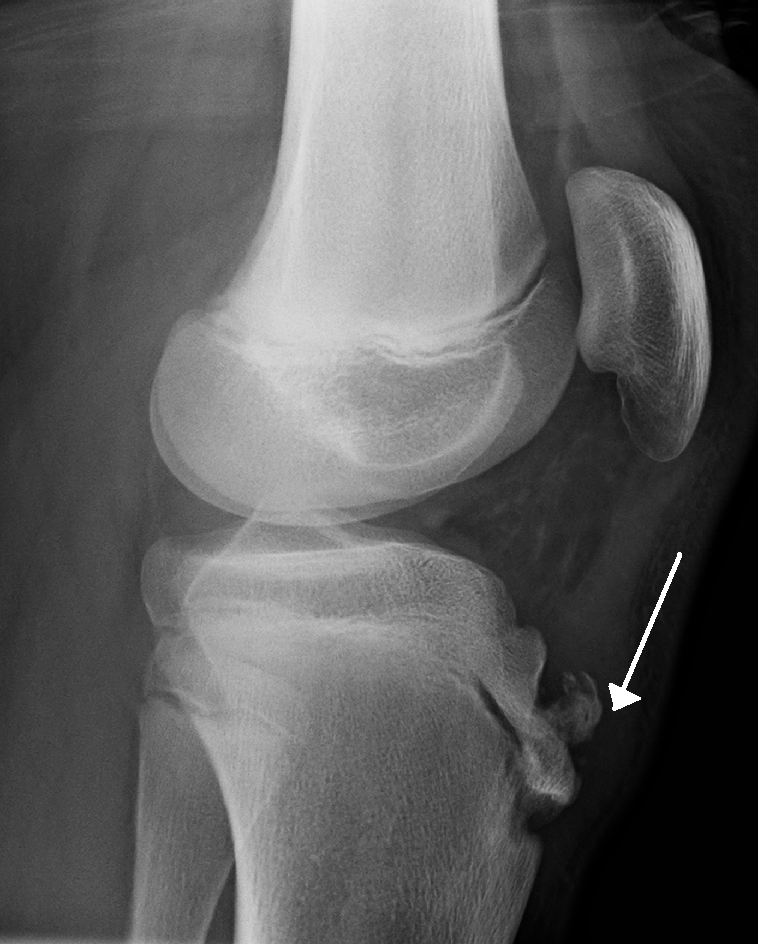
Osgood-Schlatter disease is a traction apophysitis (osteochondrosis) of the tibial tubercle seen in physically active adolescents. It’s more common in boys.
It presents with:
- Anterior knee pain
- Enlargement and tenderness of the tibial tubercle
X-ray shows fragmentation of the tibial tubercle.
Treatment is conservative with NSAIDs, rest, knee strapping, and quadriceps stretching. In severe or persistent cases, ossicle excision is required.