Apoptosis
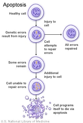
Apoptosis is programmed cell death. It can occur for physiological or pathological reasons.
Mechanism
Apoptosis is initiated through two main pathways:
- The extrinsic (death receptor) pathway
- The intrinsic (mitochondrial) pathway
Both pathways ultimately activate caspases, which carry out cell death. Caspases are cysteine-containing proteases.
-
Extrinsic or death receptor initiated pathway: This pathway begins when a ligand binds to a death receptor on the cell surface. Death receptors are members of the tumor necrosis factor family and include Fas (CD95) and the type 1 TNF receptor (TNFR1). These receptors have intracellular domains that indirectly activate caspases.
Binding of FasL (Fas ligand) to Fas leads to receptor cross-linking and activation of the cytoplasmic adaptor protein FADD (Fas-associated death domain). FADD then binds pro-caspases, which are cleaved into active caspases.
This pathway can be inhibited by FLIP, a protein used by some viruses to block Fas-mediated apoptosis.
-
Intrinsic or mitochondrial pathway: This pathway is triggered by increased mitochondrial permeability, which leads to the release of pro-apoptotic factors that activate caspases. It involves both pro-apoptotic and anti-apoptotic members of the Bcl-2 family.
Bcl-2 and Bcl-x are anti-apoptotic molecules that normally reside in the mitochondrial membrane. When the intrinsic pathway is initiated, Bcl-2 and Bcl-x are lost from the mitochondrial membrane and are replaced by pro-apoptotic molecules Bak, Bax, and Bim. This increases mitochondrial permeability and allows cyt c to escape from the mitochondria into the cytosol.
Cyt c binds to Apaf-1 (apoptosis activating factor-1), which activates caspases and leads to apoptosis. Normally, Bcl-2 and Bcl-x inhibit Apaf-1 activation. AIF (apoptosis inducing factor) enters the cytoplasm and inactivates inhibitors of apoptosis.
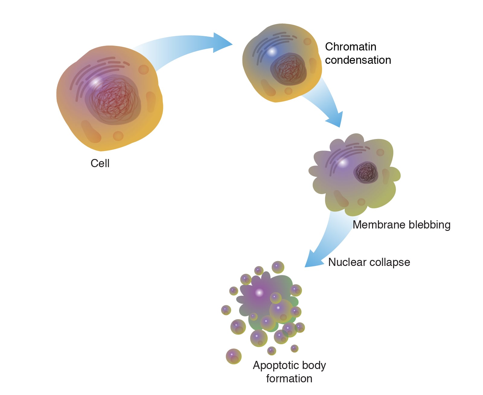
Characteristics of apoptosis
In apoptosis, cells undergo a predictable set of morphologic and biochemical changes:
- The cell shrinks.
- The cytoplasm becomes dense.
- Chromatin condenses and aggregates peripherally under the nuclear membrane.
- The cell surface forms blebs and buds.
- The cell fragments into apoptotic bodies.
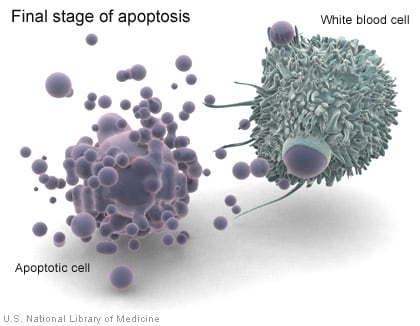
On H and E stained sections, apoptotic cells appear deeply eosinophilic. A key feature is the absence of inflammation.
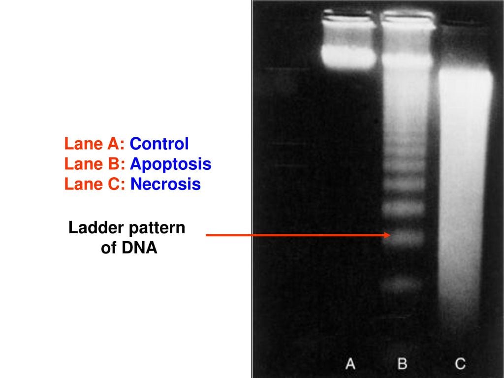
Apoptosis also produces a characteristic pattern of DNA fragmentation:
- DNA first breaks into large 50 to 300 kilobase pieces.
- It is then cleaved into 180-200 base pair segments.
These fragments can be visualized on agarose gel electrophoresis as a DNA ladder. Typically, this type of DNA ladder is not seen in necrotic cells. Apoptotic cells also display markers that allow macrophages to recognize and remove them.
Examples: Apoptosis can be initiated by lack of growth factors or hormones, by DNA damage, by cytotoxic T cells, or through tumor necrosis factor (TNF) receptors.
It can be physiological in embryogenesis, menstruation, and apoptosis of immune cells at the end of an immune response. Pathological apoptosis can result from infections like viral hepatitis and from DNA damage due to radiation or chemotherapy.
- Apoptosis due to growth factor, hormone, or cytokine deficiency occurs through the intrinsic pathway.
- DNA damage causes arrest of the cell cycle in G1 phase by p53. When the damage cannot be repaired, p53 induces apoptosis.
- FasL (CD95L) is the ligand for Fas (CD95). It is produced by immune cells and initiates the extrinsic pathway of apoptosis. It is involved in apoptosis of self-reactive T cells.
- TNF binds to TNFR1 (TNF receptor 1). This causes TRADD (TNF receptor associated death domain protein) to associate with the intracellular domain and then bind to FADD, inducing the extrinsic pathway.
Cytotoxic T lymphocytes (CTLs) have a unique method of inducing apoptosis by direct activation of caspases by granzymes secreted by the CTLs.