Plasma membrane
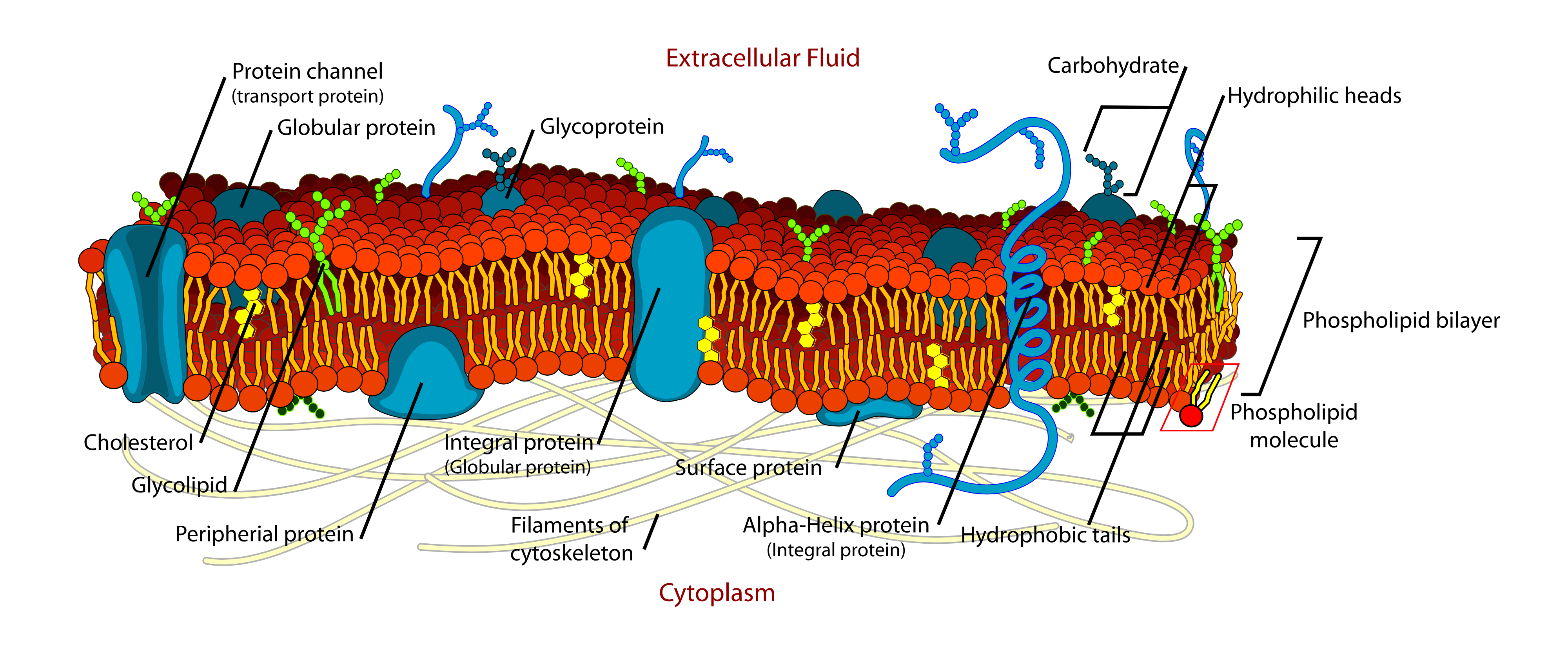
The plasma membrane is the dynamic boundary that surrounds each cell. It helps maintain the cell’s internal environment and controls how the cell interacts with its surroundings. Its core is made of phospholipids, which form a bilayer with hydrophilic heads facing outward (toward water) and hydrophobic tails facing inward (away from water).
Embedded steroids, especially cholesterol, help regulate membrane fluidity, while small amounts of waxes can add structural stability. In the fluid mosaic model, proteins move within this lipid framework and function as channels, transporters, and surface markers.
Membrane dynamics
Membrane dynamics include processes such as endocytosis and exocytosis.
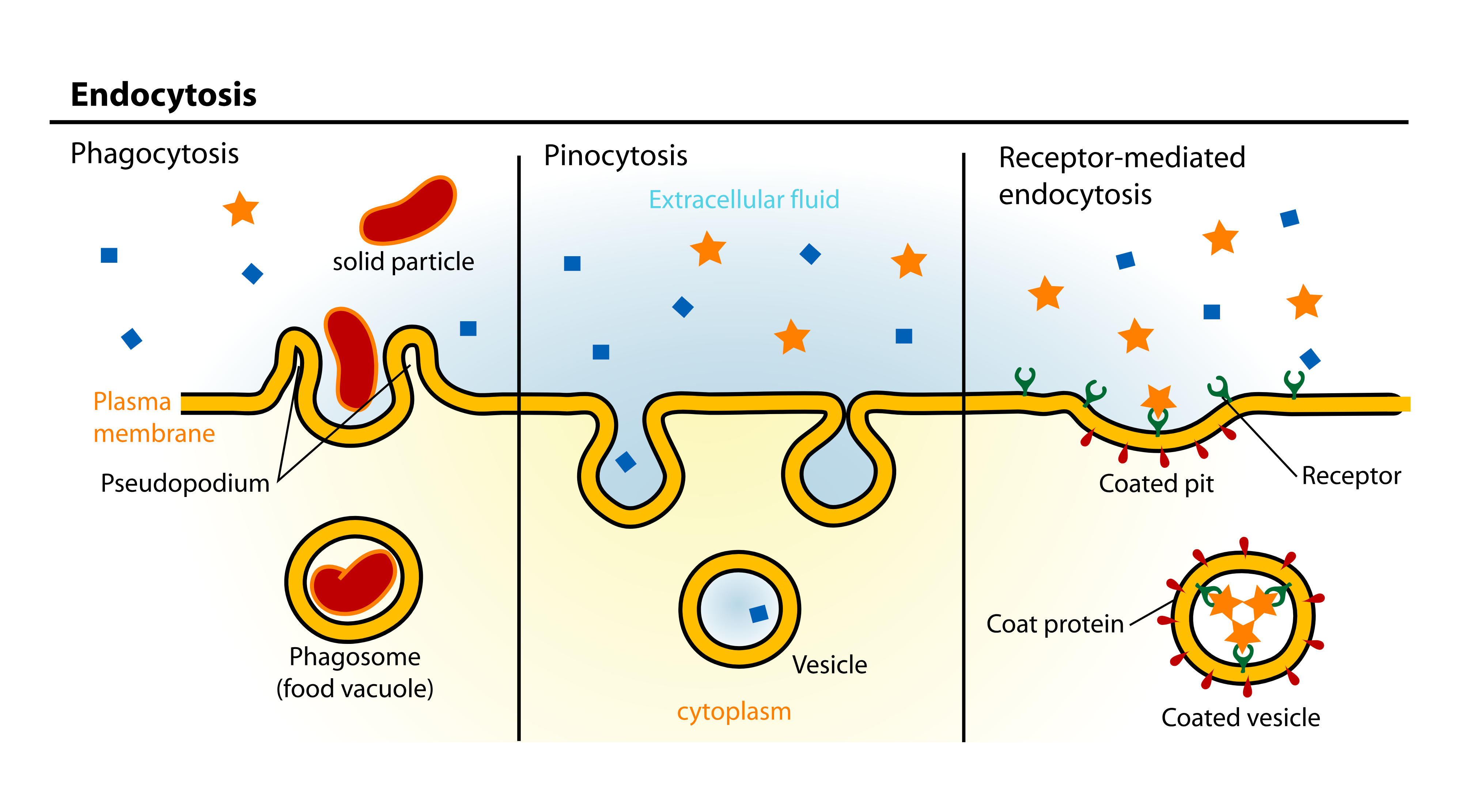
Endocytosis: the membrane invaginates to internalize particles or liquids in an envelope.
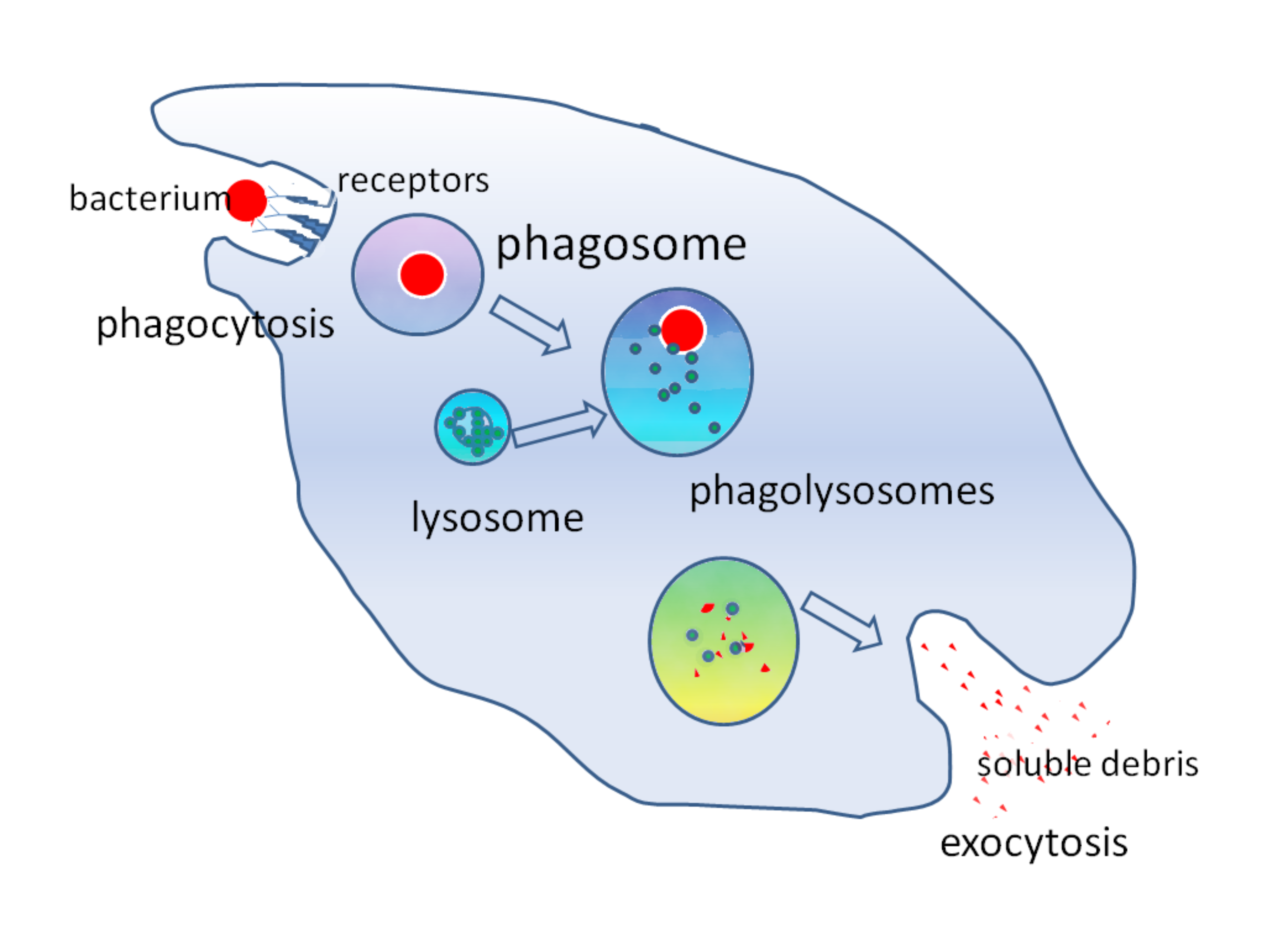
- Phagocytosis- the process by which cells engulf large particles such as bacteria or cellular debris. During this process, the plasma membrane extends around the target, often aided by a coating of clathrin, to form a vesicle that internalizes the material. This vesicle then fuses with a lysosome, creating an endosome where the engulfed substance is degraded and its nutrients extracted. Once digestion is complete, the endosome can fuse with the plasma membrane again to release any residual contents into the extracellular space.
- Pinocytosis- “cell drinking,” involves uptake of extracellular fluid and dissolved molecules into small vesicles that typically do not merge with lysosomes. A specialized form of pinocytosis, potocytosis, utilizes the protein caveolin to form distinct, receptor-rich invaginations called caveolae, which transport molecules across the cell by transcytosis or deliver them to specific organelles, such as the endoplasmic reticulum, for further processing.
- Receptor-mediated endocytosis- a highly selective form of endocytosis in which specific cell surface receptors bind targeted molecules. These receptors, often associated with clathrin coats, trigger the formation of vesicles that internalize the bound substances. This process is critical for removing compounds like low-density lipoprotein (LDL) from the bloodstream. When receptor-mediated endocytosis fails - as seen in familial hypercholesterolemia - LDL accumulates, leading to dangerously high cholesterol levels. Moreover, some pathogens and toxins can exploit this pathway by mimicking natural ligands, thereby gaining entry into the cell.
Exocytosis: releases materials from inside the cell to the extracellular environment. Waste or secretory substances are packaged into a vesicle, and the vesicle then fuses with the inner surface of the plasma membrane. The vesicle membrane merges with the plasma membrane and opens to the outside, discharging its contents into the extracellular space. Exocytosis is responsible for the secretion of extracellular matrix proteins and the release of neurotransmitters into the synaptic cleft via synaptic vesicles.
Cells can also adjust their shape for chemotaxis, often directed by the cytoskeleton. The membrane allows small nonpolar molecules to diffuse directly through the bilayer, while ions generally require membrane channels or pumps.
Membrane transport
Transport across the membrane depends on thermodynamic considerations. Mixing charged ions with the hydrophobic interior of the bilayer is unfavorable, so ions typically need assistance to cross.
Osmosis allows water to diffuse freely and can generate colligative properties that affect osmotic pressure. If osmotic pressure becomes excessive, the cell risks lysis.
Passive transport (facilitated diffusion) moves substances down a concentration gradient without ATP. Active transport (such as the sodium-potassium pump) uses ATP to move solutes against their gradients, helping maintain a negative membrane potential.
Membrane receptors and cell signaling
The membrane also contains membrane receptors that initiate cell signaling pathways by producing second messengers, which change intracellular processes. Signaling types include contact signaling, chemical signaling, and electrical signaling, such as neurotransmitter release in neurons or action potentials in muscle cells.
Intercellular junctions
In tissue organization, intercellular junctions play a key role:
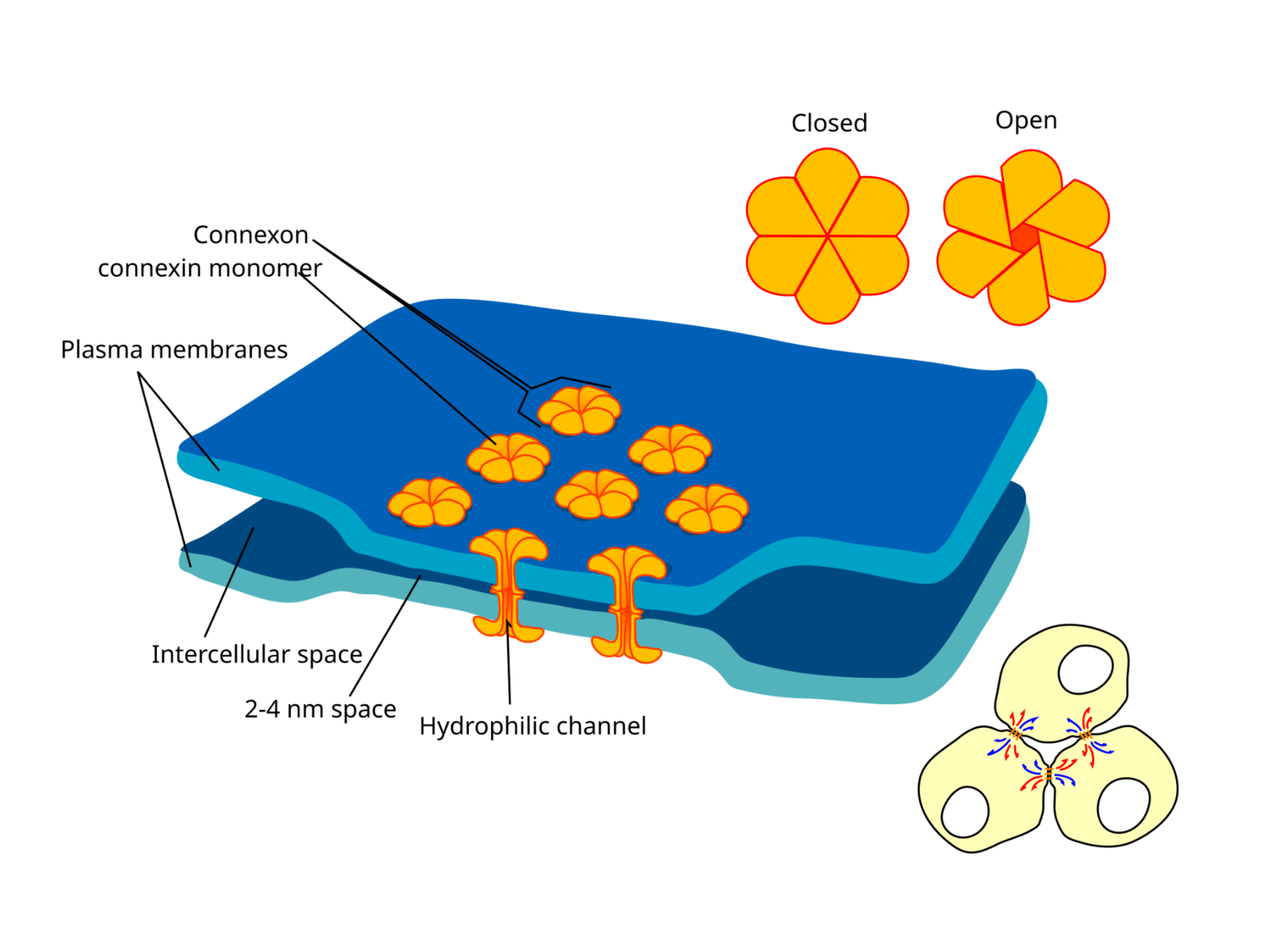
- Gap junctions permit direct exchange of small molecules between adjacent cells
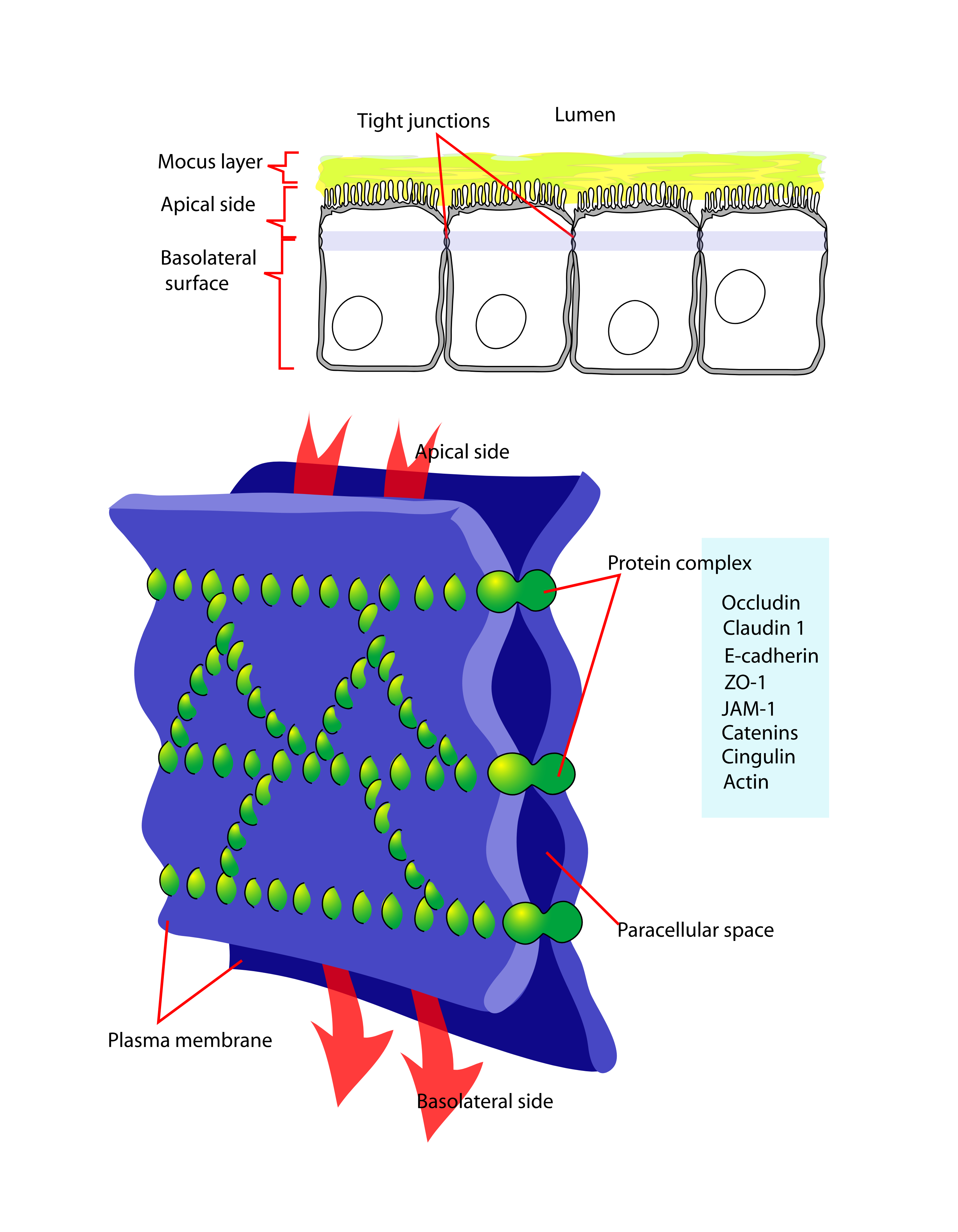
- Tight junctions form barriers that prevent leakage
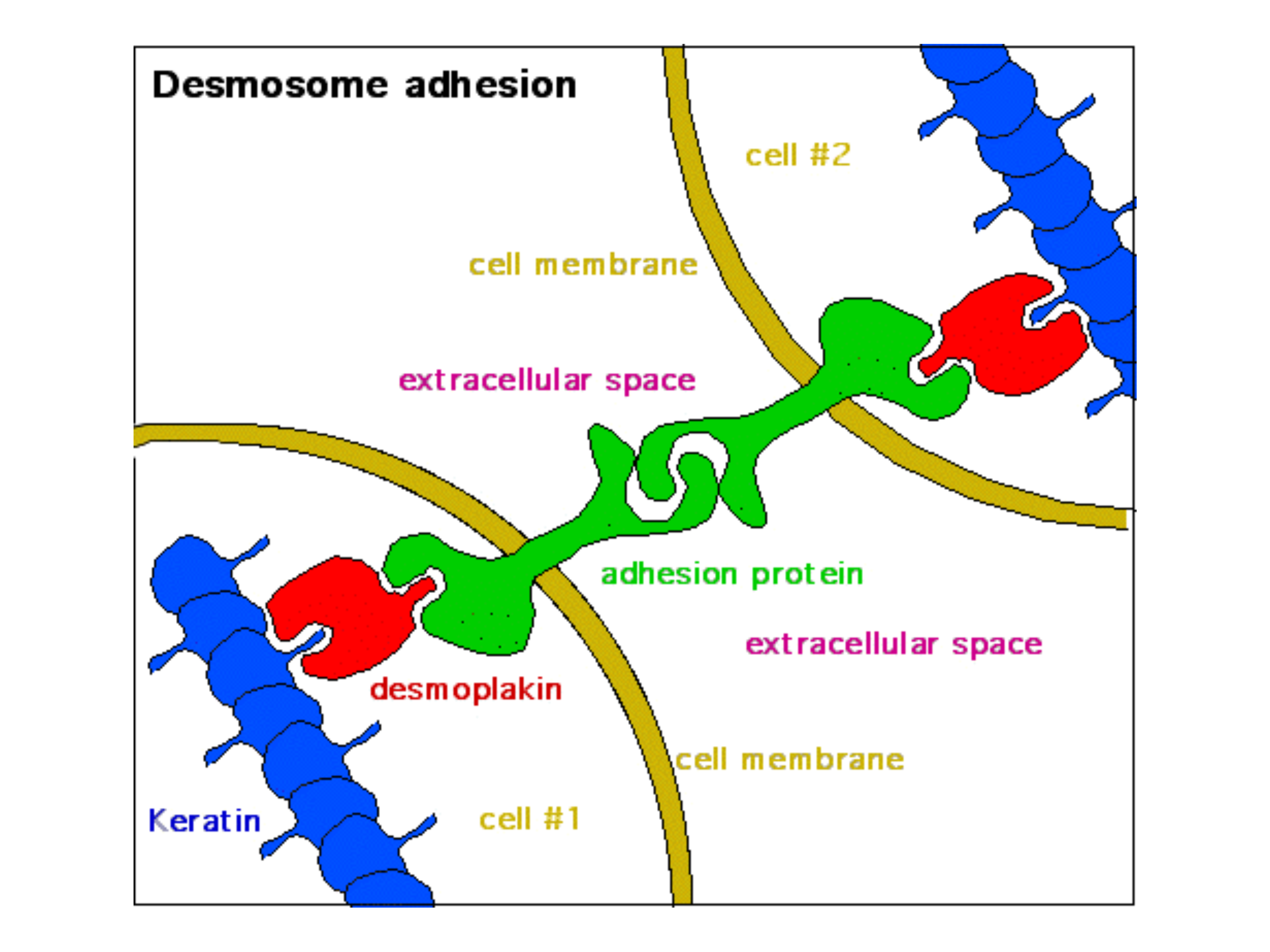
- Desmosomes anchor cells together by linking their cytoskeletons