Musculoskeletal system
The musculoskeletal system provides the body’s structural framework. It includes bones, joints, muscles, and connective tissues.
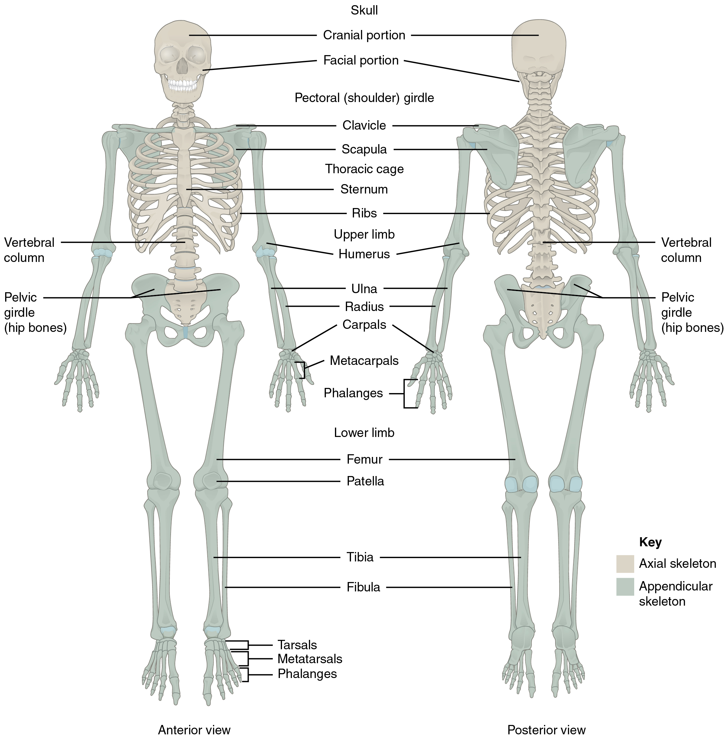
The skeleton
- Structure: The human skeleton consists of 206 bones divided into two main groups:
- Axial skeleton: Includes the skull, vertebral column, and rib cage.
- Appendicular skeleton: Includes the shoulder girdle, pelvic girdle, arms, and legs.
- Functions:
- Provides support and structure to the body.
- Protects vital organs like the brain, heart, and lungs.
- Serves as a system of levers for movement.
- Acts as a mineral reservoir, especially for calcium and phosphorus.
- Serves as the site of blood cell formation (haematopoiesis).
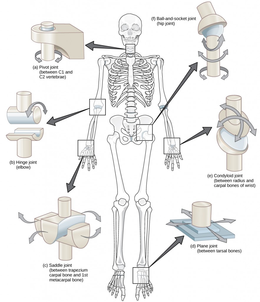
Joints
- Joints are the points where two or more bones meet. They allow movement and flexibility and are classified as:
| Type | Examples | Movement allowed |
|---|---|---|
| Fibrous joints | Sutures in the skull | Minimal |
| Cartilaginous joints | Intervertebral disks | Limited |
| Synovial joints | Elbow, knee | Considerable (multi-axial motion) |
In synovial joints, the articulating bone ends are covered with smooth hyaline cartilage. The entire joint is enclosed in a capsule that contains synovial fluid.
- Synovial joint features:
- Reduces friction and supports movement.
- Types include hinge joints (e.g., elbow) and ball-and-socket joints (e.g., hip).
Joints facilitate movement by rotating around axes. They can also be classified by the number of movement directions:
- Uniaxial joints: Rotate about one axis (e.g., elbow). The knee, often called a hinge joint, has a changing axis of rotation throughout its range of motion.
- Biaxial joints: Allow movement about two perpendicular axes (e.g., ankle, wrist).
- Multiaxial joints: Permit movement about three perpendicular axes (e.g., shoulder, hip).
The vertebral column consists of:
- 7 cervical vertebrae: Neck region.
- 12 thoracic vertebrae: Middle to upper back.
- 5 lumbar vertebrae: Lower back.
- 5 sacral vertebrae: Fused to form the rear pelvis.
- 3-5 coccygeal vertebrae: Vestigial tail extending from the pelvis.
- Intervertebral discs: Fibrocartilaginous structures located between vertebrae that provide cushioning and shock absorption.
Skeletal musculature
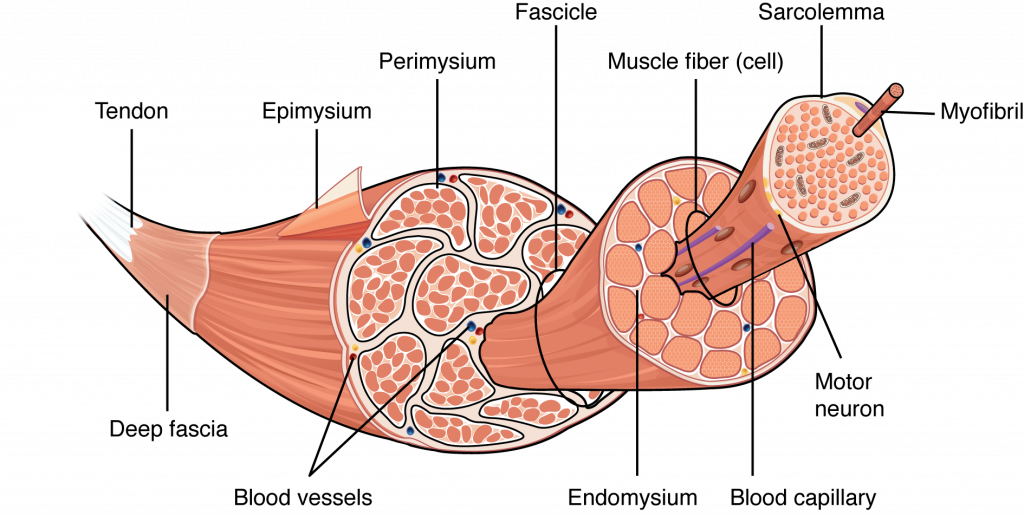
The musculoskeletal system also includes over 430 skeletal muscles. Each muscle contributes to movement by attaching to bones via tendons. When a muscle contracts, it generates a pulling force that is transmitted to bones through the body’s system of levers.
Muscle structure and function
-
Tendon attachment: Tendons connect muscles to the periosteum (a connective tissue covering bones). When the muscle contracts, it pulls on the tendon, which pulls on the bone.
- Limb muscles attachments: Proximal (closer to the trunk) and distal (farther from the trunk); referred to as origin and insertion, respectively.
- Trunk muscles attachments: Superior (closer to the head) and inferior (closer to the feet); also described as origin and insertion.
-
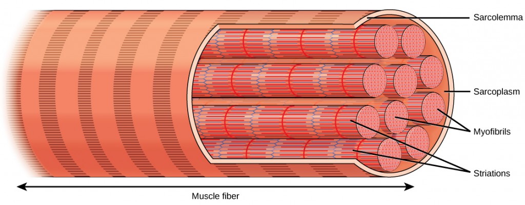
Muscle fibers: Long, cylindrical cells (50-100 µm in diameter) with multiple nuclei located at the periphery. Under magnification, they appear striated. Fibers are organized into bundles (fasciculi) surrounded by perimysium, and individual fibers are encased in endomysium.
-
Connective tissue layers: Epimysium, perimysium, and endomysium are continuous with the tendon. This continuity allows force from muscle contraction to transmit to the attached bone.
-
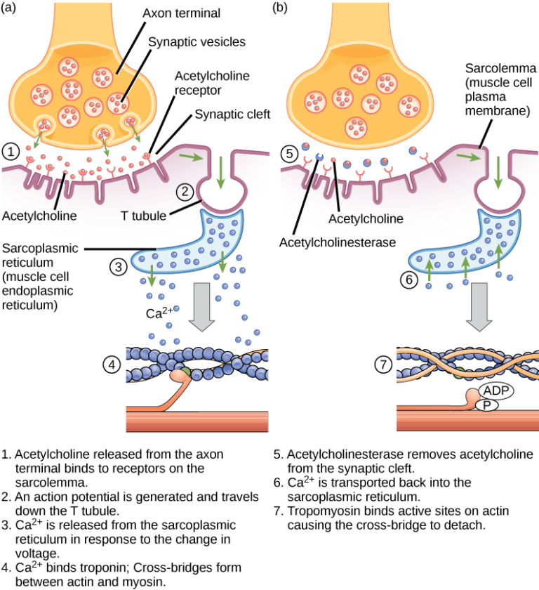
Neuromuscular junction (NMJ): The junction where a motor neuron communicates with muscle fibers. Each muscle fiber has one NMJ, but a motor neuron may innervate multiple fibers, forming a motor unit. When activated, all fibers in a motor unit contract simultaneously.
-
The sarcoplasm: Contains contractile proteins (actin and myosin), glycogen, fat particles, enzymes, mitochondria, and the sarcoplasmic reticulum (SR).
- T-tubules: Extensions of the sarcolemma that transmit action potentials deep into the muscle fiber. This helps synchronize contraction by triggering calcium release from the SR.
-
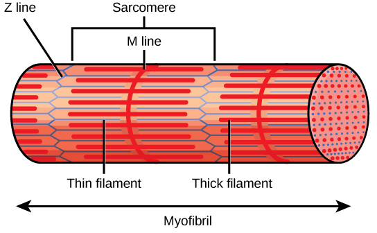
Myofibrils and sarcomeres: Myofibrils dominate the sarcoplasm and are made of repeating sarcomeres, the smallest contractile units of muscle.
- Myosin (thick filament): Contains globular heads, hinge points, and fibrous tails, forming cross-bridges with actin.
- Actin (thin filament): Arranged in a double helix and anchored at Z-lines.
- Sarcomere structure: Actin and myosin are organized longitudinally. Myosin anchors at the M-line (center of the H-zone), while actin attaches at Z-lines. Each sarcomere averages 2.5 µm in length.
-
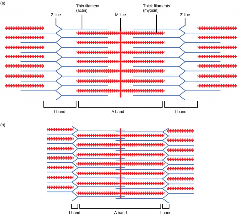
Filament arrangement: Six actin filaments surround each myosin filament, and each actin filament is surrounded by three myosin filaments. This arrangement supports efficient interaction during contraction.
-
Epimysium: A connective tissue encasing the muscle.
-
Muscle fiber: Contains many nuclei and is encased by the sarcolemma.
-
Fasciculi: Bundles of muscle fibers encased by the perimysium.
| Layer | Encases | Purpose |
|---|---|---|
| Epimysium | Entire muscle | Provides overall structural integrity |
| Perimysium | Muscle fascicles | Groups fibers for effective contraction |
| Endomysium | Individual fibers | Maintains alignment of each muscle fiber |
Muscle contraction mechanisms
The sliding filament theory explains muscle contraction by describing how actin and myosin interact within a sarcomere. During contraction, the filaments slide past each other, which shortens the sarcomere and generates force.
Structure of a sarcomere
Key components:
- Z-Line: Marks the boundary of each sarcomere.
- M-Line: Located in the center of the sarcomere, where myosin filaments anchor.
- H-Zone: Region containing only myosin filaments; decreases during contraction.
- I-Band: Area containing only actin filaments; also shortens during contraction.
- A-Band: Length of the myosin filaments; remains constant during contraction.
| Region | Contains | Changes during contraction |
|---|---|---|
| Z-Line | Actin attachment | Moves closer together |
| M-Line | Myosin attachment | Remains unchanged |
| H-Zone | Myosin only | Decreases |
| I-Band | Actin only | Shortens |
| A-Band | Myosin length | Unchanged |
Phases of muscle contraction
- Resting phase:
- Actin and myosin are not interacting; calcium ions are stored in the sarcoplasmic reticulum.
- Excitation-contraction coupling phase:
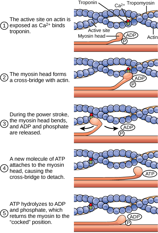
- Calcium binds to troponin, causing a shift in tropomyosin and exposing actin-binding sites for myosin crossbridges.
- Contraction phase:
- Myosin heads bind to actin, forming crossbridges. ATP hydrolysis provides energy for the power stroke, shortening the sarcomere.
- Recharge phase:
- ATP is required to detach the myosin heads and reset for another cycle.
- Relaxation phase:
- Calcium is pumped back into the sarcoplasmic reticulum, and the muscle returns to its resting state.