Cardiovascular and respiratory system
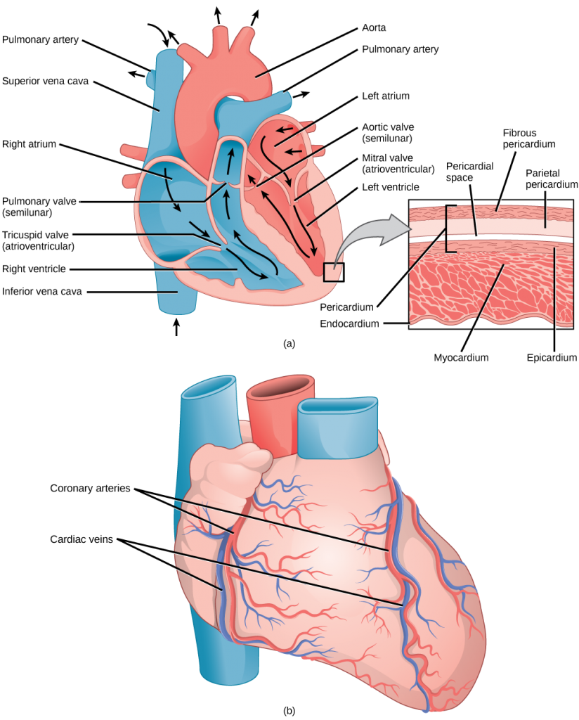
Heart
- Composed of two pumps:
- Right side: Pumps deoxygenated blood to the lungs for oxygenation.
- Left side: Pumps oxygenated blood throughout the body.
The heart is a muscular pump that generates cardiac output (CO), the amount of blood pumped per minute.
- Cardiac output (CO) = heart rate (HR) × stroke volume (SV)
The heart has two ventricles - the right ventricle and the left ventricle - that pump blood to different circulations.
- Stroke volume (SV): The amount of blood ejected from a ventricle with each beat. SV is influenced by preload, afterload, and contractility.
- Ejection fraction (EF): The percentage of the ventricle’s filled volume that is ejected with each beat. EF is a common measure of how efficiently the ventricle pumps.
- Right ventricle:
- Pumps deoxygenated blood from the right atrium to the lungs through the pulmonary arteries.
- Plays a key role in pulmonary circulation, where blood is oxygenated in the lungs.
- Generates lower pressure than the left ventricle because it only pumps blood to the lungs.
- Left ventricle:
- Pumps oxygenated blood from the left atrium to the rest of the body through the aorta.
- Plays a key role in systemic circulation, ensuring all tissues receive oxygen and nutrients.
- Has a thicker muscular wall than the right ventricle because it must generate high pressure to pump blood throughout the body.
Key differences between ventricles
| Feature | Right ventricle | Left ventricle |
|---|---|---|
| Blood type pumped | Deoxygenated | Oxygenated |
| Destination of blood | Lungs (pulmonary circulation) | Body (systemic circulation) |
| Wall thickness | Thin | Thick |
| Pressure generated | Low | High |
Valves of the heart
- Tricuspid and mitral valves: Prevent backflow into the atria during ventricular contraction.
- Pulmonary and aortic valves: Prevent backflow into the ventricles after blood has been ejected.
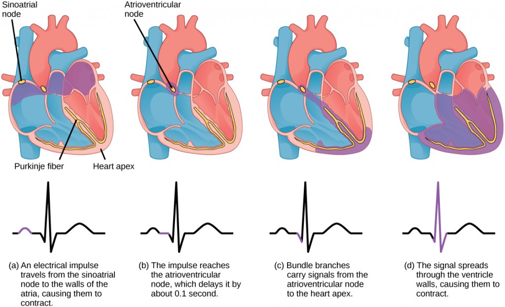
Electrical conduction of the heart
The heart’s rhythmic contractions are controlled by an intrinsic electrical system, as shown in the image below.
Conduction pathway
- Sinoatrial (SA) node: The natural pacemaker of the heart.
- Discharges at 60-80 beats per minute.
- Atrioventricular (AV) node: Delays the electrical signal so the atria can contract before the ventricles.
- Discharges at 40-60 beats per minute.
- Purkinje fibers: Rapidly conduct the signal through the ventricles, producing a coordinated ventricular contraction.
Electrocardiogram (ECG)
- A graphical representation of the heart’s electrical activity.
- Key components:
- P-wave: Atrial depolarization.
- QRS complex: Ventricular depolarization.
- T-wave: Ventricular repolarization.
- PR interval: Represents conduction time through the AV node.
- ST segment: Elevation or depression can indicate underlying pathology, such as myocardial ischemia or infarction.
Blood vessels
The circulatory system consists of arteries, capillaries, and veins. Each type of vessel is built for a specific job.
Arteries
- Carry oxygenated blood away from the heart.
- Walls are muscular and elastic to handle high pressure.
Capillaries
- Facilitate exchange of gases, nutrients, and waste between blood and tissues.
- Walls are thin and permeable to support diffusion.
Veins
- Return deoxygenated blood to the heart.
- Equipped with valves to prevent backflow.
- Operate under low pressure, relying on pressure gradients, the muscle pump, and valves to aid venous return.
Blood
Blood performs critical functions, including:
- Transporting oxygen and nutrients.
- Removing carbon dioxide and waste.
- Regulating body temperature and pH levels.
Hemoglobin
- The oxygen-carrying protein in red blood cells.
- Plays a key role in buffering blood acidity.
- Its ability to bind and release oxygen is described by the oxygen-hemoglobin dissociation curve, which shows the relationship between oxygen partial pressure (PO₂) and hemoglobin saturation. A rightward shift (caused by factors such as increased temperature, CO₂, or acidity) promotes oxygen release to working muscles, while a leftward shift favors oxygen binding in the lungs.
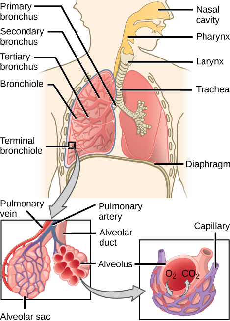
The respiratory system
The respiratory system facilitates gas exchange, ensuring the body receives oxygen and removes carbon dioxide.
Air travels through the trachea, bronchi, and bronchioles before reaching the alveoli, where gas exchange occurs. Breathing depends on pressure gradients that draw air into the lungs (inspiration) and push air out (expiration).
Key pressures include:
- Pleural pressure: The pressure in the narrow space between the lung pleura and chest wall. It’s normally negative during respiration.
- Alveolar pressure: The pressure inside the alveoli. It must drop below atmospheric pressure during inspiration to allow airflow into the lungs.
Gas exchange occurs by diffusion at the alveoli, driven by partial pressure differences of oxygen (PO₂) and carbon dioxide (PCO₂). Hemoglobin in red blood cells plays a critical role in oxygen transport and buffering blood acidity.
During exercise, respiratory muscles like the diaphragm and intercostals work harder to meet oxygen demand and remove carbon dioxide. Regular training improves respiratory efficiency and strengthens breathing muscles. The Valsalva maneuver can help stabilize the trunk during heavy lifting but also carries risks, such as increased blood pressure, making it an important consideration in resistance training.
Structure
- Upper airway: Nasal cavity, pharynx, larynx.
- Lower airway: Trachea, bronchi, bronchioles, alveoli.
Exchange of gases
- Occurs in the alveoli, where oxygen diffuses into the blood and carbon dioxide diffuses out.
Inspiration and expiration
- Inspiration: The diaphragm contracts, creating negative pressure that draws air into the lungs.
- Expiration: The diaphragm relaxes, forcing air out of the lungs.
Acute responses to exercise: Increased tidal volume, respiratory rate, and minute ventilation to meet the heightened oxygen demand.
Chronic adaptations: Enhanced capillarization of lung tissue, improved efficiency of oxygen diffusion, and stronger respiratory muscles, leading to greater overall ventilatory capacity.