


Achievable logoAchievable logo
CCMA
Sign in
Sign up
Purchase
Textbook
Practice exams
Support
How it works
Exam catalog
Mountain with a flag at the peak
Textbook
1. Medical assistant
2. Electronic records
3. Medical terminology and anatomy
4. The fundamentals of infection control
5. Introduction to vital signs
6. The patient interview and history
7. The physical examination
8. Appointment scheduling
9. Insurance billing
10. Diagnostic coding and the ICD-10-CM System
11. Procedural coding
12. Medical billing and reimbursement essentials
13. Assisting with medical specialties
14. Assisting with the musculoskeletal system
15. Assisting with the cardiovascular system
16. Assisting with the respiratory system
17. Assisting with the nervous system
18. Anatomy and physiology of the urinary system
19. Assisting in obstetrics and gynecology
20. Assisting in endocrinology
21. Assisting in ophthalmology & otolaryngology
22. Assisting in gastroenterology
23. Assisting in the immune & lymphatic systems
24. Assisting in pediatrics: the developmental stages and care
25. The medical assistant’s role in caring for the older patient
26. The role of the medical assistant in physical therapy examination and assessment
27. Preparing for minor surgery: room, solutions, and supplies
28. Introduction to the clinical laboratory
29. Urinalysis
29.1 Urine formation and specimen collection
29.2 Physical examination of urine
29.3 Chemical examination of urine
29.4 Additional CLIA-waived urine tests
29.5 Microscopic preparation of urine and cellular findings
29.6 Urine toxicology, legal issues, and professional practice
30. Blood collection
31. Analysis of blood
32. Electrocardiography and heart structure
33. The principles of pharmacology
34. Essential calculations and measurement systems
35. Solid, liquid, & solutions medication doses
36. Administering medications
37. Metabolism and core nutrient roles
38. Medical emergencies in the healthcare setting
Achievable logoAchievable logo
29.2 Physical examination of urine
Achievable CCMA
29. Urinalysis
Our CCMA course is currently in development and is a work-in-progress.

Physical examination of urine

8 min read
Font
Discuss
Share
Feedback

Routine urinalysis

The minimum volume needed for a routine UA usually is about 10 to 12 mL, but more is preferred. A complete UA is an assessment of the following:

  • Physical properties of the urine
  • Selected chemical measurements that are important for diagnosing diseases
  • Microscopic contents of the urine and its sediment

Physical examination of urine: appearance

The physical examination of urine involves observations regarding the appearance of the specimen. This does not involve chemical analysis but rather observation of the characteristics of color, turbidity (cloudiness), volume, presence of foam or odor, and specific gravity.

Color

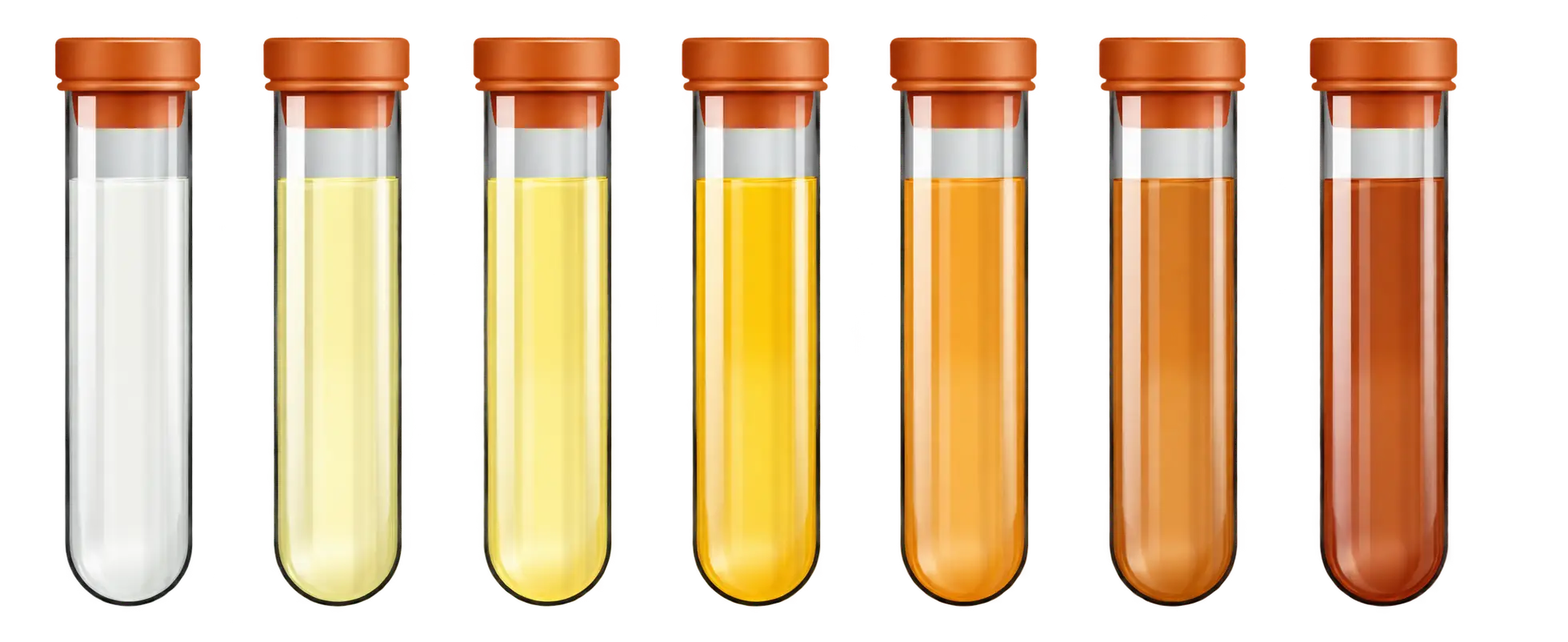
Normal urine is a shade of yellow that ranges from pale straw to yellow to amber. The color depends on the concentration of urochrome, a yellow pigment normally found in urine, and the amount of water in the specimen. A dilute specimen should be a pale straw color, and a more concentrated specimen should be a darker yellow or amber color. First morning specimens will likely be amber in color due to the concentration of the urochrome during the night. Variations in color may also be caused by diet, medication, and disease. Abnormal colors may be related to pathologic or nonpathologic factors.

Turbidity

Both normal and abnormal urine specimens may range in appearance from clear to very cloudy. Turbidity may be caused by cells, bacteria, yeast, vaginal contaminants, or crystals . It is possible for a urine specimen to be clear when voided, but as crystals form and precipitate out of the liquid, it causes the urine to become cloudy as it cools.

Range of urine color variations and clinical significance shown
Urine color variations
Wikimedia Commons
/
CC BY 3.0

Possible causes of urine colors

Color Pathologic cause Nonpathologic cause
Straw Diabetes Diuretics; high fluid intake (coffee, beer)
Amber Dehydration Concentrated first morning specimen; excessive sweating; low fluid intake
Bright yellow — Carotene, vitamins
Red Blood, porphyrins Menstruation, beets, drugs, dyes
Orange-yellow Bile, hepatitis Phenazopyridine (Pyridium, Uristat), dyes, drugs
Greenish yellow Bile, hepatitis Senna (laxative), cascara (laxative), rhubarb
Reddish brown Old blood, methemoglobin —
Brownish black Methemoglobin, melanin Carbidopa-levodopa (Sinemet, Parcopa)
Salmon pink — Amorphous urates
White (milky) Fats, pus Amorphous phosphates
Blue-green Biliverdin, infection with Pseudomonas Vitamin B, drugs, dyes, amitriptyline, indomethacin

Volume

The amount of urine is rarely measured in a random specimen. With a timed specimen such as a 24-hour collection, volume is measured by pouring the complete collection into a large, graduated cylinder . It is not accurate enough to use the markings on the side of the collection container. Once the volume has been measured and recorded, a portion of the well-mixed specimen, called an aliquot, is removed for testing. The remainder is discarded or stored, depending on the preference of the laboratory.

The normal volume of urine produced every 24 hours varies according to the age of the individual. Infants and children produce smaller volumes than adults. The normal adult volume of urine produced is approximately 800 to 2000 mL in 24 hours. Excessive production of urine is called polyuria. This is common in those who have diabetes mellitus, diabetes insipidus, and certain kidney disorders. Oliguria is an insufficient production of urine, which can be caused by dehydration, decreased fluid intake, shock, renal disease, or urinary tract infections. The absence of urine production, anuria, occurs when there is renal obstruction and renal failure.

Foam

Normally the presence of foam is not recorded, but careful observation of this property can detect a significant clue to an abnormality. Foam consists of small bubbles that persist for a long time after the specimen has been swirled to mix. Foam must not be confused with any bubbles that rapidly disappear. White foam can indicate the presence of increased protein.

Greenish-yellow foam may indicate bilirubin in the urine. Care should be taken in handling such urine specimens because the greenish-yellow color may indicate that the patient has viral hepatitis, which is highly contagious. Always observe Standard Precautions and wear appropriate personal protective equipment (PPE) when handling specimens. If foam is observed and seems significant, add a note in the Comments field of a paper or electronic test report.

Odor

As with foam, odor is not normally recorded but can be an important clue to metabolic disorders. Normal urine is said to be aromatic or having a distinct smell. Changes in the odor of urine may be caused by disease, the presence of bacteria, or diet. A patient with diabetes may pass urine that has a fruity odor if he or she is excreting ketones in the urine. An ammonia or a putrid (foul or decaying) smell in the urine can be caused by an infection. An ammonia smell may also be noticed in urine that has been at room temperature for too long before it is tested. Foods such as asparagus and garlic also can produce an abnormal odor in the urine. Urine from a child with phenylketonuria (PKU) may smell “mousy.” If the odor seems significant, add a note in the Comments field of a paper or electronic test report.

Phenylketonuria

Phenylketonuria (PKU) is a rare hereditary condition in which the amino acid phenylalanine is not properly metabolized or broken down in the body. PKU that is undiagnosed or untreated can lead to severe cognitive disabilities. An accumuation of phenylalanine in the blood and urine gives body fluids the odor of wet fur.

Specific gravity

Specific gravity is defined as the weight of a substance compared with the weight of an equal volume of distilled water. In UA, specific gravity is the approximate measurement of the concentration of substances dissolved in the urine. The specific gravity of distilled water is 1.000. The normal specific gravity of urine ranges from 1.005 to 1.030, depending on the patient’s fluid intake. Most samples fall between 1.010 and 1.025. The urine’s specific gravity indicates whether the kidneys can concentrate the urine. A change in specific gravity is one of the first indications of kidney disease. Conditions such as glomerulonephritis , chronic renal insufficiency, or diabetes may lower the specific gravity. To measure specific gravity, laboratories may use a refractometer, but most use Clinical Laboratory Improvement Amendments (CLIA)-waived chemical reagent strips.

A refractometer measures the refraction (bending) of light through solids in a liquid. The result is called the refractive index, which, for our purposes, is the same as specific gravity. The refractometer requires only a drop of urine. One drop of well-mixed urine is placed under the hinged cover of the instrument. Then the value is read directly from a scale viewed through the eyepiece. The scale on the left side of the circle shows a urine-specific gravity of 1.020. The refractometer must be calibrated daily with distilled water, which should read 1.000. Note that specific gravity carries no unit of measure after the number.

The analysis that uses a reagent strip, also called a urine dipstick, is a CLIA-waived test. A reagent strip test is the most common method used for measuring specific gravity in the POL. The pad on the strip contains a chemical that is sensitive to positively charged ions , such as sodium (Na+) and potassium (K+). The pad detects the urine’s specific gravity. Various color changes indicate values between 1.000 and 1.030.

Routine urinalysis overview

  • Minimum volume: 10–12 mL (more preferred)
  • Assesses physical, chemical, and microscopic properties
  • Key for diagnosing diseases

Physical examination of urine: appearance

  • Observes color, turbidity, volume, foam, odor, specific gravity
  • No chemical analysis in this step

Color

  • Normal: pale straw to amber (depends on urochrome concentration)
  • Abnormal colors linked to pathologic (e.g., blood, bile, infection) and nonpathologic (e.g., diet, drugs) causes
    • Table of colors: straw (diabetes, diuretics), amber (dehydration), red (blood, menstruation), etc.

Turbidity

  • Normal: clear to slightly cloudy
  • Causes: cells, bacteria, yeast, vaginal contaminants, crystals
  • Cloudiness can develop as urine cools

Volume

  • Normal adult: 800–2000 mL/24 hours
  • Polyuria: excessive urine (diabetes, kidney disorders)
  • Oliguria: low urine output (dehydration, renal disease)
  • Anuria: no urine (renal failure, obstruction)

Foam

  • Not usually recorded, but can indicate abnormalities
  • White foam: increased protein
  • Greenish-yellow foam: bilirubin (possible hepatitis, use PPE)

Odor

  • Normal: aromatic
  • Fruity odor: ketones (diabetes)
  • Ammonia/putrid: infection or old specimen
  • Foods (asparagus, garlic) can alter odor
  • PKU: “mousy” odor due to phenylalanine accumulation

Phenylketonuria (PKU)

  • Hereditary disorder: inability to metabolize phenylalanine
  • Leads to cognitive disabilities if untreated
  • Urine and body fluids have wet fur odor

Specific gravity

  • Measures urine concentration (compared to distilled water, SG = 1.000)
  • Normal range: 1.005–1.030 (most samples 1.010–1.025)
  • Low SG: glomerulonephritis, chronic renal insufficiency, diabetes
  • Measurement methods:
    • Refractometer: measures light refraction, requires calibration
    • Reagent strip (dipstick): detects Na+, K+ ions, color change indicates SG value

Sign up for free to take 11 quiz questions on this topic

Previous
Next  | 29.3 Chemical examination of urine
All rights reserved ©2016 - 2026 Achievable, Inc.

Physical examination of urine

Routine urinalysis

The minimum volume needed for a routine UA usually is about 10 to 12 mL, but more is preferred. A complete UA is an assessment of the following:

  • Physical properties of the urine
  • Selected chemical measurements that are important for diagnosing diseases
  • Microscopic contents of the urine and its sediment

Physical examination of urine: appearance

The physical examination of urine involves observations regarding the appearance of the specimen. This does not involve chemical analysis but rather observation of the characteristics of color, turbidity (cloudiness), volume, presence of foam or odor, and specific gravity.

Color

Normal urine is a shade of yellow that ranges from pale straw to yellow to amber. The color depends on the concentration of urochrome, a yellow pigment normally found in urine, and the amount of water in the specimen. A dilute specimen should be a pale straw color, and a more concentrated specimen should be a darker yellow or amber color. First morning specimens will likely be amber in color due to the concentration of the urochrome during the night. Variations in color may also be caused by diet, medication, and disease. Abnormal colors may be related to pathologic or nonpathologic factors.

Turbidity

Both normal and abnormal urine specimens may range in appearance from clear to very cloudy. Turbidity may be caused by cells, bacteria, yeast, vaginal contaminants, or crystals . It is possible for a urine specimen to be clear when voided, but as crystals form and precipitate out of the liquid, it causes the urine to become cloudy as it cools.

Possible causes of urine colors

Color Pathologic cause Nonpathologic cause
Straw Diabetes Diuretics; high fluid intake (coffee, beer)
Amber Dehydration Concentrated first morning specimen; excessive sweating; low fluid intake
Bright yellow — Carotene, vitamins
Red Blood, porphyrins Menstruation, beets, drugs, dyes
Orange-yellow Bile, hepatitis Phenazopyridine (Pyridium, Uristat), dyes, drugs
Greenish yellow Bile, hepatitis Senna (laxative), cascara (laxative), rhubarb
Reddish brown Old blood, methemoglobin —
Brownish black Methemoglobin, melanin Carbidopa-levodopa (Sinemet, Parcopa)
Salmon pink — Amorphous urates
White (milky) Fats, pus Amorphous phosphates
Blue-green Biliverdin, infection with Pseudomonas Vitamin B, drugs, dyes, amitriptyline, indomethacin

Volume

The amount of urine is rarely measured in a random specimen. With a timed specimen such as a 24-hour collection, volume is measured by pouring the complete collection into a large, graduated cylinder . It is not accurate enough to use the markings on the side of the collection container. Once the volume has been measured and recorded, a portion of the well-mixed specimen, called an aliquot, is removed for testing. The remainder is discarded or stored, depending on the preference of the laboratory.

The normal volume of urine produced every 24 hours varies according to the age of the individual. Infants and children produce smaller volumes than adults. The normal adult volume of urine produced is approximately 800 to 2000 mL in 24 hours. Excessive production of urine is called polyuria. This is common in those who have diabetes mellitus, diabetes insipidus, and certain kidney disorders. Oliguria is an insufficient production of urine, which can be caused by dehydration, decreased fluid intake, shock, renal disease, or urinary tract infections. The absence of urine production, anuria, occurs when there is renal obstruction and renal failure.

Foam

Normally the presence of foam is not recorded, but careful observation of this property can detect a significant clue to an abnormality. Foam consists of small bubbles that persist for a long time after the specimen has been swirled to mix. Foam must not be confused with any bubbles that rapidly disappear. White foam can indicate the presence of increased protein.

Greenish-yellow foam may indicate bilirubin in the urine. Care should be taken in handling such urine specimens because the greenish-yellow color may indicate that the patient has viral hepatitis, which is highly contagious. Always observe Standard Precautions and wear appropriate personal protective equipment (PPE) when handling specimens. If foam is observed and seems significant, add a note in the Comments field of a paper or electronic test report.

Odor

As with foam, odor is not normally recorded but can be an important clue to metabolic disorders. Normal urine is said to be aromatic or having a distinct smell. Changes in the odor of urine may be caused by disease, the presence of bacteria, or diet. A patient with diabetes may pass urine that has a fruity odor if he or she is excreting ketones in the urine. An ammonia or a putrid (foul or decaying) smell in the urine can be caused by an infection. An ammonia smell may also be noticed in urine that has been at room temperature for too long before it is tested. Foods such as asparagus and garlic also can produce an abnormal odor in the urine. Urine from a child with phenylketonuria (PKU) may smell “mousy.” If the odor seems significant, add a note in the Comments field of a paper or electronic test report.

Phenylketonuria

Phenylketonuria (PKU) is a rare hereditary condition in which the amino acid phenylalanine is not properly metabolized or broken down in the body. PKU that is undiagnosed or untreated can lead to severe cognitive disabilities. An accumuation of phenylalanine in the blood and urine gives body fluids the odor of wet fur.

Specific gravity

Specific gravity is defined as the weight of a substance compared with the weight of an equal volume of distilled water. In UA, specific gravity is the approximate measurement of the concentration of substances dissolved in the urine. The specific gravity of distilled water is 1.000. The normal specific gravity of urine ranges from 1.005 to 1.030, depending on the patient’s fluid intake. Most samples fall between 1.010 and 1.025. The urine’s specific gravity indicates whether the kidneys can concentrate the urine. A change in specific gravity is one of the first indications of kidney disease. Conditions such as glomerulonephritis , chronic renal insufficiency, or diabetes may lower the specific gravity. To measure specific gravity, laboratories may use a refractometer, but most use Clinical Laboratory Improvement Amendments (CLIA)-waived chemical reagent strips.

A refractometer measures the refraction (bending) of light through solids in a liquid. The result is called the refractive index, which, for our purposes, is the same as specific gravity. The refractometer requires only a drop of urine. One drop of well-mixed urine is placed under the hinged cover of the instrument. Then the value is read directly from a scale viewed through the eyepiece. The scale on the left side of the circle shows a urine-specific gravity of 1.020. The refractometer must be calibrated daily with distilled water, which should read 1.000. Note that specific gravity carries no unit of measure after the number.

The analysis that uses a reagent strip, also called a urine dipstick, is a CLIA-waived test. A reagent strip test is the most common method used for measuring specific gravity in the POL. The pad on the strip contains a chemical that is sensitive to positively charged ions , such as sodium (Na+) and potassium (K+). The pad detects the urine’s specific gravity. Various color changes indicate values between 1.000 and 1.030.

Key points

Routine urinalysis overview

  • Minimum volume: 10–12 mL (more preferred)
  • Assesses physical, chemical, and microscopic properties
  • Key for diagnosing diseases

Physical examination of urine: appearance

  • Observes color, turbidity, volume, foam, odor, specific gravity
  • No chemical analysis in this step

Color

  • Normal: pale straw to amber (depends on urochrome concentration)
  • Abnormal colors linked to pathologic (e.g., blood, bile, infection) and nonpathologic (e.g., diet, drugs) causes
    • Table of colors: straw (diabetes, diuretics), amber (dehydration), red (blood, menstruation), etc.

Turbidity

  • Normal: clear to slightly cloudy
  • Causes: cells, bacteria, yeast, vaginal contaminants, crystals
  • Cloudiness can develop as urine cools

Volume

  • Normal adult: 800–2000 mL/24 hours
  • Polyuria: excessive urine (diabetes, kidney disorders)
  • Oliguria: low urine output (dehydration, renal disease)
  • Anuria: no urine (renal failure, obstruction)

Foam

  • Not usually recorded, but can indicate abnormalities
  • White foam: increased protein
  • Greenish-yellow foam: bilirubin (possible hepatitis, use PPE)

Odor

  • Normal: aromatic
  • Fruity odor: ketones (diabetes)
  • Ammonia/putrid: infection or old specimen
  • Foods (asparagus, garlic) can alter odor
  • PKU: “mousy” odor due to phenylalanine accumulation

Phenylketonuria (PKU)

  • Hereditary disorder: inability to metabolize phenylalanine
  • Leads to cognitive disabilities if untreated
  • Urine and body fluids have wet fur odor

Specific gravity

  • Measures urine concentration (compared to distilled water, SG = 1.000)
  • Normal range: 1.005–1.030 (most samples 1.010–1.025)
  • Low SG: glomerulonephritis, chronic renal insufficiency, diabetes
  • Measurement methods:
    • Refractometer: measures light refraction, requires calibration
    • Reagent strip (dipstick): detects Na+, K+ ions, color change indicates SG value

More from Urinalysis

  • Urine formation and specimen collection
  • Chemical examination of urine
  • Additional CLIA-waived urine tests
  • Microscopic preparation of urine and cellular findings
  • Urine toxicology, legal issues, and professional practice