


Achievable logoAchievable logo
CCMA
Sign in
Sign up
Purchase
Textbook
Practice exams
Support
How it works
Exam catalog
Mountain with a flag at the peak
Textbook
1. Medical assistant
2. Electronic records
3. Medical terminology and anatomy
4. The fundamentals of infection control
5. Introduction to vital signs
6. The patient interview and history
7. The physical examination
8. Appointment scheduling
9. Insurance billing
10. Diagnostic coding and the ICD-10-CM System
11. Procedural coding
12. Medical billing and reimbursement essentials
13. Assisting with medical specialties
14. Assisting with the musculoskeletal system
15. Assisting with the cardiovascular system
16. Assisting with the respiratory system
17. Assisting with the nervous system
18. Anatomy and physiology of the urinary system
19. Assisting in obstetrics and gynecology
20. Assisting in endocrinology
21. Assisting in ophthalmology & otolaryngology
22. Assisting in gastroenterology
23. Assisting in the immune & lymphatic systems
24. Assisting in pediatrics: the developmental stages and care
25. The medical assistant’s role in caring for the older patient
26. The role of the medical assistant in physical therapy examination and assessment
27. Preparing for minor surgery: room, solutions, and supplies
28. Introduction to the clinical laboratory
29. Urinalysis
29.1 Urine formation and specimen collection
29.2 Physical examination of urine
29.3 Chemical examination of urine
29.4 Additional CLIA-waived urine tests
29.5 Microscopic preparation of urine and cellular findings
29.6 Urine toxicology, legal issues, and professional practice
30. Blood collection
31. Analysis of blood
32. Electrocardiography and heart structure
33. The principles of pharmacology
34. Essential calculations and measurement systems
35. Solid, liquid, & solutions medication doses
36. Administering medications
37. Metabolism and core nutrient roles
38. Medical emergencies in the healthcare setting
Achievable logoAchievable logo
29.3 Chemical examination of urine
Achievable CCMA
29. Urinalysis
Our CCMA course is currently in development and is a work-in-progress.

Chemical examination of urine

6 min read
Font
Discuss
Share
Feedback

Tests can be performed on urine to detect the presence of certain chemicals, which can provide valuable information to the provider. In certain situations, these chemical test results can be critical to a diagnosis. Also, some urine dipstick results will turn positive before a patient has specific symptoms of a disease or disorder. That is one reason a routine urinalysis can be a screening test for many conditions.

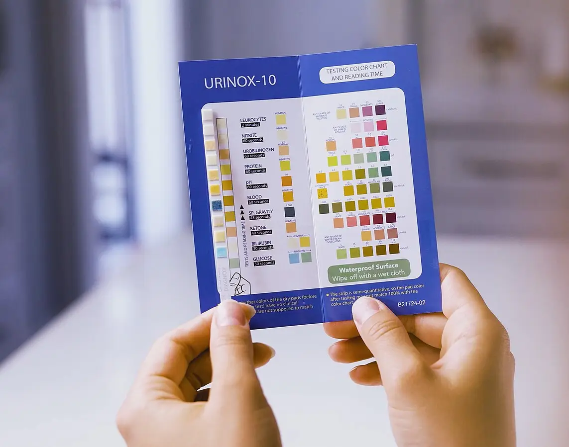
Reagent strip testing is the most widely used technique for detecting chemicals in the urine. These strip tests are available in a variety of types. Dipsticks are plastic strips with paper pads attached. The paper pads contain chemical reagents. The reagents react chemically with analytes in the urine. The chemical reaction is read by looking for a color change on each individual pad. The presence or absence of these chemicals in the urine provides information on the status of carbohydrate metabolism, liver and kidney function, and the patient’s acid-base balance.

Reagent strips are designed to be used once and then discarded in a biohazard waste container. The directions for each type of reagent strip test are included inside the package. Instructions must be followed exactly if accurate results are to be obtained. A color comparison chart is provided on the label of the container. In addition to reagent strips, a few tablet tests are available for chemically testing urine.

All strips and tablets must be kept in tightly closed containers in a cool, dry area and should be removed just before testing. To prevent contamination of the bottle, never touch a strip that has been exposed to urine against the color comparison chart on the bottle. If both a UA and C&S have been ordered for a specimen, separate the sample. The original sample in the sterile container will go to the microbiology department and be used for the C&S. Pour off an aliquot into a non sterile urine tube for the UA. If you were to introduce a reagent strip into the sterile urine sample, it would contaminate the specimen and make it unfit for C&S testing.

Urine reagent dipstick used for urinalysis
Urine reagent strip
Wikimedia Commons
/
CC BY-SA 4.0

pH

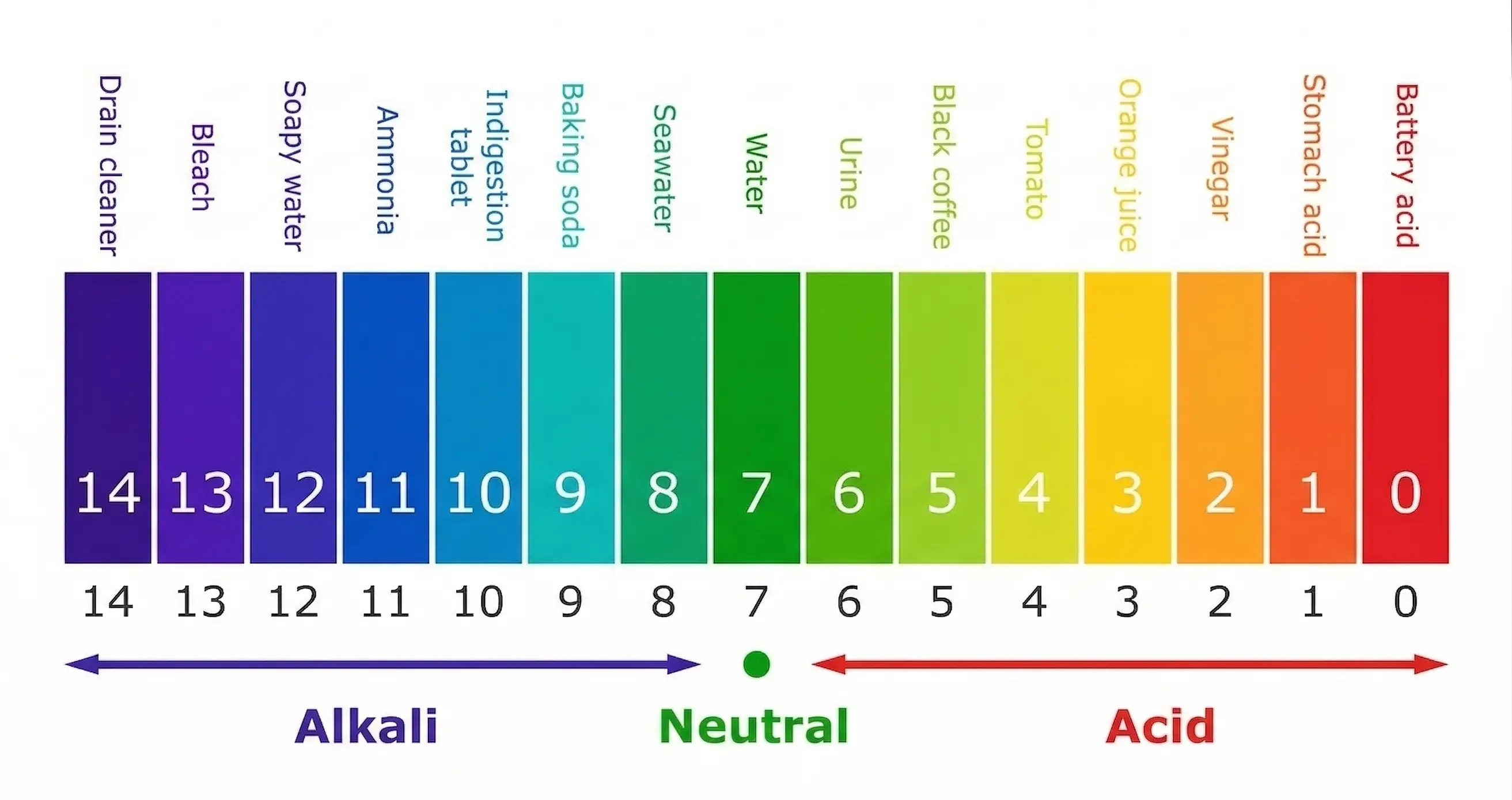
The pH is a measurement of the acidity or alkalinity of the urine. A urine specimen with a pH of 7 is neutral. A value below 7 indicates acidity and above 7 indicates alkalinity. Normal, freshly voided urine may have a pH range of 5.5 to 8. The urinary pH varies with an individual’s metabolic status, diet, drug therapy, and disease. In the case of bacteria in the urine, the urine pH is usually alkaline. Knowing the pH of the sample also helps when identifying crystals that may be found in the urine sediment.

pH scale showing acidic, neutral, and basic values
pH scale

Glucose

Glucose is filtered out of the blood in the glomerulus, and, under normal conditions, most glucose is reabsorbed in the tubules of the nephron. Detectable glycosuria (glucose in the urine) occurs when the filtered glucose in the renal tubules is so high it cannot be reabsorbed into the blood. A positive urine glucose may be the first indication that a patient is diabetic. The glucose pad on a reagent strip test is based on a specific enzyme reaction in which glucose, and no other sugar, causes a positive reaction. Because many types of bacteria use glucose for energy, specimens should be tested for glucose within an hour of collection or within 24 hours if refrigerated. This will reduce the amount of bacteria from overgrowing in the sample.

Glucose reabsorption

The reabsorption of glucose depends on its renal threshold . The renal threshold for blood glucose is below 160 mg/dL. This means that all the glucose will be reabsorbed into the blood if the blood glucose level is below 160 mg/dL. If the blood glucose level is higher than 160 mg/dL, the blood will not reabsorb the glucose. The filtered glucose then remains in the urine, which will show up as a positive test result for glucose during urinalysis.

Ketones

Ketones are the end product of fat metabolism in the body. Ketonuria (ketones in the urine) is commonly seen in patients with diabetes mellitus that is not well controlled, in persons on a very-low-carbohydrate diet (in which case the presence of ketones would be an expected finding and not necessarily considered abnormal.), or after excessive vomiting. Because ketones evaporate at room temperature, urine should be tested immediately, or the specimen should be tightly covered and refrigerated. Examples of ketones include acetoacetate and acetone.

Protein

Protein in the urine in detectable amounts is called proteinuria and is one of the first signs of renal disease. We normally excrete a very small amount of protein every day that is undetectable.

Some causes of proteinuria may include the following:

  • Orthostatic proteinuria: protein is excreted only when the patient is in an upright position.
  • Pregnancy: a common finding in pregnancy that must be monitored along with excessive weight gain and increased blood pressure (there are three possible symptoms of preeclampsia)
  • After heavy exercise

The reagent strip is highly sensitive to urinary albumin and is less sensitive to the other proteins.

Chemical examination of urine

  • Detects chemicals for diagnosis and screening
  • Reagent strip (dipstick) is most common method
  • Provides info on metabolism, liver/kidney function, acid-base balance

Reagent strip testing

  • Single-use plastic strips with reagent pads
  • Color change indicates presence/absence of analytes
  • Follow instructions exactly; store strips properly
  • Avoid contaminating sterile samples (esp. for C&S testing)

pH

  • Measures urine acidity/alkalinity (normal: 5.5–8)
  • pH < 7 = acidic; pH > 7 = alkaline
  • Influenced by metabolism, diet, drugs, disease

Glucose

  • Normally reabsorbed in kidneys; appears if blood glucose > 160 mg/dL (renal threshold)
  • Glycosuria may indicate diabetes
  • Test within 1 hour or refrigerate to prevent bacterial glucose consumption

Ketones

  • End products of fat metabolism (acetoacetate, acetone)
  • Ketonuria seen in uncontrolled diabetes, low-carb diets, vomiting
  • Test immediately or refrigerate; ketones evaporate at room temp

Protein

  • Proteinuria = detectable protein in urine, early sign of renal disease
  • Causes: orthostatic proteinuria, pregnancy (monitor for preeclampsia), heavy exercise
  • Strip most sensitive to albumin

Blood

  • Detects intact RBCs (hematuria), hemoglobin (hemoglobinuria), myoglobin (myoglobinuria)
  • Hematuria: speckled green color; causes include infection, trauma, stones, cystitis
  • Hemoglobinuria: lysed RBCs; may result from sample handling or disease
  • Myoglobinuria: muscle injury; indistinguishable from hemoglobinuria on strip
  • Confirm positive dipstick with microscopic exam

Bilirubin and urobilinogen

  • Bilirubin: not normally in urine; early sign of liver disease or bile duct obstruction
  • Protect samples from light to prevent breakdown
  • Urobilinogen: small normal amounts; elevated in RBC destruction, liver disease, bile duct obstruction

Nitrite

  • Formed when bacteria convert nitrate to nitrite
  • Positive nitrite suggests UTI (esp. E. coli)
  • Negative result does not rule out UTI

Leukocyte esterase

  • Indicates WBCs in urine (suggests UTI)
  • Requires 2 minutes to read result
  • False positives from vaginal contamination or delayed testing

Limitations of reagent strip testing

  • Errors: over-dilution, improper timing, color bleeding, misreading
  • High vitamin C intake may cause false negatives (glucose, nitrite, bilirubin, blood)
  • Automated readers improve consistency but may misread dark urine

Quality assurance and quality control in urinalysis

  • Chemical urinalysis is CLIA-waived
  • Use commercial controls (e.g., Chek-Stix) for reliability
  • Controls include positive and negative standards; compare results to manufacturer chart

Normal urine reference ranges (reagent strips)

  • Color: pale yellow to amber
  • Clarity: clear to slightly turbid
  • Specific gravity: 1.001–1.035
  • pH: 4.6–8
  • Protein, glucose, ketone, bilirubin, blood, nitrite, WBCs: NEG
  • Urobilinogen: 0.1–1 Ehrlich units

Sign up for free to take 16 quiz questions on this topic

Previous
Next  | 29.4 Additional CLIA-waived urine tests
All rights reserved ©2016 - 2026 Achievable, Inc.

Chemical examination of urine

Tests can be performed on urine to detect the presence of certain chemicals, which can provide valuable information to the provider. In certain situations, these chemical test results can be critical to a diagnosis. Also, some urine dipstick results will turn positive before a patient has specific symptoms of a disease or disorder. That is one reason a routine urinalysis can be a screening test for many conditions.

Reagent strip testing is the most widely used technique for detecting chemicals in the urine. These strip tests are available in a variety of types. Dipsticks are plastic strips with paper pads attached. The paper pads contain chemical reagents. The reagents react chemically with analytes in the urine. The chemical reaction is read by looking for a color change on each individual pad. The presence or absence of these chemicals in the urine provides information on the status of carbohydrate metabolism, liver and kidney function, and the patient’s acid-base balance.

Reagent strips are designed to be used once and then discarded in a biohazard waste container. The directions for each type of reagent strip test are included inside the package. Instructions must be followed exactly if accurate results are to be obtained. A color comparison chart is provided on the label of the container. In addition to reagent strips, a few tablet tests are available for chemically testing urine.

All strips and tablets must be kept in tightly closed containers in a cool, dry area and should be removed just before testing. To prevent contamination of the bottle, never touch a strip that has been exposed to urine against the color comparison chart on the bottle. If both a UA and C&S have been ordered for a specimen, separate the sample. The original sample in the sterile container will go to the microbiology department and be used for the C&S. Pour off an aliquot into a non sterile urine tube for the UA. If you were to introduce a reagent strip into the sterile urine sample, it would contaminate the specimen and make it unfit for C&S testing.

pH

The pH is a measurement of the acidity or alkalinity of the urine. A urine specimen with a pH of 7 is neutral. A value below 7 indicates acidity and above 7 indicates alkalinity. Normal, freshly voided urine may have a pH range of 5.5 to 8. The urinary pH varies with an individual’s metabolic status, diet, drug therapy, and disease. In the case of bacteria in the urine, the urine pH is usually alkaline. Knowing the pH of the sample also helps when identifying crystals that may be found in the urine sediment.

Glucose

Glucose is filtered out of the blood in the glomerulus, and, under normal conditions, most glucose is reabsorbed in the tubules of the nephron. Detectable glycosuria (glucose in the urine) occurs when the filtered glucose in the renal tubules is so high it cannot be reabsorbed into the blood. A positive urine glucose may be the first indication that a patient is diabetic. The glucose pad on a reagent strip test is based on a specific enzyme reaction in which glucose, and no other sugar, causes a positive reaction. Because many types of bacteria use glucose for energy, specimens should be tested for glucose within an hour of collection or within 24 hours if refrigerated. This will reduce the amount of bacteria from overgrowing in the sample.

Glucose reabsorption

The reabsorption of glucose depends on its renal threshold . The renal threshold for blood glucose is below 160 mg/dL. This means that all the glucose will be reabsorbed into the blood if the blood glucose level is below 160 mg/dL. If the blood glucose level is higher than 160 mg/dL, the blood will not reabsorb the glucose. The filtered glucose then remains in the urine, which will show up as a positive test result for glucose during urinalysis.

Ketones

Ketones are the end product of fat metabolism in the body. Ketonuria (ketones in the urine) is commonly seen in patients with diabetes mellitus that is not well controlled, in persons on a very-low-carbohydrate diet (in which case the presence of ketones would be an expected finding and not necessarily considered abnormal.), or after excessive vomiting. Because ketones evaporate at room temperature, urine should be tested immediately, or the specimen should be tightly covered and refrigerated. Examples of ketones include acetoacetate and acetone.

Protein

Protein in the urine in detectable amounts is called proteinuria and is one of the first signs of renal disease. We normally excrete a very small amount of protein every day that is undetectable.

Some causes of proteinuria may include the following:

  • Orthostatic proteinuria: protein is excreted only when the patient is in an upright position.
  • Pregnancy: a common finding in pregnancy that must be monitored along with excessive weight gain and increased blood pressure (there are three possible symptoms of preeclampsia)
  • After heavy exercise

The reagent strip is highly sensitive to urinary albumin and is less sensitive to the other proteins.

Key points

Chemical examination of urine

  • Detects chemicals for diagnosis and screening
  • Reagent strip (dipstick) is most common method
  • Provides info on metabolism, liver/kidney function, acid-base balance

Reagent strip testing

  • Single-use plastic strips with reagent pads
  • Color change indicates presence/absence of analytes
  • Follow instructions exactly; store strips properly
  • Avoid contaminating sterile samples (esp. for C&S testing)

pH

  • Measures urine acidity/alkalinity (normal: 5.5–8)
  • pH < 7 = acidic; pH > 7 = alkaline
  • Influenced by metabolism, diet, drugs, disease

Glucose

  • Normally reabsorbed in kidneys; appears if blood glucose > 160 mg/dL (renal threshold)
  • Glycosuria may indicate diabetes
  • Test within 1 hour or refrigerate to prevent bacterial glucose consumption

Ketones

  • End products of fat metabolism (acetoacetate, acetone)
  • Ketonuria seen in uncontrolled diabetes, low-carb diets, vomiting
  • Test immediately or refrigerate; ketones evaporate at room temp

Protein

  • Proteinuria = detectable protein in urine, early sign of renal disease
  • Causes: orthostatic proteinuria, pregnancy (monitor for preeclampsia), heavy exercise
  • Strip most sensitive to albumin

Blood

  • Detects intact RBCs (hematuria), hemoglobin (hemoglobinuria), myoglobin (myoglobinuria)
  • Hematuria: speckled green color; causes include infection, trauma, stones, cystitis
  • Hemoglobinuria: lysed RBCs; may result from sample handling or disease
  • Myoglobinuria: muscle injury; indistinguishable from hemoglobinuria on strip
  • Confirm positive dipstick with microscopic exam

Bilirubin and urobilinogen

  • Bilirubin: not normally in urine; early sign of liver disease or bile duct obstruction
  • Protect samples from light to prevent breakdown
  • Urobilinogen: small normal amounts; elevated in RBC destruction, liver disease, bile duct obstruction

Nitrite

  • Formed when bacteria convert nitrate to nitrite
  • Positive nitrite suggests UTI (esp. E. coli)
  • Negative result does not rule out UTI

Leukocyte esterase

  • Indicates WBCs in urine (suggests UTI)
  • Requires 2 minutes to read result
  • False positives from vaginal contamination or delayed testing

Limitations of reagent strip testing

  • Errors: over-dilution, improper timing, color bleeding, misreading
  • High vitamin C intake may cause false negatives (glucose, nitrite, bilirubin, blood)
  • Automated readers improve consistency but may misread dark urine

Quality assurance and quality control in urinalysis

  • Chemical urinalysis is CLIA-waived
  • Use commercial controls (e.g., Chek-Stix) for reliability
  • Controls include positive and negative standards; compare results to manufacturer chart

Normal urine reference ranges (reagent strips)

  • Color: pale yellow to amber
  • Clarity: clear to slightly turbid
  • Specific gravity: 1.001–1.035
  • pH: 4.6–8
  • Protein, glucose, ketone, bilirubin, blood, nitrite, WBCs: NEG
  • Urobilinogen: 0.1–1 Ehrlich units

More from Urinalysis

  • Urine formation and specimen collection
  • Physical examination of urine
  • Additional CLIA-waived urine tests
  • Microscopic preparation of urine and cellular findings
  • Urine toxicology, legal issues, and professional practice