


Achievable logoAchievable logo
CCMA
Sign in
Sign up
Purchase
Textbook
Practice exams
Support
How it works
Exam catalog
Mountain with a flag at the peak
Textbook
1. Medical assistant
2. Electronic records
3. Medical terminology and anatomy
4. The fundamentals of infection control
5. Introduction to vital signs
6. The patient interview and history
7. The physical examination
8. Appointment scheduling
9. Insurance billing
10. Diagnostic coding and the ICD-10-CM System
11. Procedural coding
12. Medical billing and reimbursement essentials
13. Assisting with medical specialties
14. Assisting with the musculoskeletal system
15. Assisting with the cardiovascular system
16. Assisting with the respiratory system
17. Assisting with the nervous system
18. Anatomy and physiology of the urinary system
19. Assisting in obstetrics and gynecology
20. Assisting in endocrinology
21. Assisting in ophthalmology & otolaryngology
22. Assisting in gastroenterology
23. Assisting in the immune & lymphatic systems
24. Assisting in pediatrics: the developmental stages and care
25. The medical assistant’s role in caring for the older patient
26. The role of the medical assistant in physical therapy examination and assessment
27. Preparing for minor surgery: room, solutions, and supplies
28. Introduction to the clinical laboratory
29. Urinalysis
30. Blood collection
31. Analysis of blood
32. Electrocardiography and heart structure
33. The principles of pharmacology
34. Essential calculations and measurement systems
35. Solid, liquid, & solutions medication doses
36. Administering medications
37. Metabolism and core nutrient roles
38. Medical emergencies in the healthcare setting
38.1 Medical emergencies in the healthcare setting
38.2 Emergency preparedness and community response
38.3 Assisting with medical emergencies
38.4 Management of emergencies (Initial assessment and unresponsive patient care)
38.5 Cardiac emergencies and choking
38.6 Common office emergencies
38.7 Pain and traumatic injury management
38.8 Special needs, metabolic, and final review
Achievable logoAchievable logo
38.7 Pain and traumatic injury management
Achievable CCMA
38. Medical emergencies in the healthcare setting
Our CCMA course is currently in development and is a work-in-progress.

Pain and traumatic injury management

9 min read
Font
Discuss
Share
Feedback

Abdominal pain

Abdominal pain is a symptom caused by many different problems, which can range from acute discomfort to life-threatening complications. The clinician should see every patient who reports abdominal pain; the question is how soon the patient should be seen. A patient with acute onset of severe, persistent abdominal pain, especially when this is accompanied by fever, should receive medical attention as soon as possible. Abdominal pain has a variety of causes, including intestinal infection, appendicitis, ectopic pregnancy, inflammation, hemorrhage, obstruction, and tumor.

Treatment in the ambulatory care office depends on the cause of the pain; however, the medical assistant should follow these general guidelines:

  • Keep the patient warm and quiet.
  • Have an emesis basin available.
  • Administer nothing by mouth (NPO).
  • Do not apply heat to the abdomen unless so instructed by the provider.
  • Administer analgesics as ordered.
  • Check and record the patient’s vital signs and follow the provider’s orders.

Screening guidelines for assessing abdominal pain

  • Assess for shock-related signs and symptoms: diaphoresis; cold, clammy skin; cyanosis or gray pallor; rapid respirations; altered state of consciousness.
  • Is the pain severe and constant, or does it come in waves?
  • Has the patient had any bloody or tarry stools?
  • Is the patient’s temperature higher than 101° F (38.3° C)?
  • Could the patient be pregnant or has she missed a menstrual period?
  • Has the patient experienced continuous vomiting or severe constipation?
  • Are any urinary symptoms present, such as frequency, hematuria, or flank pain?
  • Does the patient have chest pain, shortness of breath, or a continuous cough?
  • Does the patient have a history of serious illness, such as diabetes, heart disease, or cancer?

Sprains and strains

Sprains are tears of the ligaments that support a joint; strains are injuries to a muscle and its tendons. Both types of injury may damage surrounding soft tissues and blood vessels and nearby nerves. With a sprain, the victim develops edema and ecchymosis around the injury, and any movement of the joint, especially a twisting one, produces pain. Usually no swelling or discoloration is seen with a strain, and only mild tenderness is noted unless the injured muscle or tendon is used.

Tendon strains and ligament sprains take several weeks to heal, whereas muscle tears usually heal in 1 to 2 weeks because muscle has such a rich blood supply. These injuries are treated by elevating the affected area and applying mild compression and ice. Swelling is reduced if ice is applied within 20 to 29 minutes of the injury. After 24 to 36 hours, alternating applications of mild heat and ice usually are indicated. The patient may be advised to immobilize the part.

Fractures

A fracture is a break or crack in a bone, which can result from trauma or disease. Fractures are very painful and affect the patient’s ability to freely move the injured part. When a patient with a fracture is brought into the office, the medical assistant should make the patient as comfortable as possible. Place the patient in a position that supports the affected area at the joints above and below the suspected fracture and does not place strain on the injury. Notify the provider immediately and proceed according to the orders given. Emergency treatment for fractures includes preventing movement of the injured part through splinting, elevation of the affected extremity, application of ice, and control of any bleeding. If a patient with an open fracture (i.e., the bone is protruding through the skin) is seen in an ambulatory care office, he or she should be transported to the ED.

Burns

Burns are among the most common causes of injury in the United States. Burn injuries can result from flame, heat, scalds, electricity, chemicals, or radiation. The skin surface may be reddened, blistered, or charred. The depth and extent of a burn are the major determinants in classifying its severity. The extent of the pain is directly proportional to the extent of the surface area burned and the depth and nature of the burn.

To screen a burn injury, the medical assistant must know what caused the burn, its location and approximate size, the depth of the burn, and whether any additional injuries occurred. If the patient reports a chemical burn, it is important to have the person immediately remove all clothing that may have come into contact with the chemical and flood the affected area with running water to flush the irritant off the skin. If the chemical is not quickly flushed away or remains in the patient’s clothing, the agent will continue to burn the skin and may do very serious damage.

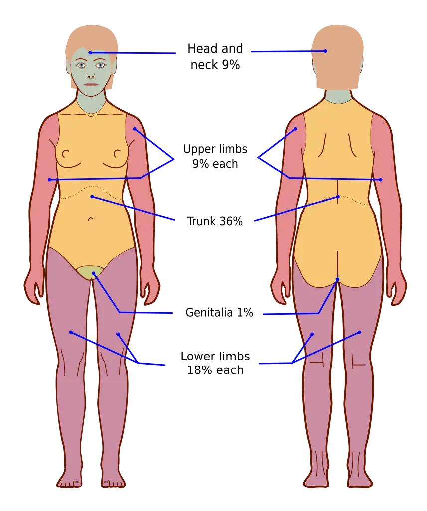
The percentage of the body surface area burned can be estimated using the Rule of Nines . With this assessment tool, the amount of burned tissue can be quickly calculated. The Rule of Nines divides the body into areas approximately equal to 9% of the total body surface area. When a burn victim is assessed, the affected regions are combined to yield an estimate of the total percentage of burned tissue. Partial-thickness burns over 15% of the total body surface and full-thickness burns of less than 2% can be treated in the ambulatory care office if the patient can be seen immediately. Patients with larger body surface area involvement or other complications should be transported immediately to a hospital, preferably one with a burn unit.

Rule of nines for estimating burn area on the body shown
The Rule of Nines
Wikimedia Commons
/
CC BY-SA 3.0

Tissue injuries

Patients may report any of several different types of wounds. A contusion is a closed wound with no evidence of injury to the skin; it typically is caused by blunt trauma, appears swollen and discolored, and is painful. A contusion results in a painful bruise, but the skin remains intact. A scrape on the surface of the skin (e.g., a skinned knee, rug burn) is called an abrasion. A deeper, jagged wound is called a laceration. Additional tissue damage may occur around a laceration; depending on its depth, the wound may need to be repaired surgically. A puncture wound occurs when an object is forced into the body (e.g., stepping on a nail). If an object is lodged in body tissues, the best course is to leave it there, stabilize it as much as possible with rolled-up material, and transport the individual to a clinic or ED. The puncture may have severed blood vessels, and if the object is removed, considerable bleeding may occur. An injury in which tissue is torn away (e.g., complete or partial removal of a finger) is known as an avulsion.

Lacerations are common presentations in a primary care provider’s office. A lacerated wound shows jagged or irregular tearing of the tissues. The severity depends on the cause of the laceration, the site and extent of the injury, and whether the area is contaminated. The injury that caused the laceration also may have damaged blood vessels, nerves, bones, joints, and organs in the body cavities.

When the patient arrives at the facility, put on gloves and notify the provider immediately. Have the patient lie down, and cover the injured area with a sterile dressing (use a dressing that is thick enough to absorb the bleeding). Reassure the patient and explain your actions as much as possible. Ask the patient when he or she last received a tetanus inoculation, and record the date in the patient’s record. If it has been longer than 10 years, the provider probably will want a booster injection given.

Wounds that are not bleeding severely and that do not involve deep tissue damage should be cleaned with antimicrobial soap and water to remove bacteria and other foreign matter. If the laceration is extremely dirty, the provider may want the area irrigated with sterile normal saline solution.

A butterfly closure strip may be used over small lacerations to hold the edges together. If the wound is superficial and has straight edges, it may be closed with a microporous tape (e.g., Steri-Strips), which eliminates the discomfort of suturing and suture removal. Another wound closure option is a tissue adhesive product (e.g., Dermabond fluid or LiquiBand), which forms a strong, flexible closure similar in strength to nylon suture material. Tissue adhesive products are very useful for closing simple lacerations in children; they provide an antimicrobial and waterproof coating to the wound site that lasts several days, even with repeated washing.

After the clinician closes the wound, the medical assistant typically applies a sterile dressing to the site. The size and thickness of the dressing depend on the type of wound.

Nosebleeds (epistaxis)

A nosebleed, or epistaxis, is a hemorrhage that usually results from the rupture of small vessels in the nose. Nosebleeds can be caused by injury, disease, hypertension, strenuous activity, high altitudes, exposure to cold, overuse of anticoagulant medications (e.g., aspirin), and nasal recreational drug use. Bleeding from the anterior nostril area usually is venous, whereas bleeding from the posterior region usually is arterial and is more difficult to stop. Treatment of epistaxis varies according to the amount of bleeding and the presence of other conditions, and whether the patient is taking anticoagulant medications.

If the bleeding is mild to moderate and from one side of the nose, the patient should sit up, lean slightly forward, and apply direct pressure to the affected nostril by pinching the nose. Constant pressure should be continued for 10 to 15 minutes to allow clotting to take place. If the bleeding cannot be controlled, insert a clean gauze pad into the nostril, and notify the provider. If the provider is not available, proceed with standard EMS protocols. Bleeding should be considered a medical emergency if it is bilateral and continuous or if it occurs in a patient who has a bleeding disorder or has been prescribed anticoagulants.

Abdominal pain

  • Many possible causes: infection, appendicitis, ectopic pregnancy, inflammation, hemorrhage, obstruction, tumor
  • Acute, severe, persistent pain (especially with fever) requires immediate attention
  • General care: keep patient warm/quiet, NPO, monitor vitals, no heat unless ordered, analgesics as directed

Screening guidelines for abdominal pain

  • Assess for shock: diaphoresis, clammy skin, cyanosis, rapid respirations, altered consciousness
  • Key questions: pain pattern, bloody/tarry stools, fever >101°F, possible pregnancy, vomiting/constipation, urinary symptoms, chest pain, serious illness history

Sprains and strains

  • Sprain: ligament tear; strain: muscle/tendon injury
  • Sprain: edema, ecchymosis, pain with movement; strain: mild tenderness, little/no swelling
  • Treatment: elevate, mild compression, ice (within 20-29 min), alternate heat/ice after 24-36 hrs, immobilize as needed

Fractures

  • Fracture = break/crack in bone (trauma/disease)
  • Emergency care: support joints above/below, prevent movement (splint), elevate, ice, control bleeding, notify provider
  • Open fractures: transport to ED

Burns

  • Causes: flame, heat, scalds, electricity, chemicals, radiation
  • Severity: determined by depth and body surface area (Rule of Nines)
  • Chemical burns: remove contaminated clothing, flush with water immediately
  • Partial-thickness burns >15% or full-thickness <2% may be treated in office if seen immediately; larger/more severe burns require hospital transfer

Tissue injuries

  • Types: contusion (bruise), abrasion (scrape), laceration (jagged wound), puncture (object forced in), avulsion (tissue torn away)
  • Laceration care: gloves, notify provider, sterile dressing, tetanus status, clean/irrigate if not severe, closure options (butterfly, Steri-Strips, tissue adhesive)
  • Do not remove embedded objects; stabilize and transport

Nosebleeds (epistaxis)

  • Causes: trauma, disease, hypertension, activity, altitude, cold, anticoagulants, drug use
  • Anterior bleeding: usually venous; posterior: arterial, harder to control
  • Treatment: sit up, lean forward, pinch nostril 10-15 min; use gauze if needed; emergency if bilateral/continuous or bleeding disorder present

Sign up for free to take 21 quiz questions on this topic

Previous
Next  | 38.8 Special needs, metabolic, and final review
All rights reserved ©2016 - 2026 Achievable, Inc.

Pain and traumatic injury management

Abdominal pain

Abdominal pain is a symptom caused by many different problems, which can range from acute discomfort to life-threatening complications. The clinician should see every patient who reports abdominal pain; the question is how soon the patient should be seen. A patient with acute onset of severe, persistent abdominal pain, especially when this is accompanied by fever, should receive medical attention as soon as possible. Abdominal pain has a variety of causes, including intestinal infection, appendicitis, ectopic pregnancy, inflammation, hemorrhage, obstruction, and tumor.

Treatment in the ambulatory care office depends on the cause of the pain; however, the medical assistant should follow these general guidelines:

  • Keep the patient warm and quiet.
  • Have an emesis basin available.
  • Administer nothing by mouth (NPO).
  • Do not apply heat to the abdomen unless so instructed by the provider.
  • Administer analgesics as ordered.
  • Check and record the patient’s vital signs and follow the provider’s orders.

Screening guidelines for assessing abdominal pain

  • Assess for shock-related signs and symptoms: diaphoresis; cold, clammy skin; cyanosis or gray pallor; rapid respirations; altered state of consciousness.
  • Is the pain severe and constant, or does it come in waves?
  • Has the patient had any bloody or tarry stools?
  • Is the patient’s temperature higher than 101° F (38.3° C)?
  • Could the patient be pregnant or has she missed a menstrual period?
  • Has the patient experienced continuous vomiting or severe constipation?
  • Are any urinary symptoms present, such as frequency, hematuria, or flank pain?
  • Does the patient have chest pain, shortness of breath, or a continuous cough?
  • Does the patient have a history of serious illness, such as diabetes, heart disease, or cancer?

Sprains and strains

Sprains are tears of the ligaments that support a joint; strains are injuries to a muscle and its tendons. Both types of injury may damage surrounding soft tissues and blood vessels and nearby nerves. With a sprain, the victim develops edema and ecchymosis around the injury, and any movement of the joint, especially a twisting one, produces pain. Usually no swelling or discoloration is seen with a strain, and only mild tenderness is noted unless the injured muscle or tendon is used.

Tendon strains and ligament sprains take several weeks to heal, whereas muscle tears usually heal in 1 to 2 weeks because muscle has such a rich blood supply. These injuries are treated by elevating the affected area and applying mild compression and ice. Swelling is reduced if ice is applied within 20 to 29 minutes of the injury. After 24 to 36 hours, alternating applications of mild heat and ice usually are indicated. The patient may be advised to immobilize the part.

Fractures

A fracture is a break or crack in a bone, which can result from trauma or disease. Fractures are very painful and affect the patient’s ability to freely move the injured part. When a patient with a fracture is brought into the office, the medical assistant should make the patient as comfortable as possible. Place the patient in a position that supports the affected area at the joints above and below the suspected fracture and does not place strain on the injury. Notify the provider immediately and proceed according to the orders given. Emergency treatment for fractures includes preventing movement of the injured part through splinting, elevation of the affected extremity, application of ice, and control of any bleeding. If a patient with an open fracture (i.e., the bone is protruding through the skin) is seen in an ambulatory care office, he or she should be transported to the ED.

Burns

Burns are among the most common causes of injury in the United States. Burn injuries can result from flame, heat, scalds, electricity, chemicals, or radiation. The skin surface may be reddened, blistered, or charred. The depth and extent of a burn are the major determinants in classifying its severity. The extent of the pain is directly proportional to the extent of the surface area burned and the depth and nature of the burn.

To screen a burn injury, the medical assistant must know what caused the burn, its location and approximate size, the depth of the burn, and whether any additional injuries occurred. If the patient reports a chemical burn, it is important to have the person immediately remove all clothing that may have come into contact with the chemical and flood the affected area with running water to flush the irritant off the skin. If the chemical is not quickly flushed away or remains in the patient’s clothing, the agent will continue to burn the skin and may do very serious damage.

The percentage of the body surface area burned can be estimated using the Rule of Nines . With this assessment tool, the amount of burned tissue can be quickly calculated. The Rule of Nines divides the body into areas approximately equal to 9% of the total body surface area. When a burn victim is assessed, the affected regions are combined to yield an estimate of the total percentage of burned tissue. Partial-thickness burns over 15% of the total body surface and full-thickness burns of less than 2% can be treated in the ambulatory care office if the patient can be seen immediately. Patients with larger body surface area involvement or other complications should be transported immediately to a hospital, preferably one with a burn unit.

Tissue injuries

Patients may report any of several different types of wounds. A contusion is a closed wound with no evidence of injury to the skin; it typically is caused by blunt trauma, appears swollen and discolored, and is painful. A contusion results in a painful bruise, but the skin remains intact. A scrape on the surface of the skin (e.g., a skinned knee, rug burn) is called an abrasion. A deeper, jagged wound is called a laceration. Additional tissue damage may occur around a laceration; depending on its depth, the wound may need to be repaired surgically. A puncture wound occurs when an object is forced into the body (e.g., stepping on a nail). If an object is lodged in body tissues, the best course is to leave it there, stabilize it as much as possible with rolled-up material, and transport the individual to a clinic or ED. The puncture may have severed blood vessels, and if the object is removed, considerable bleeding may occur. An injury in which tissue is torn away (e.g., complete or partial removal of a finger) is known as an avulsion.

Lacerations are common presentations in a primary care provider’s office. A lacerated wound shows jagged or irregular tearing of the tissues. The severity depends on the cause of the laceration, the site and extent of the injury, and whether the area is contaminated. The injury that caused the laceration also may have damaged blood vessels, nerves, bones, joints, and organs in the body cavities.

When the patient arrives at the facility, put on gloves and notify the provider immediately. Have the patient lie down, and cover the injured area with a sterile dressing (use a dressing that is thick enough to absorb the bleeding). Reassure the patient and explain your actions as much as possible. Ask the patient when he or she last received a tetanus inoculation, and record the date in the patient’s record. If it has been longer than 10 years, the provider probably will want a booster injection given.

Wounds that are not bleeding severely and that do not involve deep tissue damage should be cleaned with antimicrobial soap and water to remove bacteria and other foreign matter. If the laceration is extremely dirty, the provider may want the area irrigated with sterile normal saline solution.

A butterfly closure strip may be used over small lacerations to hold the edges together. If the wound is superficial and has straight edges, it may be closed with a microporous tape (e.g., Steri-Strips), which eliminates the discomfort of suturing and suture removal. Another wound closure option is a tissue adhesive product (e.g., Dermabond fluid or LiquiBand), which forms a strong, flexible closure similar in strength to nylon suture material. Tissue adhesive products are very useful for closing simple lacerations in children; they provide an antimicrobial and waterproof coating to the wound site that lasts several days, even with repeated washing.

After the clinician closes the wound, the medical assistant typically applies a sterile dressing to the site. The size and thickness of the dressing depend on the type of wound.

Nosebleeds (epistaxis)

A nosebleed, or epistaxis, is a hemorrhage that usually results from the rupture of small vessels in the nose. Nosebleeds can be caused by injury, disease, hypertension, strenuous activity, high altitudes, exposure to cold, overuse of anticoagulant medications (e.g., aspirin), and nasal recreational drug use. Bleeding from the anterior nostril area usually is venous, whereas bleeding from the posterior region usually is arterial and is more difficult to stop. Treatment of epistaxis varies according to the amount of bleeding and the presence of other conditions, and whether the patient is taking anticoagulant medications.

If the bleeding is mild to moderate and from one side of the nose, the patient should sit up, lean slightly forward, and apply direct pressure to the affected nostril by pinching the nose. Constant pressure should be continued for 10 to 15 minutes to allow clotting to take place. If the bleeding cannot be controlled, insert a clean gauze pad into the nostril, and notify the provider. If the provider is not available, proceed with standard EMS protocols. Bleeding should be considered a medical emergency if it is bilateral and continuous or if it occurs in a patient who has a bleeding disorder or has been prescribed anticoagulants.

Key points

Abdominal pain

  • Many possible causes: infection, appendicitis, ectopic pregnancy, inflammation, hemorrhage, obstruction, tumor
  • Acute, severe, persistent pain (especially with fever) requires immediate attention
  • General care: keep patient warm/quiet, NPO, monitor vitals, no heat unless ordered, analgesics as directed

Screening guidelines for abdominal pain

  • Assess for shock: diaphoresis, clammy skin, cyanosis, rapid respirations, altered consciousness
  • Key questions: pain pattern, bloody/tarry stools, fever >101°F, possible pregnancy, vomiting/constipation, urinary symptoms, chest pain, serious illness history

Sprains and strains

  • Sprain: ligament tear; strain: muscle/tendon injury
  • Sprain: edema, ecchymosis, pain with movement; strain: mild tenderness, little/no swelling
  • Treatment: elevate, mild compression, ice (within 20-29 min), alternate heat/ice after 24-36 hrs, immobilize as needed

Fractures

  • Fracture = break/crack in bone (trauma/disease)
  • Emergency care: support joints above/below, prevent movement (splint), elevate, ice, control bleeding, notify provider
  • Open fractures: transport to ED

Burns

  • Causes: flame, heat, scalds, electricity, chemicals, radiation
  • Severity: determined by depth and body surface area (Rule of Nines)
  • Chemical burns: remove contaminated clothing, flush with water immediately
  • Partial-thickness burns >15% or full-thickness <2% may be treated in office if seen immediately; larger/more severe burns require hospital transfer

Tissue injuries

  • Types: contusion (bruise), abrasion (scrape), laceration (jagged wound), puncture (object forced in), avulsion (tissue torn away)
  • Laceration care: gloves, notify provider, sterile dressing, tetanus status, clean/irrigate if not severe, closure options (butterfly, Steri-Strips, tissue adhesive)
  • Do not remove embedded objects; stabilize and transport

Nosebleeds (epistaxis)

  • Causes: trauma, disease, hypertension, activity, altitude, cold, anticoagulants, drug use
  • Anterior bleeding: usually venous; posterior: arterial, harder to control
  • Treatment: sit up, lean forward, pinch nostril 10-15 min; use gauze if needed; emergency if bilateral/continuous or bleeding disorder present

More from Medical emergencies in the healthcare setting

  • Medical emergencies in the healthcare setting
  • Emergency preparedness and community response
  • Assisting with medical emergencies
  • Management of emergencies (Initial assessment and unresponsive patient care)
  • Cardiac emergencies and choking