



Respiration and oxygen saturation
Respiration
The purpose of respiration is to provide for the exchange of oxygen and carbon dioxide among the atmosphere, the blood, and the body cells. Oxygen is taken into the body to be used for life-sustaining body processes, and carbon dioxide is released as a waste product.
One complete inspiration and expiration is called a respiration. During the inspiratory phase, the diaphragm contracts and drops down and the intercostal muscles pull the ribs up and outward; this causes the lungs to expand and fill with air. During the expiratory phase, the diaphragm returns to its normal elevated position and the intercostal muscles relax; this causes the lungs to expel the waste air back into the atmosphere.
Respirations should be assessed immediately after the radial pulse, without alerting the patient that you’re doing so - see the counting technique below for why this matters.
Respiration is both internal and external. External respiration is the exchange of oxygen and carbon dioxide in the lungs. Internal respiration occurs at the cellular level, when oxygen in the bloodstream is transferred into the cells for energy, and carbon dioxide is released as a waste product and transported back to the lungs for exhalation.
The respiratory center in the medulla oblongata, which is the lowest part of the brainstem, where it meets the spinal cord, is sensitive to changes in blood oxygen and carbon dioxide levels. Rising carbon dioxide is the primary trigger for breathing: as CO2 levels climb, the respiratory center signals the diaphragm and intercostal muscles to contract. This makes respiration primarily an involuntary function controlled by the nervous system, though a person can also control it voluntarily to a limited extent - which is why breath-holding is only possible for a short time before rising CO2 forces breathing to resume.
Characteristics of respirations
Normally, a person’s breathing is relaxed, automatic, and silent. However, respiratory disease or chronic conditions can influence the characteristics of an individual’s respirations. Dyspnea occurs in patients with pneumonia, asthma, or chronic obstructive pulmonary disease (COPD). It also occurs after physical exertion or at very high altitudes. Other alterations in breathing are bradypnea, apnea, tachypnea, and hyperpnea. Hyperpnea usually is accompanied by hyperventilation and often occurs when the patient is extremely anxious or in pain. Orthopnea frequently occurs in patients with congestive heart failure (CHF) and COPD. Wheezing signals difficulty breathing in patients with asthma.
When assessing a patient’s respirations, you must note three important characteristics: rate, rhythm, and depth.
- Rate: The rate of respiration is the number of respirations per minute and is described as normal, rapid, or slow. Typically, a ratio of four pulse beats to one respiration is seen. As a rule, both the pulse and respiratory rates respond to exercise or emotional upset.
Approximate age-related respiration ranges:
| Age | Range (breaths/min) | Average |
| Newborn | 30-50 | 40 |
| 1-3 years | 20-30 | 25 |
| 4-6 years | 18-26 | 22 |
| 7-11 years | 16-22 | 19 |
| Adolescence to adulthood | 12-20 | 16 |
- Rhythm: Rhythm refers to the breathing pattern. A regular breathing pattern is normal in adults; however, the breathing pattern for infants varies. Automatic interruptions, such as sighing, are also considered normal.
- Depth: The depth of respiration is the amount of air inhaled and exhaled. When a patient is at rest, normal respirations have a consistent depth, which can be noted as you watch the rise and fall of the chest. Rapid, shallow breathing at rest occurs with some diseases, such as asthma and emphysema. An alteration in the depth and sometimes the rate of breathing is also seen in Cheyne-Stokes respirations.
Normally, no noticeable breath sounds occur during the breathing process, except during snoring. Noticeable breath sounds are a sign of certain diseases, such as pneumonia, asthma, and pulmonary edema. After auscultating breath sounds with a stethoscope, the provider can describe the characteristics of breath sounds using specific terminology.
When an individual cannot inspire enough oxygen to supply all body cells with oxygenated blood, normal skin coloring, particularly around the mouth and the nail beds, changes to a bluish, dusky color. This coloration, called cyanosis, indicates a low level of oxygen in the blood (an increased amount of deoxygenated hemoglobin). The patient also may have other signs and symptoms, such as vertigo, chest pain (angina), and numbness in the fingers and toes.
Counting respirations
Because most people are unaware of their breathing, do not mention that you will be counting the person’s respirations. The respiratory rate is easily controlled, and patients self-consciously alter their breathing rate when they know they are being watched. Therefore, count the respirations while appearing to count the radial pulse. Keep your eyes alternately on the patient’s chest and your watch while you count the pulse rate; then, without removing your fingers from the pulse site, determine the respiratory rate. If the patient is supine, the arm on which you are taking the radial pulse may be crossed over the chest so that respirations can be felt with the rise and fall of the chest. Another way of observing respirations is to watch the movement of the patient’s shoulders with each inspiration. Count the respirations for 30 seconds and multiply the number by 2. Do not use the 15-second interval because this count can vary by a factor of ±4, which is significant when dealing with such a small number. Note any variation or irregularity in the rate. Record the respiratory count in the health record.
Oxygen saturation
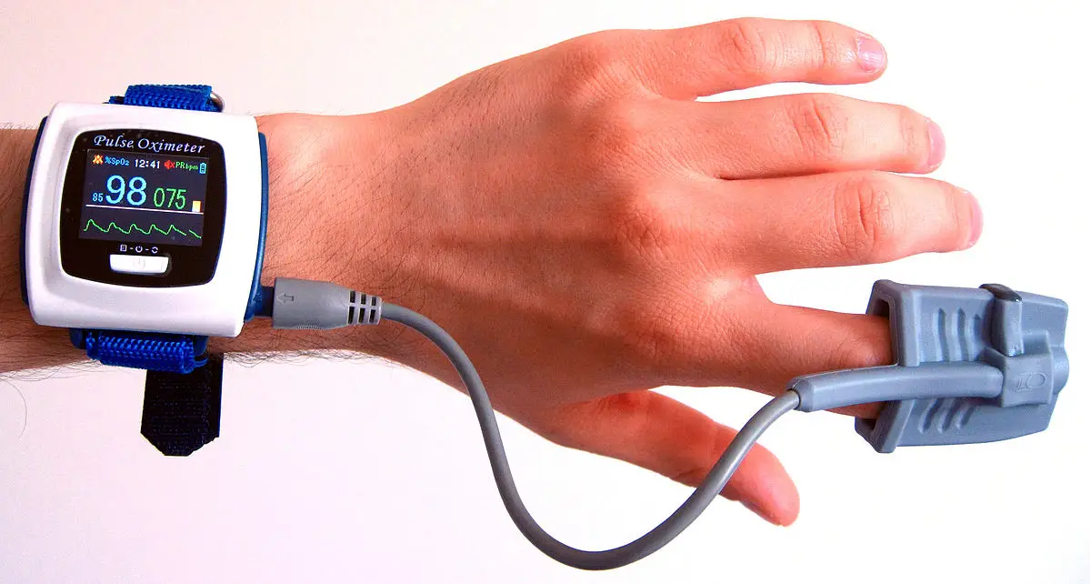
Pulse oximetry is a noninvasive test that measures the pulse electronically and the saturation of oxygen in a patient’s arterial blood. The pulse oximeter includes a sensor and a monitoring device. The sensor is placed on a patient’s finger, earlobe, toe, or bridge of nose.
When using these devices, be certain to attach the clip firmly to the finger, lobe, or nose. There must be a good blood supply at the site where the sensor is placed to detect the oxygen level. For the fingers or toes, you may need to check the capillary refill to determine if the blood supply is adequate. The finger clip also works best when no nail polish is present on the patient’s finger. When using a nose bridge pulse oximeter, make sure there is good skin contact. The nose bridge pulse oximeter should only be used with patients who have good peripheral circulation.
Factors that may affect the measurement of oxygen saturation
Several factors can impact oxygen saturation measurements:
- Poor circulation: If the blood is not reaching the fingertips, the measurement will be low.
- Skin temperature: Cold skin can have less blood flow than warm skin, resulting in a low measurement.
- Tobacco use: The use of tobacco can cause a low measurement.
- Fingernail polish: Nail polish, especially darker colors, affect the infrared light passing through the finger to the sensor on the other side.
Normal pulse oximeter readings range from 95 to 100 %. Values under 90% are considered low. Low blood oxygen is known as hypoxemia. During the pulse oximetry procedure, if the oxygen level is less than 90%, the patient should be asked to take deep breaths to increase their oxygen level. An oxygen level below 90% that does not improve with deep breaths should be reported to the provider.