


Achievable logoAchievable logo
CCMA
Sign in
Sign up
Purchase
Textbook
Practice exams
Support
How it works
Exam catalog
Mountain with a flag at the peak
Textbook
1. Medical assistant
2. Electronic records
3. Medical terminology and anatomy
4. The fundamentals of infection control
5. Introduction to vital signs
5.1 Introduction to vital signs
5.2 Pulse sites and characteristics
5.3 Fever and temperature management
5.4 Respiration and oxygen saturation
5.5 Measuring blood pressure: equipment and techniques
6. The patient interview and history
7. The physical examination
8. Appointment scheduling
9. Insurance billing
10. Diagnostic coding and the ICD-10-CM System
11. Procedural coding
12. Medical billing and reimbursement essentials
13. Assisting with medical specialties
14. Assisting with the musculoskeletal system
15. Assisting with the cardiovascular system
16. Assisting with the respiratory system
17. Assisting with the nervous system
18. Anatomy and physiology of the urinary system
19. Assisting in obstetrics and gynecology
20. Assisting in endocrinology
21. Assisting in ophthalmology & otolaryngology
22. Assisting in gastroenterology
23. Assisting in the immune & lymphatic systems
24. Assisting in pediatrics: the developmental stages and care
25. The medical assistant’s role in caring for the older patient
26. The role of the medical assistant in physical therapy examination and assessment
27. Preparing for minor surgery: room, solutions, and supplies
28. Introduction to the clinical laboratory
29. Urinalysis
30. Blood collection
31. Analysis of blood
32. Electrocardiography and heart structure
33. The principles of pharmacology
34. Essential calculations and measurement systems
35. Solid, liquid, & solutions medication doses
36. Administering medications
37. Metabolism and core nutrient roles
38. Medical emergencies in the healthcare setting
Achievable logoAchievable logo
5.2 Pulse sites and characteristics
Achievable CCMA
5. Introduction to vital signs
Our CCMA course is currently in development and is a work-in-progress.

Pulse sites and characteristics

10 min read
Font
Discuss
Share
Feedback

Pulse

A patient’s pulse rate reflects the palpable beat of the arteries throughout the body as they expand in response to contraction of the heart. With every beat, the heart pumps an amount of blood, known as the stroke volume, into the aorta. Arteries branch off the aorta as it travels down through the center of the abdomen, transferring the pulse beat throughout the body. To measure the pulse, an artery is used that is close to the body surface and can be pushed against a bone. Palpating a** peripheral** pulse gives the rate and rhythm of the heartbeat and local information about the condition of the artery used.

Pulse and respiration are related because the circulatory and respiratory systems work together. Pulse is measured as the number of times the heart beats in one minute. Respiration is the number of times a patient breathes in one minute. One breath, or respiration, equals one inhalation and one exhalation. Usually if either the pulse or respiration rate is high or low, the other is also. Additionally, adult female rates tend to be faster than those for males.

Pulse sites

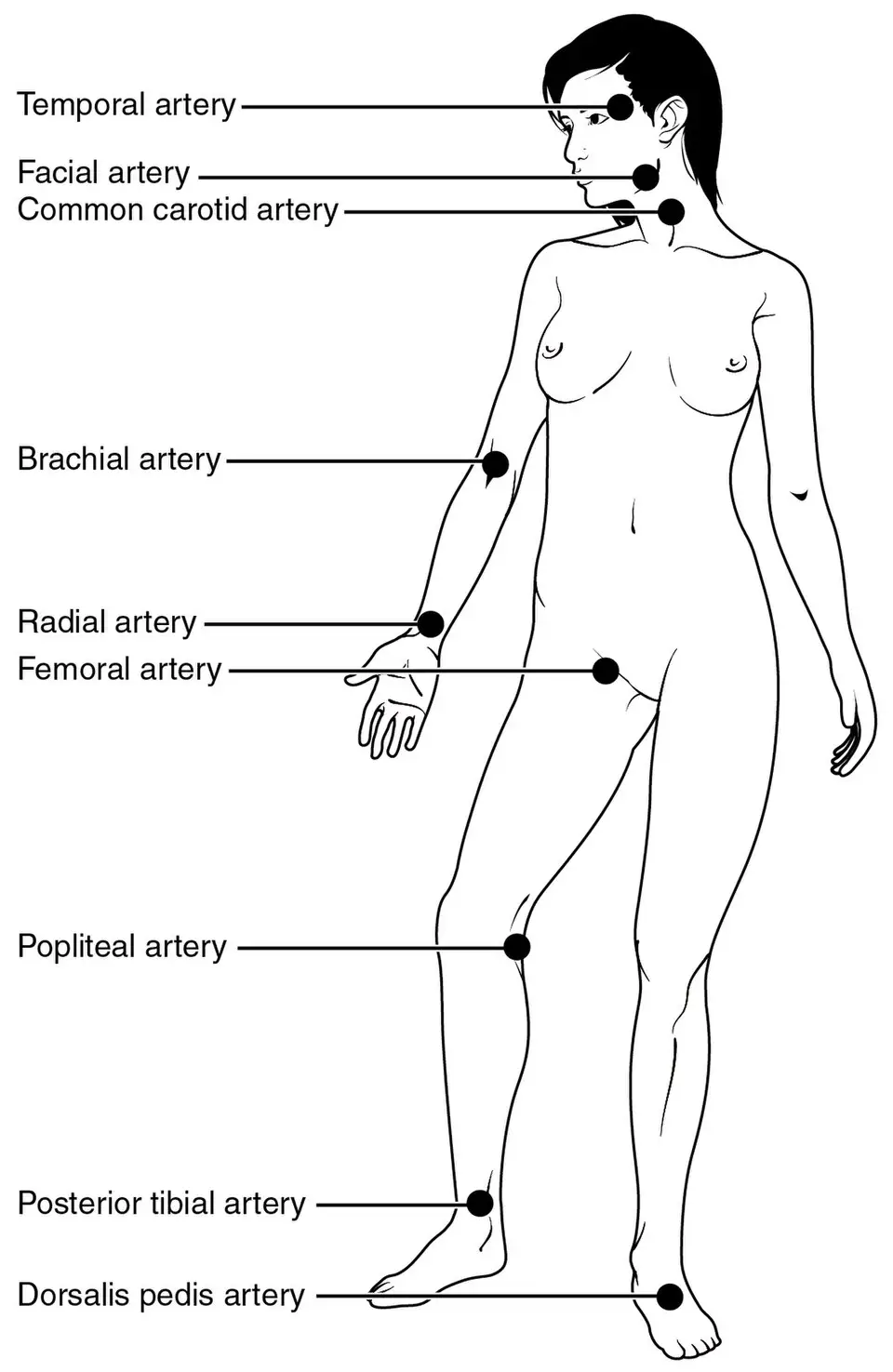
A pulse rate may be counted anyplace an artery is near the surface of the body and the vessel can be pressed against a bone. The most common pulse sites are the temporal, carotid, apical, brachial, radial, femoral, popliteal, and dorsalis pedis arteries.

Common pulse sites on the human body labeled
Pulse sites
Wikimedia Commons
/
CC BY 3.0
The temporal pulse is located in the temple area of the skull, parallel and lateral to the eyes. It is seldom used as a pulse site but may be used as a pressure point to help control bleeding from a head injury.

The carotid artery is located between the larynx and the sternocleidomastoid muscle in the front and to the side of the neck. It most frequently is used in emergencies and to check the pulse during cardiopulmonary resuscitation (CPR). It can be felt by pushing the muscle to the side and pressing against the larynx.

The apical heart rate, or the heartbeat at the apex of the heart, is heard with a stethoscope. It is used for infants and young children because the radial pulse is difficult to palpate in young patients. Apical rates are also recorded on adult patients who have irregular or difficult to feel radial pulses just to make sure you are recording an accurate heart rate. An apical count may be requested if the patient is taking cardiac drugs or has bradycardia or tachycardia. To determine the presence of a pulse deficit, the provider may listen to the apical beat while the medical assistant counts the pulse at another site (usually the radial pulse). The stethoscope is placed at the apex of the heart which is located in the left fifth intercostal space on the midclavicular line, that is, between the fifth and sixth ribs on a line with the midpoint of the left clavicle. The pulse should be counted for 1 full minute and should be documented with (AP) beside the recorded count. Listening for the apical heart rate with a stethoscope is known as auscultation.

The brachial pulse is felt at the inner (antecubital) aspect of the elbow. This is the artery that is felt and heard when blood pressure is measured. It also can be felt in the groove between the biceps and triceps muscles on the inner surface of the middle upper arm. This is the pulse that is checked on infants and young children receiving CPR.

The radial artery is the most frequently used site for counting the pulse rate. It is best found on the thumb side of the wrist, 1 inch below the base of the thumb.

The femoral pulse is located at the site where the femoral artery passes through the groin. The examiner must press deeply below the inguinal ligament to palpate this pulse.

The popliteal pulse is found at the back of the leg behind the knee. Palpation of this pulse requires the patient to be in a recumbent position with the knee slightly flexed. The popliteal artery is deep and difficult to feel. It is palpated and also monitored with a stethoscope when a leg blood pressure reading is necessary. The provider checks blood flow through the popliteal artery if a circulatory system problem, such as a blood clot, is suspected in the lower leg.

The dorsalis pedis (pedal) artery is felt across the arch of the foot, just slightly lateral to the midline, beside the extensor tendon of the great toe. This pulse may be congenitally absent in some patients. Because a good pulse rate at this site is an indicator of normal lower limb circulation and arterial sufficiency, the provider checks the pedal pulses in patients with peripheral vascular problems, such as patients with diabetes mellitus.

Characteristics of a pulse

When measuring a pulse, you must note three important characteristics: rate, rhythm, and volume. These characteristics vary with the size and elasticity of the artery and the strength and regularity of the heart’s contractions. A patient’s pulse may reveal valuable information about the cardiovascular system.

Rate

The pulse rate is a measure of the number of heartbeats felt from the movement of blood through an artery. When the heart contracts, pressure throughout the arteries is increased, and the arteries expand. When the heart relaxes, arterial pressure is decreased, and the arteries relax. Each contraction and relaxation of the heart muscle is a heartbeat, and each resulting expansion and relaxation of the arteries is the pulse rate. Normally, the heartbeat (rate) and the pulse rate are the same. The rate of the pulse is the number of heartbeats (pulsations) that occur in 1 minute. Because the body must balance heat loss by increasing circulation (a faster heart rate), the pulse rate is proportionate to the size of the heart. The smaller the body, the greater the heat loss and the faster the heart must pump to compensate. Therefore, infants and children normally have a faster pulse than adults; as aging progresses, the pulse rate declines.

Pulse rates normally vary as a result of a person’s age, body size, gender, and health status. The rate is affected by an individual’s activities and psychological state, and by certain medications. It usually is faster in women (70 to 80 beats per minute) than in men (60 to 70 beats per minute). Children tend to have more rapid pulse rates than adults. The rate is more rapid when a person is sitting than when he or she is lying down, and it increases when an individual stands, walks, or runs. During sleep or rest, the pulse rate may drop to as low as 45 to 50 beats per minute. Well-conditioned athletes tend to have pulse rates of 50 to 60 beats per minute because consistent aerobic exercise strengthens the heart muscle (the myocardium) so that each heart contraction ejects an increased volume of blood into the arterial system.

Approximate age-related pulse ranges

AGE RANGE (beats/min) AVERAGE
Newborn 120-160 140
1-2 years 80-140 120
3-6 years 75-120 100
7-11 years 75-110 95
Adolescence to adulthood 60-100 80

Rhythm

The pulse rhythm is the time between pulse beats. A normal rhythm pattern has an even tempo, which indicates that the intervals between the beats are of equal duration. An abnormal rhythm, or arrhythmia, is described according to the rhythm pattern detected. An intermittent pulse may occur in healthy individuals during exercise or after drinking a beverage containing caffeine. A common irregularity found in children and young adults is sinus arrhythmia, in which the heart rate varies with the respiratory cycle, speeding up at the peak of inspiration and slowing to normal with expiration. If beats are frequently skipped or if the beats are markedly irregular, the provider should be advised, because this may indicate heart disease. If an irregular rhythm is detected, the apical pulse should be measured for a full minute to ensure accuracy, and the rate should be recorded for the provider’s review. A note also should be made that the patient’s pulse was irregular. For example: P-86 irregular.

Volume

The volume (pulse amplitude) reflects the strength of the heart when it contracts. Volume can be assessed by feeling the strength of the pulse as blood flows through the vessel. The force of each pulse beat is described as bounding, or full; strong, or normal; or thready, or weak. The force of the heartbeat and the condition of the arterial wall (whether hard or soft) influence the volume. The pulse may vary only in intensity and otherwise may be perfectly regular. This condition also can indicate heart disease. The pulse volume is recorded using a three-point scale.

Three-point scale for measuring pulse volume:

3+ Full bounding pulse Pulsation is very strong and does not disappear with moderate pressure.
2+ Normal pulse Pulsation is easily felt but disappears with moderate pressure.
1+ Weak thready pulse Pulsation is not easily felt and disappears with slight pressure.

Radial and apical pulse rates

To record an accurate radial pulse, you must have the patient in a comfortable position with the artery to be used at the same level as or lower than the heart. The limb should be well supported and relaxed. The patient may be lying down or sitting. As with all pulse readings, the pads of the first two or three fingers are placed over the artery. Never use your thumb to determine the pulse rate; the thumb has its own pulse, and your pulse rate may be confused with the patient’s rate. Push the radial artery against the bone until the strongest pulsation is felt. The pulse should be counted for 1 full minute. A 15- or 30-second interval may be used once you become proficient at performing the skill.

Femoral, popliteal, and pedal pulses

Pulses in the lower extremities may be difficult to find and equally difficult to hear. A Doppler unit, which is an ultrasound unit that magnifies the pulsation, may be used to locate and count these pulses accurately. A Doppler unit is battery operated and can be attached to a stethoscope so that only the provider can hear the beat, or it can be set so that both the provider and the patient can hear the pulsations.

Pulse

  • Palpable beat of arteries from heart contractions (stroke volume)
  • Pulse and respiration rates are related; both measured per minute
  • Adult females usually have faster rates than males

Pulse sites

  • Common sites: temporal, carotid, apical, brachial, radial, femoral, popliteal, dorsalis pedis
  • Radial: most frequently used; thumb side of wrist
  • Apical: heard with stethoscope at heart apex; used for infants, irregular pulses, cardiac drugs
    • Auscultation = listening with stethoscope
  • Carotid: used in emergencies/CPR; side of neck
  • Brachial: inner elbow; used for BP and infant CPR
  • Femoral: groin area; deep palpation
  • Popliteal: behind knee; deep, hard to feel
  • Dorsalis pedis: top of foot; checks lower limb circulation

Characteristics of a pulse

  • Three key characteristics: rate, rhythm, volume
  • Reflects heart strength, regularity, and artery condition

Rate

  • Number of heartbeats (pulsations) per minute
  • Faster in infants/children; declines with age
  • Influenced by age, size, gender, activity, health, medications
  • Normal adult range: 60–100 bpm; women usually 70–80 bpm, men 60–70 bpm
  • Athletes may have lower resting rates (50–60 bpm)

Approximate age-related pulse ranges

  • Newborn: 120–160 bpm (avg 140)
  • 1–2 years: 80–140 bpm (avg 120)
  • 3–6 years: 75–120 bpm (avg 100)
  • 7–11 years: 75–110 bpm (avg 95)
  • Adolescence to adult: 60–100 bpm (avg 80)

Rhythm

  • Even tempo = normal rhythm
  • Arrhythmia = abnormal rhythm (irregular intervals)
    • Sinus arrhythmia: rate varies with respiration (common in youth)
  • Irregular rhythm: measure apical pulse for 1 minute, note irregularity

Volume

  • Reflects pulse strength (amplitude)
  • Three-point scale:
    • 3+: full, bounding pulse (very strong)
    • 2+: normal pulse (easily felt)
    • 1+: weak, thready pulse (barely felt)
  • Influenced by heart force and arterial wall condition

Radial and apical pulse rates

  • Patient comfortable, artery at/below heart level
  • Use first two/three fingers (not thumb)
  • Count for 1 full minute for accuracy

Femoral, popliteal, and pedal pulses

  • Lower extremity pulses may be difficult to find
  • Doppler ultrasound may be used to locate/count pulses accurately

Sign up for free to take 18 quiz questions on this topic

Previous
Next  | 5.3 Fever and temperature management
All rights reserved ©2016 - 2026 Achievable, Inc.

Pulse sites and characteristics

Pulse

A patient’s pulse rate reflects the palpable beat of the arteries throughout the body as they expand in response to contraction of the heart. With every beat, the heart pumps an amount of blood, known as the stroke volume, into the aorta. Arteries branch off the aorta as it travels down through the center of the abdomen, transferring the pulse beat throughout the body. To measure the pulse, an artery is used that is close to the body surface and can be pushed against a bone. Palpating a** peripheral** pulse gives the rate and rhythm of the heartbeat and local information about the condition of the artery used.

Pulse and respiration are related because the circulatory and respiratory systems work together. Pulse is measured as the number of times the heart beats in one minute. Respiration is the number of times a patient breathes in one minute. One breath, or respiration, equals one inhalation and one exhalation. Usually if either the pulse or respiration rate is high or low, the other is also. Additionally, adult female rates tend to be faster than those for males.

Pulse sites

A pulse rate may be counted anyplace an artery is near the surface of the body and the vessel can be pressed against a bone. The most common pulse sites are the temporal, carotid, apical, brachial, radial, femoral, popliteal, and dorsalis pedis arteries.

The temporal pulse is located in the temple area of the skull, parallel and lateral to the eyes. It is seldom used as a pulse site but may be used as a pressure point to help control bleeding from a head injury.

The carotid artery is located between the larynx and the sternocleidomastoid muscle in the front and to the side of the neck. It most frequently is used in emergencies and to check the pulse during cardiopulmonary resuscitation (CPR). It can be felt by pushing the muscle to the side and pressing against the larynx.

The apical heart rate, or the heartbeat at the apex of the heart, is heard with a stethoscope. It is used for infants and young children because the radial pulse is difficult to palpate in young patients. Apical rates are also recorded on adult patients who have irregular or difficult to feel radial pulses just to make sure you are recording an accurate heart rate. An apical count may be requested if the patient is taking cardiac drugs or has bradycardia or tachycardia. To determine the presence of a pulse deficit, the provider may listen to the apical beat while the medical assistant counts the pulse at another site (usually the radial pulse). The stethoscope is placed at the apex of the heart which is located in the left fifth intercostal space on the midclavicular line, that is, between the fifth and sixth ribs on a line with the midpoint of the left clavicle. The pulse should be counted for 1 full minute and should be documented with (AP) beside the recorded count. Listening for the apical heart rate with a stethoscope is known as auscultation.

The brachial pulse is felt at the inner (antecubital) aspect of the elbow. This is the artery that is felt and heard when blood pressure is measured. It also can be felt in the groove between the biceps and triceps muscles on the inner surface of the middle upper arm. This is the pulse that is checked on infants and young children receiving CPR.

The radial artery is the most frequently used site for counting the pulse rate. It is best found on the thumb side of the wrist, 1 inch below the base of the thumb.

The femoral pulse is located at the site where the femoral artery passes through the groin. The examiner must press deeply below the inguinal ligament to palpate this pulse.

The popliteal pulse is found at the back of the leg behind the knee. Palpation of this pulse requires the patient to be in a recumbent position with the knee slightly flexed. The popliteal artery is deep and difficult to feel. It is palpated and also monitored with a stethoscope when a leg blood pressure reading is necessary. The provider checks blood flow through the popliteal artery if a circulatory system problem, such as a blood clot, is suspected in the lower leg.

The dorsalis pedis (pedal) artery is felt across the arch of the foot, just slightly lateral to the midline, beside the extensor tendon of the great toe. This pulse may be congenitally absent in some patients. Because a good pulse rate at this site is an indicator of normal lower limb circulation and arterial sufficiency, the provider checks the pedal pulses in patients with peripheral vascular problems, such as patients with diabetes mellitus.

Characteristics of a pulse

When measuring a pulse, you must note three important characteristics: rate, rhythm, and volume. These characteristics vary with the size and elasticity of the artery and the strength and regularity of the heart’s contractions. A patient’s pulse may reveal valuable information about the cardiovascular system.

Rate

The pulse rate is a measure of the number of heartbeats felt from the movement of blood through an artery. When the heart contracts, pressure throughout the arteries is increased, and the arteries expand. When the heart relaxes, arterial pressure is decreased, and the arteries relax. Each contraction and relaxation of the heart muscle is a heartbeat, and each resulting expansion and relaxation of the arteries is the pulse rate. Normally, the heartbeat (rate) and the pulse rate are the same. The rate of the pulse is the number of heartbeats (pulsations) that occur in 1 minute. Because the body must balance heat loss by increasing circulation (a faster heart rate), the pulse rate is proportionate to the size of the heart. The smaller the body, the greater the heat loss and the faster the heart must pump to compensate. Therefore, infants and children normally have a faster pulse than adults; as aging progresses, the pulse rate declines.

Pulse rates normally vary as a result of a person’s age, body size, gender, and health status. The rate is affected by an individual’s activities and psychological state, and by certain medications. It usually is faster in women (70 to 80 beats per minute) than in men (60 to 70 beats per minute). Children tend to have more rapid pulse rates than adults. The rate is more rapid when a person is sitting than when he or she is lying down, and it increases when an individual stands, walks, or runs. During sleep or rest, the pulse rate may drop to as low as 45 to 50 beats per minute. Well-conditioned athletes tend to have pulse rates of 50 to 60 beats per minute because consistent aerobic exercise strengthens the heart muscle (the myocardium) so that each heart contraction ejects an increased volume of blood into the arterial system.

Approximate age-related pulse ranges

AGE RANGE (beats/min) AVERAGE
Newborn 120-160 140
1-2 years 80-140 120
3-6 years 75-120 100
7-11 years 75-110 95
Adolescence to adulthood 60-100 80

Rhythm

The pulse rhythm is the time between pulse beats. A normal rhythm pattern has an even tempo, which indicates that the intervals between the beats are of equal duration. An abnormal rhythm, or arrhythmia, is described according to the rhythm pattern detected. An intermittent pulse may occur in healthy individuals during exercise or after drinking a beverage containing caffeine. A common irregularity found in children and young adults is sinus arrhythmia, in which the heart rate varies with the respiratory cycle, speeding up at the peak of inspiration and slowing to normal with expiration. If beats are frequently skipped or if the beats are markedly irregular, the provider should be advised, because this may indicate heart disease. If an irregular rhythm is detected, the apical pulse should be measured for a full minute to ensure accuracy, and the rate should be recorded for the provider’s review. A note also should be made that the patient’s pulse was irregular. For example: P-86 irregular.

Volume

The volume (pulse amplitude) reflects the strength of the heart when it contracts. Volume can be assessed by feeling the strength of the pulse as blood flows through the vessel. The force of each pulse beat is described as bounding, or full; strong, or normal; or thready, or weak. The force of the heartbeat and the condition of the arterial wall (whether hard or soft) influence the volume. The pulse may vary only in intensity and otherwise may be perfectly regular. This condition also can indicate heart disease. The pulse volume is recorded using a three-point scale.

Three-point scale for measuring pulse volume:

3+ Full bounding pulse Pulsation is very strong and does not disappear with moderate pressure.
2+ Normal pulse Pulsation is easily felt but disappears with moderate pressure.
1+ Weak thready pulse Pulsation is not easily felt and disappears with slight pressure.

Radial and apical pulse rates

To record an accurate radial pulse, you must have the patient in a comfortable position with the artery to be used at the same level as or lower than the heart. The limb should be well supported and relaxed. The patient may be lying down or sitting. As with all pulse readings, the pads of the first two or three fingers are placed over the artery. Never use your thumb to determine the pulse rate; the thumb has its own pulse, and your pulse rate may be confused with the patient’s rate. Push the radial artery against the bone until the strongest pulsation is felt. The pulse should be counted for 1 full minute. A 15- or 30-second interval may be used once you become proficient at performing the skill.

Femoral, popliteal, and pedal pulses

Pulses in the lower extremities may be difficult to find and equally difficult to hear. A Doppler unit, which is an ultrasound unit that magnifies the pulsation, may be used to locate and count these pulses accurately. A Doppler unit is battery operated and can be attached to a stethoscope so that only the provider can hear the beat, or it can be set so that both the provider and the patient can hear the pulsations.

Key points

Pulse

  • Palpable beat of arteries from heart contractions (stroke volume)
  • Pulse and respiration rates are related; both measured per minute
  • Adult females usually have faster rates than males

Pulse sites

  • Common sites: temporal, carotid, apical, brachial, radial, femoral, popliteal, dorsalis pedis
  • Radial: most frequently used; thumb side of wrist
  • Apical: heard with stethoscope at heart apex; used for infants, irregular pulses, cardiac drugs
    • Auscultation = listening with stethoscope
  • Carotid: used in emergencies/CPR; side of neck
  • Brachial: inner elbow; used for BP and infant CPR
  • Femoral: groin area; deep palpation
  • Popliteal: behind knee; deep, hard to feel
  • Dorsalis pedis: top of foot; checks lower limb circulation

Characteristics of a pulse

  • Three key characteristics: rate, rhythm, volume
  • Reflects heart strength, regularity, and artery condition

Rate

  • Number of heartbeats (pulsations) per minute
  • Faster in infants/children; declines with age
  • Influenced by age, size, gender, activity, health, medications
  • Normal adult range: 60–100 bpm; women usually 70–80 bpm, men 60–70 bpm
  • Athletes may have lower resting rates (50–60 bpm)

Approximate age-related pulse ranges

  • Newborn: 120–160 bpm (avg 140)
  • 1–2 years: 80–140 bpm (avg 120)
  • 3–6 years: 75–120 bpm (avg 100)
  • 7–11 years: 75–110 bpm (avg 95)
  • Adolescence to adult: 60–100 bpm (avg 80)

Rhythm

  • Even tempo = normal rhythm
  • Arrhythmia = abnormal rhythm (irregular intervals)
    • Sinus arrhythmia: rate varies with respiration (common in youth)
  • Irregular rhythm: measure apical pulse for 1 minute, note irregularity

Volume

  • Reflects pulse strength (amplitude)
  • Three-point scale:
    • 3+: full, bounding pulse (very strong)
    • 2+: normal pulse (easily felt)
    • 1+: weak, thready pulse (barely felt)
  • Influenced by heart force and arterial wall condition

Radial and apical pulse rates

  • Patient comfortable, artery at/below heart level
  • Use first two/three fingers (not thumb)
  • Count for 1 full minute for accuracy

Femoral, popliteal, and pedal pulses

  • Lower extremity pulses may be difficult to find
  • Doppler ultrasound may be used to locate/count pulses accurately

More from Introduction to vital signs

  • Introduction to vital signs
  • Fever and temperature management
  • Respiration and oxygen saturation