


Achievable logoAchievable logo
CCMA
Sign in
Sign up
Purchase
Textbook
Practice exams
Support
How it works
Exam catalog
Mountain with a flag at the peak
Textbook
1. Medical assistant
2. Electronic records
3. Medical terminology and anatomy
4. The fundamentals of infection control
5. Introduction to vital signs
6. The patient interview and history
7. The physical examination
8. Appointment scheduling
9. Insurance billing
10. Diagnostic coding and the ICD-10-CM System
11. Procedural coding
12. Medical billing and reimbursement essentials
13. Assisting with medical specialties
14. Assisting with the musculoskeletal system
15. Assisting with the cardiovascular system
16. Assisting with the respiratory system
17. Assisting with the nervous system
18. Anatomy and physiology of the urinary system
19. Assisting in obstetrics and gynecology
20. Assisting in endocrinology
21. Assisting in ophthalmology & otolaryngology
22. Assisting in gastroenterology
22.1 Assisting in gastroenterology
22.2 The anatomy of the gastrointestinal tract and accessory organs
22.3 Chronic intestinal disorders, accessory organ diseases, and GI cancers
22.4 Additional digestive system diseases and disorders
22.5 Diseases and disorders of the upper GI tract and acute intestinal conditions
22.6 The medical assistant’s role in examinations, procedures, and treatments
23. Assisting in the immune & lymphatic systems
24. Assisting in pediatrics: the developmental stages and care
25. The medical assistant’s role in caring for the older patient
26. The role of the medical assistant in physical therapy examination and assessment
27. Preparing for minor surgery: room, solutions, and supplies
28. Introduction to the clinical laboratory
29. Urinalysis
30. Blood collection
31. Analysis of blood
32. Electrocardiography and heart structure
33. The principles of pharmacology
34. Essential calculations and measurement systems
35. Solid, liquid, & solutions medication doses
36. Administering medications
37. Metabolism and core nutrient roles
38. Medical emergencies in the healthcare setting
Achievable logoAchievable logo
22.1 Assisting in gastroenterology
Achievable CCMA
22. Assisting in gastroenterology
Our CCMA course is currently in development and is a work-in-progress.

Assisting in gastroenterology

9 min read
Font
Discuss
Share
Feedback

Gastroenterology is the healthcare specialty that deals with most digestive diseases and disorders. A gastroenterologist is a specialist involved in the diagnosis, treatment, and prevention of disorders of the digestive organs and liver. A proctologist is a subspecialist who treats disorders of the rectum and anus. Hepatology is a subspecialty that deals with liver disorders. A hepatologist is a specialist who focuses only on the liver.

Gastrointestinal system disorders are common in the ambulatory care facility. Besides the gastroenterology department, patients with digestive concerns are seen in primary care, pediatrics, internal medicine, and urgent care settings.

The gastrointestinal (GI) system is also called the digestive system. It is made up of the gastrointestinal tract and the accessory organs, which are discussed in the following sections.

Anatomy of the gastrointestinal tract

The gastrointestinal tract is also called the digestive tract and the alimentary canal. It consists of a large, muscular tube that, with the help of hormones and enzymes, digests food. The GI tract starts at the mouth and extends to the anus. It includes the mouth, pharynx (throat), esophagus, stomach, small intestine, and large intestine. Each of these structures is discussed in depth.

Mouth

The cheeks, lips, tongue, hard palate, and soft palate form the mouth (also called the oral cavity or buccal cavity). The roof of the mouth is created by the anterior hard palate and the posterior soft palate. The uvula is a fleshy structure that hangs above the throat, at the back of the soft palate. Adults have 32 permanent teeth that are set in the gums (also called gingivae). Saliva from the salivary glands helps to lubricate the food, making it easier to swallow. The salivary glands will be discussed later in the chapter.

Pharynx

When food or liquid is swallowed, it moves into the pharynx (FAIR inks), or throat. The pharynx is divided into three sections:

  • Nasopharynx: Located behind the nasal cavity
  • Oropharynx: Located behind the mouth; part of the respiratory and digestive systems
  • Laryngopharynx: Located between the epiglottis and the esophagus

Esophagus

The esophagus connects the pharynx to the stomach. This muscular tube runs behind the trachea and the heart. The esophagus is lined with a mucous membrane that secretes mucus, helping the mass of food, or bolus, pass into the stomach. Peristalsis, or the muscular contractions of the esophagus, helps to move the food into the stomach.

A sphincter is located at the top and bottom of the esophagus. When the upper esophageal sphincter (UES) constricts, it prevents air from entering the esophagus. The lower esophageal sphincter (LES), or the cardiac sphincter, is located between the esophagus and the stomach. When the LES constricts, it prevents the stomach contents from moving up the esophagus. When a person swallows, the LES relaxes, allowing the bolus to move into the stomach.

Stomach

The stomach serves as a reservoir for food. The stomach is divided into three sections:

  • Fundus: Top of the stomach, sits just below the diaphragm
  • Body: Main part of the stomach
  • Pylorus: Bottom of the stomach, between the body and the small intestine

The stomach wall contains rugae, which, when unfolded, allow for greater expansion of the stomach size.

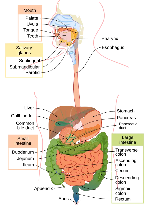
Organs of the digestive system labeled
Anatomy of digestive system
Wikimedia Commons
/
Public Domain

Tiny glands in the mucous membrane lining of the stomach produce digestive enzymes, intrinsic factor, hydrochloric acid, mucus, and bicarbonate. These make up the gastric juice. Gastric juice is continually being made, but the amount made at certain times varies. Certain things trigger more gastric juice to be made, including thoughts of eating, the smell of food, and the presence of food in the mouth and stomach. The smooth muscles in the stomach provide churning and mixing action, which helps combine the gastric juice with the food ingested, creating a mixture called chyme. A continuous coating of mucus protects the stomach and the rest of the digestive system from the acidic nature of chyme and gastric juices.

Besides being a food reservoir and secreting gastric juices, the stomach has additional roles:

  • Producing gastrin, a hormone that regulates the digestive functions
  • Producing ghrelin, which increases the appetite
  • Protecting the body by killing disease-causing bacteria in food
  • Absorbing alcohol, some water, certain drugs, and some fatty acids

The pyloric sphincter is located between the pylorus and the small intestine and regulates the passage of food into the small intestine. When the pyloric sphincter relaxes, the chyme empties out of the stomach and into the small intestine.

Small intestine

The small intestine has about a 1-inch lumen and is about 20 feet long. It loops around and fills most of the abdominal cavity. The small intestine is made up of three parts:

  • Duodenum: Smallest part of the small intestine; connected to the stomach by the pyloric sphincter.
  • Jejunum: Second largest part of the small intestine; connected to the duodenum and the ileum.
  • Ileum: Largest part of the small intestine; connects to the jejunum and the large intestine. It joins the cecum at the ileocecal valve.

The lining of the small intestine has plicae (folds), which contain small projections called villi. The villi are covered by microvilli (epithelial cells that have a brushlike shape). The villi and microvilli increase the surface area of the small intestine and make absorption of nutrients more efficient. Each villus contains a lacteal (lymph vessel) that absorbs lipids (fats) and fat-soluble vitamins. The blood capillary absorbs glucose and amino acids.

Each of the three sections of the small intestine has important roles:

  • Duodenum: Receives chyme from the stomach. Pancreatic enzymes, bile from the liver, and bicarbonate mix with the chyme. Pancreatic enzymes break down chyme. Bile helps break down and absorb fat. Bicarbonate neutralizes the acid from the stomach.
  • Jejunum: Contains larger villi; thus, absorption is the primary function of this section. Sugars, fatty acids, and amino acids are absorbed.
  • Ileum: Limited absorption occurs in this section. Bile acids and vitamin B12 are most often absorbed for reuse in the body.

Chyme from the stomach empties into the duodenum. Peristalsis moves the chyme through the duodenum, jejunum, and the ileum. The ileocecal valve is found between the ileum (of the small intestine) and the cecum (of the large intestine). The ileocecal valve controls the passage of chyme into the cecum and prevents the backflow of chyme into the small intestine.

Large intestine

The large intestine or colon has about a 3-inch lumen and is about 5 feet long. The large intestine is made up of these six sections:

  • Cecum: A 2- to 3-inch pouch or tubelike structure in the lower right abdomen that is considered the first section of the large intestine. The main roles of the cecum include receiving chyme from the small intestine and absorbing fluids and salts. Mucus is also mixed with the chyme in the cecum. Attached to the cecum is the vermiform appendix. The function of the appendix is not entirely known, though research suggests that it harbors “good bacteria,” which will repopulate the intestines after an illness.
  • Ascending colon: The second part of the large intestine, which extends vertically from the cecum to just below the liver.
  • Transverse colon: Extends horizontally from the ascending colon to the descending colon.
  • Descending colon: Extends vertically on the left side of the abdomen from the transverse colon to the sigmoid colon.
  • Sigmoid colon: Forms an S-shaped curve; attaches to the descending colon and the rectum.
  • Rectum: Stores the stool until defecation, or a bowel movement (BM), occurs, and the stool is released through the anus.
  • The watery waste products move from the small intestine into the large intestine. The primary functions of the large intestine include the following:
  • Reabsorbing water and electrolytes: The large intestine has no villi for nutrient absorption but can reabsorb water and electrolytes (e.g., sodium and potassium). The longer the stool is in the colon, the more water is absorbed. The quicker the stool passes through the large intestine, the more water it contains.
  • Making vitamin K: The bacteria in the large intestine make vitamin K, which is used for blood coagulation, bone mineralization, and cardiovascular health.
  • Eliminating waste products from the body.

Bacteria in the colon

The bacteria in the colon can help with the digestion of some materials. This digestive process can cause gas to be formed, and when it is released through the anus, it is called flatus. The bacteria in the colon produce vitamin K and biotin (vitamin B7).

The majority of the colon bacteria are nonpathogenic. The bacteria colonized in the colon (also called gut flora or microbiota) are important for our maturation, immune system development, and metabolism. Cardiovascular disease, metabolic diseases (e.g., diabetes and obesity), and inflammatory bowel disease are some diseases that have been associated with changes in the intestinal bacteria. Antibiotic therapy can also change the delicate microbiota balance in the gut.

Usually, the microbiota prevents Clostridium difficile colonization. With some antibiotic therapies, C. difficile may be allowed to grow, causing intestinal inflammation, diarrhea, and, in some cases, death. C. difficile is spread by the fecal-oral route and is shed in the stool of infected patients. The C. difficile spores have a long survival and are difficult to kill. They are not destroyed by alcohol-based hand sanitizers. C. difficile infections are treated with antibiotics, and the risk of reinfection is high.

Gastroenterology Overview

  • Specialty for digestive system and liver disorders
  • Subspecialties:
    • Proctology: rectum and anus
    • Hepatology: liver
  • GI disorders common in multiple healthcare settings

Anatomy of the Gastrointestinal Tract

  • GI tract = digestive tract = alimentary canal
  • Structures: mouth, pharynx, esophagus, stomach, small intestine, large intestine
  • Accessory organs discussed separately

Mouth

  • Formed by cheeks, lips, tongue, hard/soft palate, uvula
  • 32 permanent teeth set in gums (gingivae)
  • Saliva lubricates food for swallowing

Pharynx

  • Three sections:
    • Nasopharynx: behind nasal cavity
    • Oropharynx: behind mouth; part of respiratory and digestive systems
    • Laryngopharynx: between epiglottis and esophagus

Esophagus

  • Connects pharynx to stomach; behind trachea and heart
  • Lined with mucous membrane; secretes mucus
  • Peristalsis moves food (bolus) to stomach
  • Sphincters:
    • UES: prevents air entry
    • LES (cardiac sphincter): prevents reflux from stomach

Stomach

  • Divided into fundus, body, pylorus
  • Rugae allow expansion
  • Produces gastric juice (enzymes, acid, mucus, bicarbonate)
  • Functions:
    • Churns/mixes food into chyme
    • Produces gastrin (regulates digestion) and ghrelin (stimulates appetite)
    • Kills bacteria, absorbs alcohol, some drugs, fatty acids
  • Pyloric sphincter controls chyme entry into small intestine

Small Intestine

  • About 20 feet long; 1-inch lumen
  • Three parts:
    • Duodenum: receives chyme, mixes with pancreatic enzymes, bile, bicarbonate
    • Jejunum: primary absorption (sugars, fatty acids, amino acids)
    • Ileum: absorbs bile acids, vitamin B12
  • Lining features:
    • Plicae, villi, microvilli increase surface area for absorption
    • Villi contain lacteals (absorb fats) and blood capillaries (absorb glucose, amino acids)
  • Ileocecal valve regulates chyme passage to large intestine

Large Intestine

  • About 5 feet long; 3-inch lumen
  • Sections: cecum (with appendix), ascending colon, transverse colon, descending colon, sigmoid colon, rectum
  • Functions:
    • Reabsorbs water and electrolytes
    • Produces vitamin K (by bacteria)
    • Eliminates waste (feces)
  • Appendix may help repopulate “good bacteria” after illness

Bacteria in the Colon

  • Aid in digestion, produce gas (flatus), synthesize vitamin K and biotin
  • Gut flora important for immune system, metabolism, disease prevention
  • Disruption (e.g., antibiotics) can lead to C. difficile infection
    • C. difficile: causes diarrhea, inflammation; spread fecal-orally; resistant to alcohol sanitizers
    • Treated with antibiotics; high reinfection risk

Sign up for free to take 20 quiz questions on this topic

Previous
Next  | 22.2 The anatomy of the gastrointestinal tract and accessory organs
All rights reserved ©2016 - 2026 Achievable, Inc.

Assisting in gastroenterology

Gastroenterology is the healthcare specialty that deals with most digestive diseases and disorders. A gastroenterologist is a specialist involved in the diagnosis, treatment, and prevention of disorders of the digestive organs and liver. A proctologist is a subspecialist who treats disorders of the rectum and anus. Hepatology is a subspecialty that deals with liver disorders. A hepatologist is a specialist who focuses only on the liver.

Gastrointestinal system disorders are common in the ambulatory care facility. Besides the gastroenterology department, patients with digestive concerns are seen in primary care, pediatrics, internal medicine, and urgent care settings.

The gastrointestinal (GI) system is also called the digestive system. It is made up of the gastrointestinal tract and the accessory organs, which are discussed in the following sections.

Anatomy of the gastrointestinal tract

The gastrointestinal tract is also called the digestive tract and the alimentary canal. It consists of a large, muscular tube that, with the help of hormones and enzymes, digests food. The GI tract starts at the mouth and extends to the anus. It includes the mouth, pharynx (throat), esophagus, stomach, small intestine, and large intestine. Each of these structures is discussed in depth.

Mouth

The cheeks, lips, tongue, hard palate, and soft palate form the mouth (also called the oral cavity or buccal cavity). The roof of the mouth is created by the anterior hard palate and the posterior soft palate. The uvula is a fleshy structure that hangs above the throat, at the back of the soft palate. Adults have 32 permanent teeth that are set in the gums (also called gingivae). Saliva from the salivary glands helps to lubricate the food, making it easier to swallow. The salivary glands will be discussed later in the chapter.

Pharynx

When food or liquid is swallowed, it moves into the pharynx (FAIR inks), or throat. The pharynx is divided into three sections:

  • Nasopharynx: Located behind the nasal cavity
  • Oropharynx: Located behind the mouth; part of the respiratory and digestive systems
  • Laryngopharynx: Located between the epiglottis and the esophagus

Esophagus

The esophagus connects the pharynx to the stomach. This muscular tube runs behind the trachea and the heart. The esophagus is lined with a mucous membrane that secretes mucus, helping the mass of food, or bolus, pass into the stomach. Peristalsis, or the muscular contractions of the esophagus, helps to move the food into the stomach.

A sphincter is located at the top and bottom of the esophagus. When the upper esophageal sphincter (UES) constricts, it prevents air from entering the esophagus. The lower esophageal sphincter (LES), or the cardiac sphincter, is located between the esophagus and the stomach. When the LES constricts, it prevents the stomach contents from moving up the esophagus. When a person swallows, the LES relaxes, allowing the bolus to move into the stomach.

Stomach

The stomach serves as a reservoir for food. The stomach is divided into three sections:

  • Fundus: Top of the stomach, sits just below the diaphragm
  • Body: Main part of the stomach
  • Pylorus: Bottom of the stomach, between the body and the small intestine

The stomach wall contains rugae, which, when unfolded, allow for greater expansion of the stomach size.

Tiny glands in the mucous membrane lining of the stomach produce digestive enzymes, intrinsic factor, hydrochloric acid, mucus, and bicarbonate. These make up the gastric juice. Gastric juice is continually being made, but the amount made at certain times varies. Certain things trigger more gastric juice to be made, including thoughts of eating, the smell of food, and the presence of food in the mouth and stomach. The smooth muscles in the stomach provide churning and mixing action, which helps combine the gastric juice with the food ingested, creating a mixture called chyme. A continuous coating of mucus protects the stomach and the rest of the digestive system from the acidic nature of chyme and gastric juices.

Besides being a food reservoir and secreting gastric juices, the stomach has additional roles:

  • Producing gastrin, a hormone that regulates the digestive functions
  • Producing ghrelin, which increases the appetite
  • Protecting the body by killing disease-causing bacteria in food
  • Absorbing alcohol, some water, certain drugs, and some fatty acids

The pyloric sphincter is located between the pylorus and the small intestine and regulates the passage of food into the small intestine. When the pyloric sphincter relaxes, the chyme empties out of the stomach and into the small intestine.

Small intestine

The small intestine has about a 1-inch lumen and is about 20 feet long. It loops around and fills most of the abdominal cavity. The small intestine is made up of three parts:

  • Duodenum: Smallest part of the small intestine; connected to the stomach by the pyloric sphincter.
  • Jejunum: Second largest part of the small intestine; connected to the duodenum and the ileum.
  • Ileum: Largest part of the small intestine; connects to the jejunum and the large intestine. It joins the cecum at the ileocecal valve.

The lining of the small intestine has plicae (folds), which contain small projections called villi. The villi are covered by microvilli (epithelial cells that have a brushlike shape). The villi and microvilli increase the surface area of the small intestine and make absorption of nutrients more efficient. Each villus contains a lacteal (lymph vessel) that absorbs lipids (fats) and fat-soluble vitamins. The blood capillary absorbs glucose and amino acids.

Each of the three sections of the small intestine has important roles:

  • Duodenum: Receives chyme from the stomach. Pancreatic enzymes, bile from the liver, and bicarbonate mix with the chyme. Pancreatic enzymes break down chyme. Bile helps break down and absorb fat. Bicarbonate neutralizes the acid from the stomach.
  • Jejunum: Contains larger villi; thus, absorption is the primary function of this section. Sugars, fatty acids, and amino acids are absorbed.
  • Ileum: Limited absorption occurs in this section. Bile acids and vitamin B12 are most often absorbed for reuse in the body.

Chyme from the stomach empties into the duodenum. Peristalsis moves the chyme through the duodenum, jejunum, and the ileum. The ileocecal valve is found between the ileum (of the small intestine) and the cecum (of the large intestine). The ileocecal valve controls the passage of chyme into the cecum and prevents the backflow of chyme into the small intestine.

Large intestine

The large intestine or colon has about a 3-inch lumen and is about 5 feet long. The large intestine is made up of these six sections:

  • Cecum: A 2- to 3-inch pouch or tubelike structure in the lower right abdomen that is considered the first section of the large intestine. The main roles of the cecum include receiving chyme from the small intestine and absorbing fluids and salts. Mucus is also mixed with the chyme in the cecum. Attached to the cecum is the vermiform appendix. The function of the appendix is not entirely known, though research suggests that it harbors “good bacteria,” which will repopulate the intestines after an illness.
  • Ascending colon: The second part of the large intestine, which extends vertically from the cecum to just below the liver.
  • Transverse colon: Extends horizontally from the ascending colon to the descending colon.
  • Descending colon: Extends vertically on the left side of the abdomen from the transverse colon to the sigmoid colon.
  • Sigmoid colon: Forms an S-shaped curve; attaches to the descending colon and the rectum.
  • Rectum: Stores the stool until defecation, or a bowel movement (BM), occurs, and the stool is released through the anus.
  • The watery waste products move from the small intestine into the large intestine. The primary functions of the large intestine include the following:
  • Reabsorbing water and electrolytes: The large intestine has no villi for nutrient absorption but can reabsorb water and electrolytes (e.g., sodium and potassium). The longer the stool is in the colon, the more water is absorbed. The quicker the stool passes through the large intestine, the more water it contains.
  • Making vitamin K: The bacteria in the large intestine make vitamin K, which is used for blood coagulation, bone mineralization, and cardiovascular health.
  • Eliminating waste products from the body.

Bacteria in the colon

The bacteria in the colon can help with the digestion of some materials. This digestive process can cause gas to be formed, and when it is released through the anus, it is called flatus. The bacteria in the colon produce vitamin K and biotin (vitamin B7).

The majority of the colon bacteria are nonpathogenic. The bacteria colonized in the colon (also called gut flora or microbiota) are important for our maturation, immune system development, and metabolism. Cardiovascular disease, metabolic diseases (e.g., diabetes and obesity), and inflammatory bowel disease are some diseases that have been associated with changes in the intestinal bacteria. Antibiotic therapy can also change the delicate microbiota balance in the gut.

Usually, the microbiota prevents Clostridium difficile colonization. With some antibiotic therapies, C. difficile may be allowed to grow, causing intestinal inflammation, diarrhea, and, in some cases, death. C. difficile is spread by the fecal-oral route and is shed in the stool of infected patients. The C. difficile spores have a long survival and are difficult to kill. They are not destroyed by alcohol-based hand sanitizers. C. difficile infections are treated with antibiotics, and the risk of reinfection is high.

Key points

Gastroenterology Overview

  • Specialty for digestive system and liver disorders
  • Subspecialties:
    • Proctology: rectum and anus
    • Hepatology: liver
  • GI disorders common in multiple healthcare settings

Anatomy of the Gastrointestinal Tract

  • GI tract = digestive tract = alimentary canal
  • Structures: mouth, pharynx, esophagus, stomach, small intestine, large intestine
  • Accessory organs discussed separately

Mouth

  • Formed by cheeks, lips, tongue, hard/soft palate, uvula
  • 32 permanent teeth set in gums (gingivae)
  • Saliva lubricates food for swallowing

Pharynx

  • Three sections:
    • Nasopharynx: behind nasal cavity
    • Oropharynx: behind mouth; part of respiratory and digestive systems
    • Laryngopharynx: between epiglottis and esophagus

Esophagus

  • Connects pharynx to stomach; behind trachea and heart
  • Lined with mucous membrane; secretes mucus
  • Peristalsis moves food (bolus) to stomach
  • Sphincters:
    • UES: prevents air entry
    • LES (cardiac sphincter): prevents reflux from stomach

Stomach

  • Divided into fundus, body, pylorus
  • Rugae allow expansion
  • Produces gastric juice (enzymes, acid, mucus, bicarbonate)
  • Functions:
    • Churns/mixes food into chyme
    • Produces gastrin (regulates digestion) and ghrelin (stimulates appetite)
    • Kills bacteria, absorbs alcohol, some drugs, fatty acids
  • Pyloric sphincter controls chyme entry into small intestine

Small Intestine

  • About 20 feet long; 1-inch lumen
  • Three parts:
    • Duodenum: receives chyme, mixes with pancreatic enzymes, bile, bicarbonate
    • Jejunum: primary absorption (sugars, fatty acids, amino acids)
    • Ileum: absorbs bile acids, vitamin B12
  • Lining features:
    • Plicae, villi, microvilli increase surface area for absorption
    • Villi contain lacteals (absorb fats) and blood capillaries (absorb glucose, amino acids)
  • Ileocecal valve regulates chyme passage to large intestine

Large Intestine

  • About 5 feet long; 3-inch lumen
  • Sections: cecum (with appendix), ascending colon, transverse colon, descending colon, sigmoid colon, rectum
  • Functions:
    • Reabsorbs water and electrolytes
    • Produces vitamin K (by bacteria)
    • Eliminates waste (feces)
  • Appendix may help repopulate “good bacteria” after illness

Bacteria in the Colon

  • Aid in digestion, produce gas (flatus), synthesize vitamin K and biotin
  • Gut flora important for immune system, metabolism, disease prevention
  • Disruption (e.g., antibiotics) can lead to C. difficile infection
    • C. difficile: causes diarrhea, inflammation; spread fecal-orally; resistant to alcohol sanitizers
    • Treated with antibiotics; high reinfection risk

More from Assisting in gastroenterology

  • The anatomy of the gastrointestinal tract and accessory organs
  • Chronic intestinal disorders, accessory organ diseases, and GI cancers
  • Additional digestive system diseases and disorders
  • Diseases and disorders of the upper GI tract and acute intestinal conditions
  • The medical assistant’s role in examinations, procedures, and treatments