



Diseases and disorders of the upper GI tract and acute intestinal conditions
Diseases & disorders of the digestive system
Disorders can impact the structures of the mouth, including the gums, hard and soft palate, tissues, tongue, teeth, and lips.
This chapter covers the mouth, the esophagus, and the stomach. The acute intestinal conditions, appendicitis among them, are covered in the previous chapter, Additional digestive system diseases and disorders.
Orofacial clefts
Orofacial clefts are one of the most common birth defects in the United States. Orofacial clefts include the following congenital disorders:
- Cleft lip: An opening in the upper lip caused by the lip tissues not completely joining before birth.
- Cleft palate: The tissue that makes up the palate (roof of the mouth) does not completely join before birth. The hard palate makes up the front section, and the soft palate makes up the back section. Cleft palate can affect one or both palates.
Children can have a cleft lip, a cleft palate, or both. Children with clefts can have problems with feeding, speaking, hearing, and ear infections. In most cases, the etiology is unknown, but researchers believe it involves genetics and environmental factors. Research studies have shown that women who smoke, have diabetes, or use certain medications (e.g., valproic acid) during pregnancy have an increased risk of having a baby with an orofacial cleft. The signs of an orofacial cleft are visible at birth. The child may have a split in the lip or the roof of the mouth. The cleft can affect one or both sides of the face.
The provider will identify the cleft right after birth. Sometimes the cleft is detected during a prenatal ultrasound. Treatment involves surgery to improve the child’s ability to eat, speak, and hear. A team of specialists is involved with the treatment of the related complications.
Additional mouth diseases and disorders
Additional diseases and disorders of the mouth include the following:
Esophageal and stomach diseases and disorders
Certain esophageal and stomach diseases and disorders can occur in early childhood, whereas others affect older people. The following sections describe common esophageal and stomach diseases and disorders.
Gastroesophageal reflux disease
Gastroesophageal reflux (GER) occurs when the stomach contents back up into the esophagus, causing acid reflux or heartburn. Gastroesophageal reflux disease (GERD) is more serious and longer lasting than GER. GERD can occur in adults and infants. The presentation in infants is different from that in adults.
The cause of GERD is a weakened or abnormal lower esophageal sphincter. The sphincter may relax when it should not, causing stomach contents to back up into the esophagus. The risk for GERD and GER in adults increases with pregnancy, being overweight, certain medications, and smoking. The most common symptom of GERD in adults is heartburn. Other common GERD symptoms in adults include bad breath, nausea, vomiting, chest pain, upper abdominal pain, respiratory problems, and erosion of the teeth.
A provider will do a history and physical examination. Diagnostic procedures include an upper endoscopy, esophageal manometry, ambulatory acid probe test, and an upper-digestive system x-ray. Treatment is based on the severity of the symptoms and can include lifestyle changes, medications, or surgery. Usually, patients are encouraged to reduce their intake of foods and drinks that make the symptoms worse.
GER and GERD in infants
Infants can have gastroesophageal reflux (GER) and gastroesophageal reflux disease (GERD). The cause of GER and GERD in infants is an immature (not fully developed) lower esophageal sphincter. The sphincter becomes weak or relaxed, and stomach contents back up into the esophagus.
The main sign of GERD in infants is spitting up more than normal. Additional signs include the following:
- Arching of the back during or right after feedings
- Colic, coughing, trouble breathing, wheezing, gagging, and projectile or forceful vomiting
- Poor feeding and refusal to feed
- Poor growth and weight gain; malnutrition
Treatment for GERD focuses on feeding changes and medications. Rice cereal may be added to the milk. Holding the baby upright for 30 minutes after feeding and avoiding overfeeding the child may be encouraged. H2 blockers or proton-pump inhibitors may be given to reduce acid production. Surgery may be recommended in severe cases. Most infant GERD resolves by 9 to 18 months of age.
Hiatal hernia
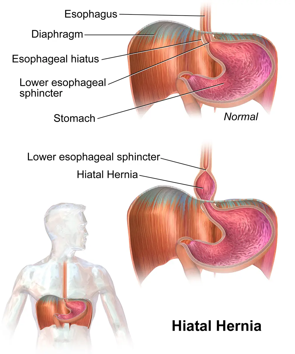
A hiatal hernia occurs when a section of the upper stomach pushes through an opening of the diaphragm into the chest.
The cause is unknown. The risk of hiatal hernias increases with age, obesity, and smoking. Often people over 50 years of age experience this condition. The signs and symptoms include chest pain, heartburn, and difficulty swallowing.
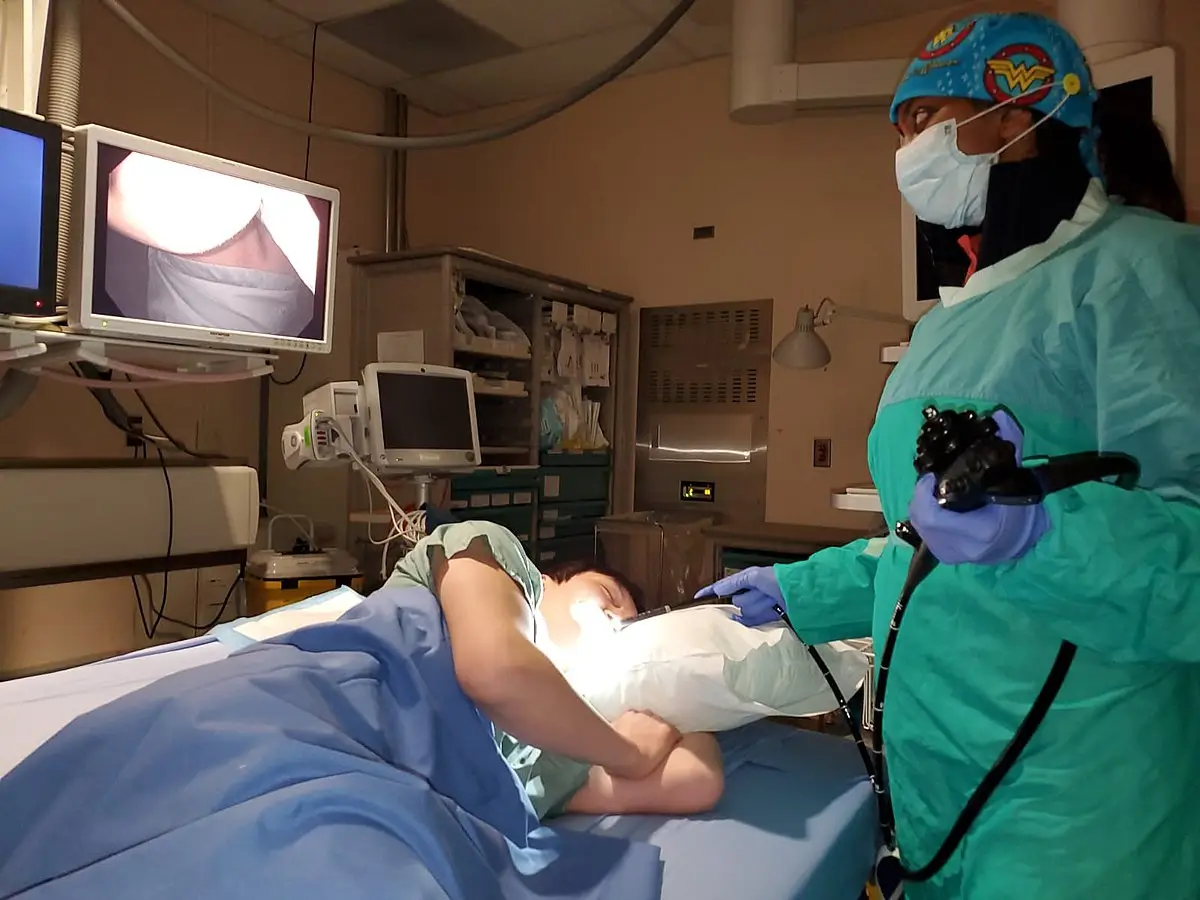
The provider will order an upper GI (UGI) series (also called a barium swallow) and an esophagogastroduodenoscopy (EGD) to help diagnose the condition. The goal of treatment is to relieve symptoms and prevent complications. Treatment includes medications for acid reflux and surgery to repair the hernia. The person should avoid alcohol and use medications with care.
Peptic ulcers
Peptic ulcers are also known as duodenal, gastric, and stomach ulcers. A peptic ulcer is a sore or breakdown in the lining of the stomach or duodenum. Gastric ulcers occur in the stomach, and duodenal ulcers occur in the first section of the small intestine.
The most common etiology for peptic ulcers is an infection of the stomach by Helicobacter pylori (H. pylori) bacteria. The risk for peptic ulcers may increase with drinking too much alcohol, regular use of aspirin or nonsteroidal anti-inflammatory drugs (NSAIDs) (e.g., ibuprofen and naproxen), using cigarettes and chewing tobacco, radiation treatments, and stress. Zollinger-Ellison syndrome can also cause peptic ulcers. Signs and symptoms include upper abdominal pain at night or when the stomach is empty (1 to 3 hours after a meal). Additional symptoms include nausea, melena, chest pain, fatigue, vomiting and hematemesis, and weight loss.
After a physical exam, the provider may order an upper GI (UGI) endoscopy or a UGI series to visibly examine the lining of the esophagus, stomach, and duodenum. Testing for H. pylori requires a biopsy of the stomach lining or a urea breath test. A stool occult blood test and a hemoglobin test may also be done. Treatment is based on the reasons for the ulcer. If H. pylori bacteria are present, the patient may need to take medication, such as antibiotics and an H2 blocker or a proton-pump inhibitor (PPI) (e.g., omeprazole [Prilosec], lansoprazole [Prevacid], or esomeprazole [Nexium]). Additional ulcer treatment may include a lining protectant, such as sucralfate.
Pyloric stenosis
Pyloric stenosis is the narrowing of the pylorus, the muscular opening between the stomach and the small intestine. It occurs more often in males than females and in infants younger than 6 months.
The exact etiology is unknown, but there is a genetic link. Other risk factors include taking certain antibiotics, hyperacidity in the duodenum, and diseases such as type 1 diabetes mellitus. Vomiting is the most common sign. It can occur after feedings, may be projectile (forceful), and usually starts around 3 weeks of age. Other signs and symptoms include weight loss, constant hunger, dehydration, burping, abdominal pain, and a wavelike motion of the abdomen just before vomiting. Upon physical exam, the provider may feel an olive-sized mass in the upper abdomen.
The provider will order an abdominal ultrasound and possibly a UGI series to detect the stenosis, in addition to blood tests to identify electrolyte imbalances. Treatment depends on the severity of the stenosis and may include surgery to widen the pylorus (pyloromyotomy). If surgery cannot be done, an endoscope is inserted in the upper GI tract, and a balloon is inflated to widen the pylorus. Medications or a feeding tube may also be used to relax the pylorus.